Axa Assurance Maroc - Insurer Innovation Award 2024
Bio lab act1
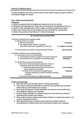
1. Activity on MITOTIC CELLS
In this investigation, you will examine cells of onion (Allium cepa) root tips to observe
the different stages of mitosis.
Part I. Observing Dividing Cells
Procedure:
1. Obtain a prepared slide of longitudinal sections of an onion root tip.
2. Using the scanning objective, locate the zone of division preceding the root cap.
3. Look for cells undergoing the various stages of division using the key below.
4. Show your teacher some of the stages you have identified for confirmation.
5. Answer the questions that follow on a sheet of pad paper.
KEY TO IDENTIFYING CELL DIVISION STAGE
A. Nuclear membrane and nucleolus visible
B. Chromosomes not visible
C. Cell is of normal size. Interphase
C. Two adjacent cells, smaller in size than
most of the other cells, separated by a thin line. 2– daughter cell phase
B. Chromosomes are visible as long and slender threads. Early prophase
A. Nuclear membrane and nucleolus lacking.
D. cell contains one set of chromosomes
E. Chromosomes loosely arranged in the center of the cell. Late prophase
E. Chromosomes are arranged on the equatorial plate. Metaphase
D. cell contains two sets of chromosomes
F. Two sets of chromatids widely separated, nearer to
either pole of the cell, farther from the equator.
G. No visible partial line between the two sets of Late anaphase
widely separated chromatids.
G. Center of the cell shows the beginning of a Telophase
cell plate (partial line); nuclear membrane starting
to reform.
F. Two sets of chromatids only slightly separated Early Anaphase
Analyze and Conclude:
1. Why were plant root tip cells used for viewing cell division?
2. If you were to observe cell division of animals, what specific type of animal
cells would you use? Explain your reasons.
3. How would cell division in plant cells differ from that of animal cells? Use a
Venn diagram organize your response.
4. At what stage of mitosis is it easiest to determine the kind and number of
chromosomes? Explain your reasons.
5. What evidence did you observe that shows mitosis is a continuous process, not
a series of separate events?
6. What specific process in mitosis ensures that the normal number of
chromosomes is restored after each cell division? Explain your response.
MITOTIC CELLS Activity biology 3 /page 1
2. Part II. Determine the Frequency of Cell Division
Procedure:
1. Count 20 adjacent cells in the meristematic region of the onion root and record
whether the cells are in interphase or division phase. On a sheet of pad paper,
record the number of cells in interphase and the number of cells that are
actively dividing.
2. Calculate the percentage of cells that are dividing using the following formula:
Number of cells dividing × 100
= ___% dividing
Total number of cells counted
3. Create a circle graph showing the percentage of cells in division phase and
percentage of cells in interphase. Label the diagrams appropriately.
Part III. Creating a Cell-Division Clock
Procedure:
1. Under HPO, locate 50 onion root cells that are dividing. Do not include cells that are
between divisions. Identify the phase of mitosis each cell is in. Record the
number of cells in each phase.
2. Calculate the percentage of cells that are in each of these four phases: prophase,
metaphase, anaphase, and telophase.
3. Construct a circle graph showing the percentage of cells in each phase of mitosis.
Include labels and titles.
4. If it takes 12 h for onions to complete one cycle of mitosis, determine the time
spent in each phase. Include this information in your circle graphs.
Synthesize and Conclude
1. The number of animal cells in each phase of mitosis is recorded in the table
below. If the time taken to complete one cycle of mitosis was 15h, create a
cell-division clock to represent the data.
Mitotic phase Number of cells in phase
prophase 15
metaphase 20
anaphase 10
telophase 5
2. Do your observations on onion root tip cells indicate that there were more cells
in some phases than in others? Identify the most common phase/s and
explain what these differences in numbers might mean.
MITOTIC CELLS Activity biology 3 /page 2