
Recommended
More Related Content
Similar to BILT 215 Gram staining and other Classification Criteria.pdf
Similar to BILT 215 Gram staining and other Classification Criteria.pdf (20)
Recently uploaded
Recently uploaded (20)
BILT 215 Gram staining and other Classification Criteria.pdf
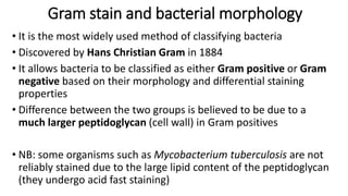
- 1. Gram stain and bacterial morphology • It is the most widely used method of classifying bacteria • Discovered by Hans Christian Gram in 1884 • It allows bacteria to be classified as either Gram positive or Gram negative based on their morphology and differential staining properties • Difference between the two groups is believed to be due to a much larger peptidoglycan (cell wall) in Gram positives • NB: some organisms such as Mycobacterium tuberculosis are not reliably stained due to the large lipid content of the peptidoglycan (they undergo acid fast staining)
- 2. Gram staining - Principles ▪ Gram-positive organisms contain a highly cross-linked layer of peptidoglycan ▪ They retain the primary dye, crystal violet (CV), following the application of the mordant, iodine (I). ▪ The iodine and crystal violet form a complex within the peptidoglycan. ▪ When decolourizer is applied to the cells, the CV-I complex remains within the cell, making it appear dark purple to blue
- 4. Gram staining - Principles ▪ The gram-negative organisms do not contain a thick cross-linked layer of peptidoglycan ▪ The peptidoglycan is loosely distributed between the inner cell and the outer cell membranes ▪ Following the application of the crystal violet and iodine, the CV-I complexes are not trapped within the peptidoglycan ▪ Application of the acid-alcohol decolourizer dehydrates the outer cellular membrane, leaving holes in the membrane and effectively washing or removing the CV-I complex from the cells ▪ The cells appear colourless. ▪ To make the colourless cells visible, a secondary stain, safranin, is applied, leaving the gram-negative cells pink
- 5. Dyes employed in Gram-staining • Crystal violet –violet in colour • Grams Iodine or Lugol’s iodine (mordant) • Decolourizer- (Alcohol/Acetone) • Counterstain –red in colour (Safranin/Fuchsin)
- 6. Gram Staining Steps • Crystal violet acts as the primary stain. • Gram’s iodine acts as a mordant (Helps to fix the primary dye to the cell wall) •Decolorizer is used next to remove the primary stain (crystal violet) from Gram Negative bacteria • Decolorizer is composed of an organic solvent, such as, acetone or ethanol or a combination of both.) • Finally, a counter stain (Safranin), is applied to stain those cells (Gram Negative) that have lost the primary stain as a result of decolorization
- 7. Procedure • Crystal violet – 1 min - wash. • Iodine – 1 min – wash. • Acetone add drop by drop and watch out colour comes out – wash immediately. • Safranine/dilute carbol fuchsin – 1 min- wash. • Allow to dry – examine under microscope. • Note: Results should be confirmed only with 100x
- 9. Video illustration of Gram Staining
- 11. Gram positive cocci in clusters
- 12. Gram positive cocci in chains
- 16. Examples • Gram positive cocci in clusters: • 1. Staphylococci species. • Gram negative cocci in chains: • 1. Streptococci species. • Gram negative cocci: • 1. Neisseria species. • Gram negative bacilli: • 1. Escherichia coli • 2. Klebsiella pneumoniae • Gram positive bacilli: • 1. Clostridium species. • 2. Corynebacterium species. • 3. Bacillus anthracis.
- 17. Classification of bacteria based on growth requirements pH, Temperature, salt concentration and oxygen
- 18. pH • Many bacteria grow best at neutral pH (pH 7.2-7.6) • Some specialized bacteria can survive and even grow in acid or alkaline conditions. • T.B. (Tubercule Bacillus) pH 6.5-6.8 • V. cholerae pH 8.4-9.2 • Neutrophiles ( pH 6-8) • Acidophiles ( pH 1-5) • Alkalophiles ( pH 9-11) 1/30/2024 R. YEBOAH 18
- 19. 30/01/2024 R. YEBOAH 19
- 20. Four categories of microbes based on temperature ranges for growth Psychrophiles Mesophiles Thermophiles Hyperthermophiles Growth rate Temperature (°C)
- 21. • Halophilic organisms thrive in high salt concentrations. • Halophilic organisms can be divided into: - Obligate – requiring a high salt concentration - Extreme – requiring very high salt concentration - Facultative – can grow either with or without high salt levels Salt concentration 30/01/2024 R. YEBOAH 21
- 22. Oxygen requirements Obligate aerobes: can only survive in the presence of oxygen Microaerophiles: grow better in low concentrations of oxygen Aerotolerant anaerobes: can survive and grow in the absence of oxygen, but can also tolerate the presence of oxygen Facultative anaerobes: These organisms grow equally well in aerobic or anaerobic environments. In the absence of oxygen, they can switch to anaerobic respiration Obligate anaerobes: can only survive in the absence of oxygen *Capnophilic (or carbon dioxide–loving) bacteria require increased concentration of carbon dioxide (5% – 10%) and approximately 15% oxygen
- 23. Oxygen requirements • Aerobes • Bacillus subtilis, Pseudomonas aeruginosa, Mycobacteri um tuberculosis e • Anaerobes • Clostridium, Bacteroides • Facultative anaerobes • E. coli, S. aureus • Aerotolerant anaerobes • Lactobacilli, streptococci, campylobacter jejuni • Microaerophiles • Borrelia burgdorferi 30/01/2024 R. YEBOAH 23
- 24. Aerobic and anaerobic bacteria can be identified by growing them in liquid culture: 1: Obligate aerobic bacteria gather at top of test tube to absorb maximal amount of oxygen 2: Obligate anaerobic bacteria gather at bottom to avoid oxygen 3: Facultative anaerobes gather mostly at the top, since aerobic respiration is most beneficial; but as lack of oxygen does not hurt them, they can be found all along the test tub 4: Microaerophiles gather at upper part of test tube, not at top. Require O2, but at low concentration 5: Aerotolerant bacteria are not affected by oxygen, and they are evenly spread along the test tube. 30/01/2024 R. YEBOAH 24 Oxygen requirements
- 25. Biochemical reactions • Microbiologists can identify a pathogen in a sample, purify the microorganism by plating a single colony of the microorganism on a separate plate, and then perform a series of biochemical studies to identify the bacterial species • Examples include: • Catalase test • Citrate utilisation test • Coagulase test • Oxidase test • Indole test • Urease test • CAMP test, etc
- 26. Serologic systems • Selected antisera can be used to classify different bacterial species • This may be based on either carbohydrate or protein antigens from the bacterial cell wall or the capsular polysaccharide • (Group A streptococcal M proteins or O and H polysaccharide antigens of salmonella)
- 27. Environmental Reservoirs • Environmental reservoirs are generally divided into those that are endogenous (i.e., on or within the human body) and • Exogenous (somewhere in the environment). • The human body provides bacteria with the following: • Optimum osmotic pressure • Optimum temperature range • Optimum pH range • The human body is therefore an excellent incubator for pathogens.
- 28. Genotypic systems • Genotypic bacterial typing is a method of identifying and characterizing bacterial strains based on their genetic makeup • This can be done by analyzing DNA sequences of specific genes, such as the 16S rRNA gene, or by comparing the entire genome of the bacteria • There are several genotypic typing methods available, including • Pulsed-field gel electrophoresis (PFGE) • Multilocus sequence typing (MLST) • Whole genome sequencing (WGS)
- 29. Open System vs. Closed System • Open System – Organisms that grow in nature. – Nutrients replenished and wastes removed. • Closed System – In the lab (i.e. agar plates, broth tubes). – Nutrients will run out and wastes are not removed. 30/01/2024 R. YEBOAH 29