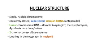
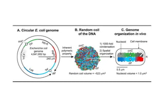
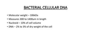
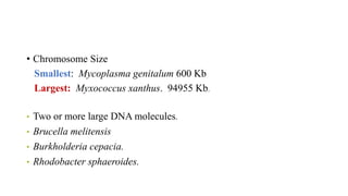
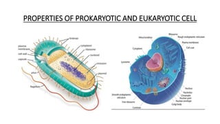
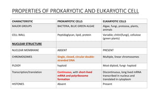
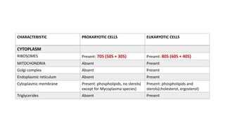
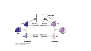
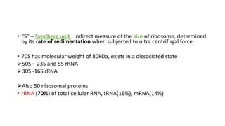
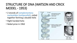
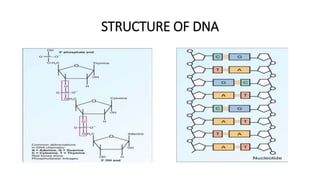
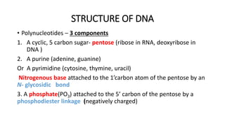
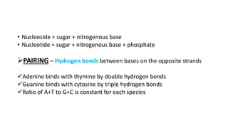
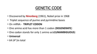
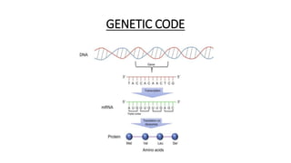
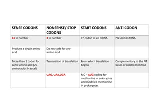
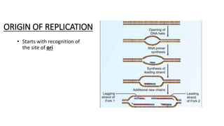
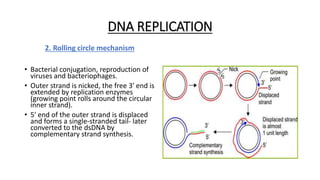
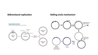
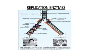
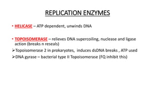
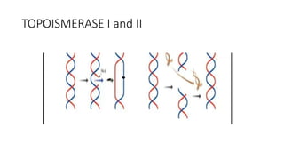
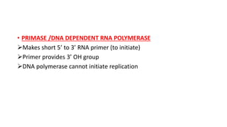
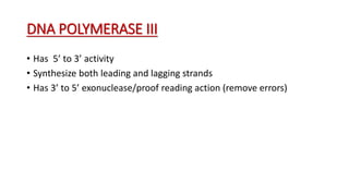
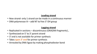
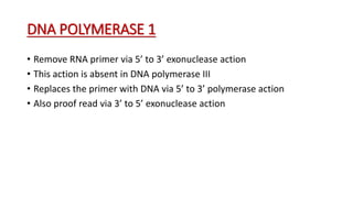
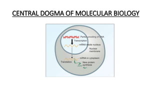
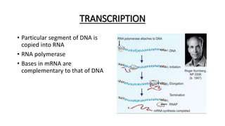
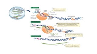
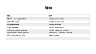
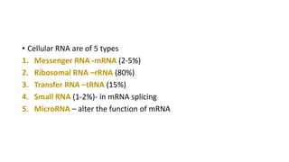
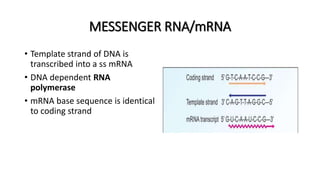
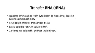
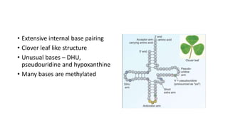
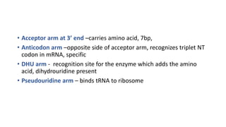
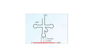
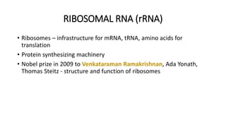
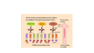
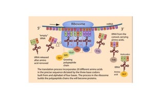
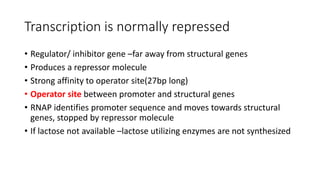
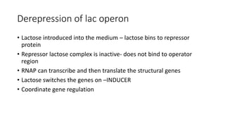
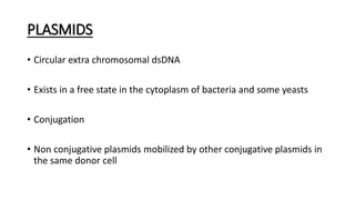
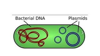
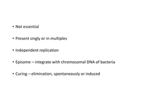
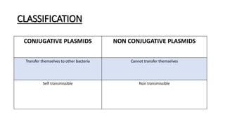
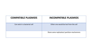
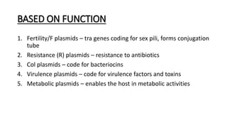
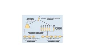
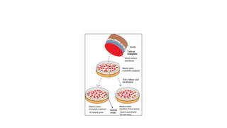
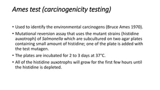
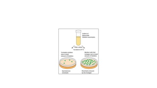
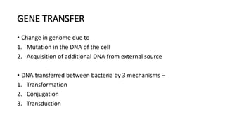
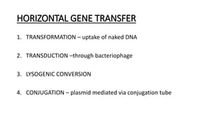
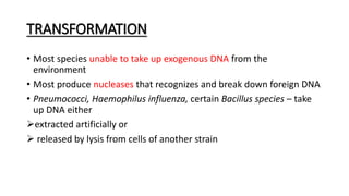
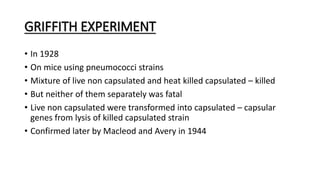
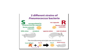
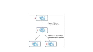
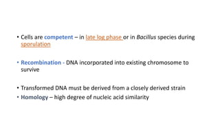
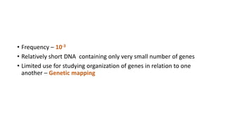
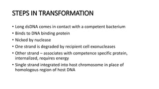
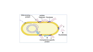
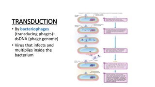
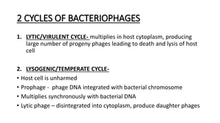
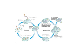
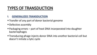
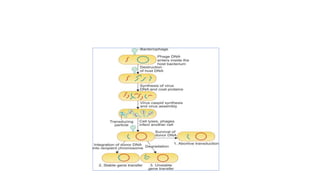
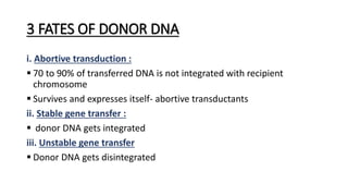
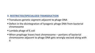
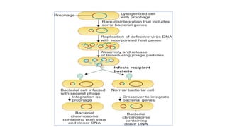
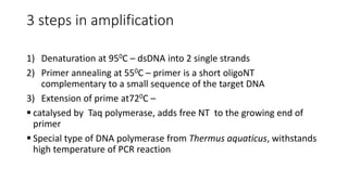
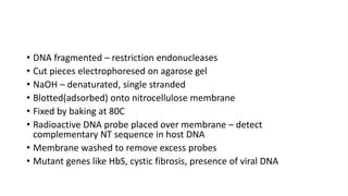
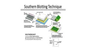
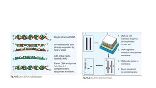
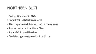
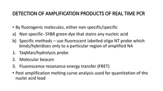
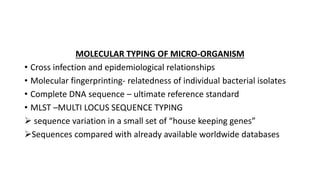
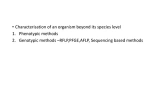
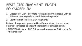
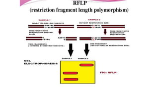
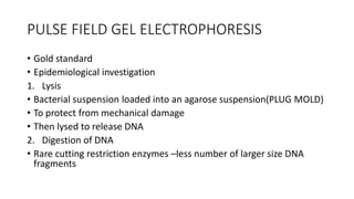
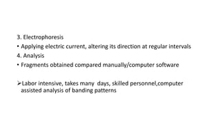
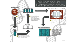
BACTERIAL GENETICS
NUCLEAR STRUCTURE
• Single, haploid chromosome
• covalently closed, supercoiled, circular dsDNA (anti parallel)
• Linear chromosomal DNA – Borrelia burgdorferi, the streptomyces,
Agrobacterium tumefaciens
• 2 chromosomes- Vibrio cholerae
• Lies free in the cytoplasm in nucleoid
BACTERIAL CELLULAR DNA
• Molecular weight – 106kDa
• Measures 300 to 1400um in length
• Nucleoid – 10% of cell volume
• DNA – 2% to 3% of dry weight of the cell
• Chromosome Size
Smallest: Mycoplasma genitalum 600 Kb
Largest: Myxococcus xanthus. 94955 Kb.
• Two or more large DNA molecules.
• Brucella melitensis
• Burkholderia cepacia.
• Rhodobacter sphaeroides.
PROPERTIES OF PROKARYOTIC AND EUKARYOTIC CELL
CHARACTERISTIC PROKARYOTIC CELLS EUKARYOTIC CELLS
MAJOR GROUPS BACTERIA, BLUE GREEN ALGAE Algae, fungi, protozoa, plants,
animals
CELL WALL Peptidoglycan, lipid, protein Variable; chitin(fungi), cellulose
(green plants)
NUCLEAR STRUCTURE
NUCLEAR MEMBRANE ABSENT PRESENT
CHROMOSOMES Single, closed, circular double-
stranded DNA
Multiple, linear chromosomes
PLOIDY haploid Most diploid, fungi- haploid
Transcription/translation Continuous, with short-lived
mRNA and polyribosome
formation
Discontinuous, long lived mRNA
transcribed in nucleus and
translated in cytoplasm
HISTONES Absent Present
CHARACTERISTIC PROKARYOTIC CELLS EUKARYOTIC CELLS
CYTOPLASM
RIBOSOMES Present: 70S (50S + 30S) Present: 80S (60S + 40S)
MITOCHONDRIA Absent Present
Golgi complex Absent Present
Endoplasmic reticulum Absent Present
Cytoplasmic membrane Present: phospholipids, no sterols(
except for Mycoplasma species)
Present: phospholipids and
sterols(cholesterol, ergosterol)
Triglycerides Absent Present
• “S” – Svedberg unit : indirect measure of the size of ribosome, determined
by its rate of sedimentation when subjected to ultra centrifugal force
• 70S has molecular weight of 80kDa, exists in a dissociated state
50S – 23S and 5S rRNA
30S -16S rRNA
Also 50 ribosomal proteins
• rRNA (70%) of total cellular RNA, tRNA(16%), mRNA(14%)
STRUCTURE OF DNA (WATSON AND CRICK
MODEL - 1953)
• 2 strands of complementary
nucleotides (antiparallel), coiled
together forming a double helix
• Right handed helix
• Nobel prize in 1962
STRUCTURE OF DNA
STRUCTURE OF DNA
• Polynucleotides – 3 components
1. A cyclic, 5 carbon sugar- pentose (ribose in RNA, deoxyribose in
DNA )
2. A purine (adenine, guanine)
Or A pyrimidine (cytosine, thymine, uracil)
Nitrogenous base attached to the 1’carbon atom of the pentose by an
N- glycosidic bond
3. A phosphate(PO3) attached to the 5’ carbon of the pentose by a
phosphodiester linkage (negatively charged)
• Nucleoside = sugar + nitrogenous base
• Nucleotide = sugar + nitrogenous base + phosphate
PAIRING – Hydrogen bonds between bases on the opposite strands
Adenine binds with thymine by double hydrogen bonds
Guanine binds with cytosine by triple hydrogen bonds
Ratio of A+T to G+C is constant for each species
GENETIC CODE
• Discovered by Nirenberg (1961), Nobel prize in 1968
• Triplet sequence of purine and pyrimidine bases
• On mRNA - TRIPLET CODON
• One amino acid has more than 1 codon (DEGENERATE)
• One codon stands for only 1 amino acid(UNAMBIGUOUS)
• Universal
• 64 (43 )in total
GENETIC CODE
SENSE CODONS NONSENSE/ STOP
CODONS
START CODONS ANTI CODON
61 in number 3 in number 1st codon of an mRNA Present on tRNA
Produce a single amino
acid
Do not code for any
amino acid
More than 1 codon for
same amino acid (20
amino acids in total)
Termination of translation From which translation
begins
Complementary to the NT
bases of codon on mRNA
UAG, UAA,UGA MC – AUG coding for
methionine in eukaryotes
and modified methionine
in prokaryotes
DNA REPLICATION
1. Bidirectional replication
• begins at a single point, the origin.
• DNA helix unwinds at replication fork- DNA
synthesis, individual strands are replicated.
• Two replication forks move outwards from
the origin - until they have copied the whole
replicon and a structure shaped like the
Greek letter theta (θ) is formed.
• Since the forks meet on the other side, the
two chromosomes are separated.
• e.g. in E.coli
ORIGIN OF REPLICATION
• Starts with recognition of
the site of ori
DNA REPLICATION
2. Rolling circle mechanism
• Bacterial conjugation, reproduction of
viruses and bacteriophages.
• Outer strand is nicked, the free 3′ end is
extended by replication enzymes
(growing point rolls around the circular
inner strand).
• 5′ end of the outer strand is displaced
and forms a single-stranded tail- later
converted to the dsDNA by
complementary strand synthesis.
Bidirectional replication Rolling circle mechanism
REPLICATION ENZYMES
REPLICATION ENZYMES
• HELICASE – ATP dependent, unwinds DNA
• TOPOISOMERASE – relieves DNA supercoiling, nuclease and ligase
action (breaks n reseals)
Topoisomerase 2 in prokaryotes, induces dsDNA breaks , ATP used
DNA gyrase – bacterial type II Topoisomerase (FQ inhibit this)
TOPOISMERASE I and II
• PRIMASE /DNA DEPENDENT RNA POLYMERASE
Makes short 5’ to 3’ RNA primer (to initiate)
Primer provides 3’ OH group
DNA polymerase cannot initiate replication
DNA POLYMERASE III
• Has 5’ to 3’ activity
• Synthesize both leading and lagging strands
• Has 3’ to 5’ exonuclease/proof reading action (remove errors)
Leading strand
• New strand- only 1 strand can be made in a continuous manner
• DNA polymerase III – add NT to free 3’ OH group
Lagging strand
• Replicated in sections – discontinuous (OKAZAKI fragments) ,
• Synthesized on 5’ to 3’ parent strand
• 3’ end is not available for primer synthesis
• Takes open 3’ end for primer synthesis
• Annealed by DNA ligase by making phosphodiester bond
DNA POLYMERASE 1
• Remove RNA primer via 5’ to 3’ exonuclease action
• This action is absent in DNA polymerase III
• Replaces the primer with DNA via 5’ to 3’ polymerase action
• Also proof read via 3’ to 5’ exonuclease action
CENTRAL DOGMA OF MOLECULAR BIOLOGY
TRANSCRIPTION
• Particular segment of DNA is
copied into RNA
• RNA polymerase
• Bases in mRNA are
complementary to that of DNA
RNA
RNA DNA
Mainly seen in cytoplasm Mostly inside nucleus
100-5000 bases Millions of base pairs
Single stranded Double stranded
Sugar is ribose Sugar is deoxyribose
Purines – adenine, guanine
Pyrimidines – uracil, cytosine
Purines – adenine, guanine
Pyrimidines – thymine, cytosine
Easily destroyed by alkali Alkali resistant
• Cellular RNA are of 5 types
1. Messenger RNA -mRNA (2-5%)
2. Ribosomal RNA –rRNA (80%)
3. Transfer RNA –tRNA (15%)
4. Small RNA (1-2%)- in mRNA splicing
5. MicroRNA – alter the function of mRNA
MESSENGER RNA/mRNA
• Template strand of DNA is
transcribed into a ss mRNA
• DNA dependent RNA
polymerase
• mRNA base sequence is identical
to coding strand
Transfer RNA (tRNA)
• Transfer amino acids from cytoplasm to ribosomal protein
synthesizing machinery
• RNA polymerase III transcribes tRNA
• Easily soluble –sRNA/ soluble RNA
• 73 to 93 NT in length, shorter than mRNA
• Extensive internal base pairing
• Clover leaf like structure
• Unusual bases – DHU,
pseudouridine and hypoxanthine
• Many bases are methylated
• Acceptor arm at 3’ end –carries amino acid, 7bp,
• Anticodon arm –opposite side of acceptor arm, recognizes triplet NT
codon in mRNA, specific
• DHU arm - recognition site for the enzyme which adds the amino
acid, dihydrouridine present
• Pseudouridine arm – binds tRNA to ribosome
RIBOSOMAL RNA (rRNA)
• Ribosomes – infrastructure for mRNA, tRNA, amino acids for
translation
• Protein synthesizing machinery
• Nobel prize in 2009 to Venkataraman Ramakrishnan, Ada Yonath,
Thomas Steitz - structure and function of ribosomes
TRANSLATION
• mRNA is decoded by ribosome to produce a specific amino acid
chain/polypeptide
• In cytoplasm
• 4 phases
1. Initiation –ribosome assembles around target of mRNA, 1st tRNA is
attached at the start codon of mRNA
2. Elongation – tRNA transfers an aminoacid to the adjacent tRNA,
corresponding to next codon
3. Translocation – ribosome moves to the next mRNA codon to
continue the process, creating amino acid chain
4. Termination – when stop codon is reached, ribosome releases the
polypeptide
OPERON CONCEPT OF GENE REGULATION
• Francois Jacob and Jacques Monod – 1961
• Nobel prize in 1965
• Lactose metabolism in E.coli
• Cells grown in glucose medium do not contain beta galactosidase
(lactase)
• When cells transferred to medium containing lactose- enzyme level
increases several threshold
• Lactose metabolism regulated by induction/derepression of genes
Lac operon
Lac operon
• Operon – unit of gene expression, includes structural genes, control
elements, regulator/inhibitor gene, promoter and operator areas
• Z gene – encodes beta-galactosidase (hydrolyses lactose to galactose and
glucose)
• Y gene – produce permease, transports lactose and galactose into cell
• A gene – code for thiogalactoside transacetylase
• Z, Y, A- Structural genes, contiguous segments of DNA
• Transcription starts from PROMOTER(P), transcribes these structural genes
as a single mRNA
Transcription is normally repressed
• Regulator/ inhibitor gene –far away from structural genes
• Produces a repressor molecule
• Strong affinity to operator site(27bp long)
• Operator site between promoter and structural genes
• RNAP identifies promoter sequence and moves towards structural
genes, stopped by repressor molecule
• If lactose not available –lactose utilizing enzymes are not synthesized
Derepression of lac operon
• Lactose introduced into the medium – lactose bins to repressor
protein
• Repressor lactose complex is inactive- does not bind to operator
region
• RNAP can transcribe and then translate the structural genes
• Lactose switches the genes on –INDUCER
• Coordinate gene regulation
PLASMIDS
• Circular extra chromosomal dsDNA
• Exists in a free state in the cytoplasm of bacteria and some yeasts
• Conjugation
• Non conjugative plasmids mobilized by other conjugative plasmids in
the same donor cell
• Not essential
• Present singly or in multiples
• Independent replication
• Episome – integrate with chromosomal DNA of bacteria
• Curing – elimination, spontaneously or induced
CLASSIFICATION
CONJUGATIVE PLASMIDS NON CONJUGATIVE PLASMIDS
Transfer themselves to other bacteria Cannot transfer themselves
Self transmissible Non transmissible
COMPATIBLE PLASMIDS INCOMPATIBLE PLASMIDS
Can exist in a bacterial cell Either one would be lost from the cell
Share same replication/ partition mechanisms
BASED ON FUNCTION
1. Fertility/F plasmids – tra genes coding for sex pili, forms conjugation
tube
2. Resistance (R) plasmids – resistance to antibiotics
3. Col plasmids – code for bacteriocins
4. Virulence plasmids – code for virulence factors and toxins
5. Metabolic plasmids – enables the host in metabolic activities
MUTATION
• Bacteria reproduce by asexual binary fission
• Progeny having identical genome
• For evolution – occasional variations in gene replication
• Heritable mutations passed on stably to subsequent generations
• Eg: acquired antibiotic resistance
• Frequency – between one per 104 and one per 1010 cell divisions
• Random, undirected heritable variation caused by a change in the
nucleotide sequence of the genome of the cell
OCCURENCE OF MUTATIONS
• 2 ways
1. Spontaneous mutations - naturally, without adding any mutagen
2. Induced mutations – when exposed to a mutagen
a. Physical agents : UV radiations
b. Chemical agents : alkylating agents, acridine dyes, 5 bromo uracil
CLASSIFICATION OF MUTATIONS
1. SUBSTITUTION – most common
a) Transition – purine to purine/pyrimidine to pyrimidine
b) Transversion – purine to pyrimidine and vice versa
2. DELETION
a) Large gene deletion- alpha thalassemia(entire), thalassemia (partial)
b) Codon – cystic fibrosis (one amino acid 508th phenyl alanine missing)
c) Single base – frame shift mutations
3. INSERTION
a) Single base – frameshift mutations
b) Trinucleotide expansions – Huntington’s chorea(CAG)
c) Duplications
TYPES OF MUTATIONS
PHENOTYPIC VARIATION
• Phenotype – properties of a bacterial cell at a particular time
• Depends on genome (genotype) and the environment
• Expression of genes is changed in response to environment
• Not a form of mutation
• Synthesis of flagella, expression of certain enzymes, etc.
GENOTYPIC MUTATIONS PHENOTYPIC VARIATIONS
Heritable Reversible
Maintained through changes in
environment
Depends on environmental
conditions
TYPES OF MUTATIONS
MULTISITE MUTATIONS POINT MUTATIONS
Extensive chromosomal rearrangements One or very few nucleotides
Inversions, duplications, deletions Substitution ,deletion, insertion
EFFECTS OF MUTATIONS
AT CODON LEVEL
Silent mutation New codon codes for same amino acid
Neutral mutation New codon forms different but functionally same
amino acid
Missense mutation New codon for a different amino acid
Nonsense mutation New codon is a stop codon causing termination
Frame shift mutations
Conditional mutations
FRAMESHIFT MUTATIONS
• Addition/deletion of bases
• Reading frame shifts
• Garbled( completely irrelevant
protein ) with altered amino acid
REVERSE MUTATIONS
• 2nd mutation that nullifies the effect of 1st mutation
• Gain back function of the wild phenotype
1. True reversion –mutant to wild type
2. Equivalent reversion – different codon for the same amino acid of
wild type
3. Suppressor mutation – 2nd mutation in a different gene that reverts
the phenotypic effects of an already existing mutation
MANIFESTATIONS OF MUTATIONS
• LETHAL MUTATIONS – incompatible with life
Eg: 4 gene deletion leads to IUD
• SILENT MUTATIONS
• BENEFICIAL MUTATIONS – rare
• CARCINOGENIC EFFECT – all carcinogens are mutagens
EFFECTS OF MUTATIONS
• Sensitivity to bacteriophages
• Loss of ability to produce capsule or flagella/ virulence
• Alteration in colony morphology/ pigment production
• Drug susceptibility
• Biochemical reactions
• Antigenic structure
DETECTION AND ISOLATION OF MUTANTS
• Gene sequencing
• Phenotypic methods
1. Fluctuation test – spontaneous mutations
2. Replica plating method – auxotrophic mutants
3. Ames test- carcinogenicity of a mutant
Fluctuation test
• Demonstrates the spontaneous mutation in bacteria (Luria and
Delbruck -1943).
• When bacterial suspension is subjected to selective pressure by
subculturing on to agar plate containing a growth limiting substance
(e.g. streptomycin or bacteriophage etc.),they undergo spontaneous
mutation.
• Rate of mutation vary widely(some bacteria mutate early, some late)
which leads to fluctuations.
• Fluctuations in mutation are wide when small volume sub cultures
are made (more frequent mutations) as compared to large volume
subcultures (less frequent mutations).
This experiment was not widely appreciated, probably due to the
complicated statistical evaluation.
Replica plating method
• Detects auxotrophic mutants (Lederberg -1952).
• Differentiates between the normal strains from auxotrophic mutants
• E.g. Lysine auxotroph will grow on lysine-supplemented media but not
on a medium lacking lysine.
Ames test (carcinogenicity testing)
• Used to identify the environmental carcinogens (Bruce Ames 1970).
• Mutational reversion assay that uses the mutant strains (histidine
auxotroph) of Salmonella which are subcultured on two agar plates
containing small amount of histidine; one of the plate is added with
the test mutagen.
• The plates are incubated for 2 to 3 days at 37°C.
• All of the histidine auxotrophs will grow for the first few hours until
the histidine is depleted.
GENE TRANSFER
• Change in genome due to
1. Mutation in the DNA of the cell
2. Acquisition of additional DNA from external source
• DNA transferred between bacteria by 3 mechanisms –
1. Transformation
2. Conjugation
3. Transduction
HORIZONTAL GENE TRANSFER
1. TRANSFORMATION – uptake of naked DNA
2. TRANSDUCTION –through bacteriophage
3. LYSOGENIC CONVERSION
4. CONJUGATION – plasmid mediated via conjugation tube
TRANSFORMATION
• Most species unable to take up exogenous DNA from the
environment
• Most produce nucleases that recognizes and break down foreign DNA
• Pneumococci, Haemophilus influenza, certain Bacillus species – take
up DNA either
extracted artificially or
 released by lysis from cells of another strain
GRIFFITH EXPERIMENT
• In 1928
• On mice using pneumococci strains
• Mixture of live non capsulated and heat killed capsulated – killed
• But neither of them separately was fatal
• Live non capsulated were transformed into capsulated – capsular
genes from lysis of killed capsulated strain
• Confirmed later by Macleod and Avery in 1944
• Cells are competent – in late log phase or in Bacillus species during
sporulation
• Recombination - DNA incorporated into existing chromosome to
survive
• Transformed DNA must be derived from a closely derived strain
• Homology – high degree of nucleic acid similarity
• Frequency – 10-3
• Relatively short DNA containing only very small number of genes
• Limited use for studying organization of genes in relation to one
another – Genetic mapping
STEPS IN TRANSFORMATION
• Long dsDNA comes in contact with a competent bacterium
• Binds to DNA binding protein
• Nicked by nuclease
• One strand is degraded by recipient cell exonucleases
• Other strand – associates with competence specific protein,
internalized, requires energy
• Single strand integrated into host chromosome in place of
homologous region of host DNA
TRANSDUCTION
• By bacteriophages
(transducing phages)–
dsDNA (phage genome)
• Virus that infects and
multiplies inside the
bacterium
2 CYCLES OF BACTERIOPHAGES
1. LYTIC/VIRULENT CYCLE- multiplies in host cytoplasm, producing
large number of progeny phages leading to death and lysis of host
cell
2. LYSOGENIC/TEMPERATE CYCLE-
• Host cell is unharmed
• Prophage - phage DNA integrated with bacterial chromosome
• Multiplies synchronously with bacterial DNA
• Lytic phage – disintegrated into cytoplasm, produce daughter phages
TYPES OF TRANSDUCTION
I. GENERALIZED TRANSDUCTION
 Transfer of any part of donor bacterial genome
 Defective assembly
 Packaging errors – part of host DNA incorporated into daughter
bacteriophages
 Transducing phage injects donor DNA into another bacterial cell but
doesn’t initiate a lytic cycle
3 FATES OF DONOR DNA
i. Abortive transduction :
 70 to 90% of transferred DNA is not integrated with recipient
chromosome
 Survives and expresses itself- abortive transductants
ii. Stable gene transfer :
 donor DNA gets integrated
iii. Unstable gene transfer
 Donor DNA gets disintegrated
II. RESTRICTED/SPECIALIZED TRANSDUCTION
 Transduces genetic segment adjacent to phage DNA
 Defect in the disintegration of lysogenic phage DNA from bacterial
chromosome
 Lambda phage of E.coli
 When prophage leaves host chromosome – portions of bacterial
chromosome adjacent to phage DNA gets wrongly excised along with
it
• Transfer of donor DNA takes place in 2 ways:
a. Entire transducing genome( phage DNA + donor DNA) acts as
prophage – gets integrated to the recipients chromosome, occurs if
already infected with another helper bacteriophage
b. Cross over between donor DNA and part of recipients DNA
ROLE OF TRANSDUCTION
• Drug resistance – plasmid coded penicillin resistance in staphylococci
• Method of genetic engineering for treatment of some inborn
metabolic defects
LYSOGENIC CONVERSION
• Temperate/lysogenic life cycle- phage DNA acts as prophage
• Additional chromosomal element encoding for new characters –
transferred to daughter cells
• Toxigenicity to bacteria – PHAGE CODED TOXINS
1. Diphtheria toxin
2. Cholera toxin
3. Verocytotoxin of E.coli
4. Streptococcal pyrogenic exotoxin(SPE) – A and C
5. Botulinum toxin – C and D
CONJUGATION
• Donor/male cell makes contact with recipient/female cell
• DNA is transferred directly
• Lederberg and Tatum in 1946
F+×F- MATING
• F+ - donor, contains a plasmid – F factor/ fertility factor
• F- - recipient, lacks F factor
• F factor- conjugative plasmid, encodes for sex pilus and self plasmid
transfer
• Brings donor and recipient cells closer, forms conjugation tube
• Plasmid DNA replicates by rolling circle mechanism
• Copy moves to recipient through conjugation tube, produces F factor
• Infectious/ transmissible
• Chromosomal genes rarely transferred
HFR CONJUGATION
• F factor integrate with bacterial chromosome- episome
• Transfer chromosomal DNA with high frequency
• Hfr cells- high frequency of recombination
• Only few genes get transferred – connection breaks
• F- does not become F+
F’ CONJUGATION
• Conversion of a F+ into a Hfr cell is reversible
• F factor carrying some chromosomal DNA
• F’ × F- - transfers host DNA, recipients becomes F ’
• Sexduction
• Transfer of plasmids coding for anti bacterial drug resistance transfer
factor(RTF), bacteriocin production
COLICINOGENIC (Col) FACTOR
• Plasmids called col factors
• Bacteriocins -Antibiotic like substances produced by one bacteria that
inhibit other bacteria
• Colicin – bacteriocin produced by coliforms
FATE OF DONOR DNA
• Gets degraded by host nucleases
• May integrate with recipients chromosome – recombination
• Either as a replacement piece or as an extra piece
TRANSPOSITION
• Bacterial genes capable of intra cellular transfer
• Between chromosomes, plasmids, chromosome to plasmid and vice
versa -Transposition
• Jumping genes/ mobile genetic elements
• No DNA homology required unlike recombination
• Not self replicating – chromosomal/ plasmid dependent for
replication
• 1940s- Barbara McClintock studied on maize genetics
• Also discovered in virus, eukaryotic genome
TYPES OF TRANSPOSONS
 INSERTION SEQUENCE TRANSPOSON
• Simplest, 1 to 2kbp in length
• Transposase gene flanked at both ends by inverted repeat sequences
of NT
• Each strand of transposon forms a single stranded loop carrying the
gene and ds stem formed by hydrogen bonding between terminal IRS
COMPOSITE TRANSPOSON
• Larger, additional genes present
• Ends flanked by IS which are identical/ similar in sequence
GENETIC ENGINEERING
• Modification of an organism’s genetic information
• By directly altering its nucleic acid genome
• By recombinant DNA technology
• Gene coding for a desired protein isolated and then inserted into a
suitable vector
• Followed by cloning leading to formation of a specific desired protein
VECTORS
• To introduce human gene into bacteria, gene is transferred to a
carrier
1. PLASMIDS –most common, accept 6-10kbp long foreign DNA
2. BACTERIOPHAGES – accept 10-20kbp
3. COSMIDS – accept 20 to 50kbp, plasmids that contain DNA
sequences (cos sites) for packaging of lambda DNA into phage
4. ARTIFICIAL CHROMOSOMES
RECOMBINANT DNA TECHNOLOGY
1. RESTRICTION ENZYMES –DNA extracted , cleaved by restriction
endonucleases producing mixture of DNA fragments
2. SOUTHERN BLOT
3. RECOMBINATION WITH A VECTOR – by DNA ligase
4. INTRODUCTING VECTOR INTO BACTERIA: by transformation usually
5. CLONING – culture of bacteria followed by expression of gene
products yields large quantity of desired protein
APPLICATIONS OF MOLECULAR GENETICS
1. SPECIFIC GENE PROBES
• Unique DNA sequence identified using specific labelled DNA probe
• detect pathogens that cannot be cultured in vitro
• Detect specific antibiotic resistance genes to determine AST
2. NUCLEIC ACID AMPLIFICATION TECHNOLOGY
• PCR- thermostable DNA polymerase and 2 specific oligonucleotide
primers used to produce multiple copies of specific nucleic acid
regions
• Specificity depends on oligonucleotide primers
• Target sequence is amplified million fold within few hours
• To detect the amplified product by size-electrophoresis and migration
on an agarose gel
• Sequence identified by specific hybridization tests
1. PCR
2. rtPCR
3. Loop mediated isothermal amplification(LAMP)
4. Automated PCR –Biofire FilmArray
5. Automated real time PCR -CBNAAT
POLYMERASE CHAIN REACTION -PCR
• Amplify a single/few copies of DNA to generate millions of copies of
DNA
• Kary Mullis(1983)
• Nobel Prize in Chemistry for him and Michael Smith in 1993
PRINCIPLES – 3 BASIC STEPS
1. DNA extraction from the organism
2. Amplification of extracted DNA
3. Gel electrophoresis of amplified product
DNA EXTRACTION
Lysis of organism and release of DNA
Boiling, adding enzymes like lysozyme, proteinase K
Commercial kits available
AMPLIFICATION OF EXTRACTED DNA
• In thermocycler
• Repeated cycles (30 to 35 numbers) of amplification of extracted
DNA- 3 to 4 hours
• 3 steps in amplication
1) Denaturation at 950C – dsDNA into 2 single strands
2) Primer annealing at 550C – primer is a short oligoNT
complementary to a small sequence of the target DNA
3) Extension of prime at720C -
3 steps in amplification
1) Denaturation at 950C – dsDNA into 2 single strands
2) Primer annealing at 550C – primer is a short oligoNT
complementary to a small sequence of the target DNA
3) Extension of prime at720C –
 catalysed by Taq polymerase, adds free NT to the growing end of
primer
 Special type of DNA polymerase from Thermus aquaticus, withstands
high temperature of PCR reaction
GEL ELECTROPHORESIS OF AMPLIFIED
PRODUCT
• Electrophoretically migrated according to their molecular size
• By agarose gel electrophoresis
• Amplified DNA forms clear band
• Visualized under UV light
APPLICATIONS OF PCR
• More sensitive
• More specific – use of primers
• Amplify DNA either 1.)directly from sample or to 2.)confirm organism
grown in culture
• To detect fastidious organisms
• Genes responsible for drug resistance
• Detect genetic diseases
Disadvantages
• Qualitative- detect presence or absence of DNA
• Viability – cannot differentiate between viable or non viable
organisms
• False positive amplification – contamination with environmental DNA.
Strict asepsis should be followed
• False negative amplification – PCR inhibitors such as blood/feces may
inhibit amplification
MODIFICATIONS OF PCR
1. REVERSE TRANSCRIPTASE PCR (RT-PCR)
• For amplifying RNA
• After RNA extraction, addition of reverse transcriptase enzyme
coverts RNA to DNA
• Detect RNA viruses/16SrRNA genes of the organism
2. NESTED PCR
• 2 rounds of PCR amplification using 2 primers targeted against 2
different DNA sequences of the same organism
• More sensitive and more specific
• Detecting MTB targeting IS6110 gene
• More chances of contamination of PCR tubes leading to false positive
results
3. MULTIPLEX PCR
• Uses more than 1 primer to detect many DNA sequences of several
organisms in one reaction
• SYNDROMIC APPROACH- diagnosis of infectious diseases
• Risk of contamination with environmental DNA
BLOT TECHNIQUES
SOUTHERN BLOT
• EM. Southern in 1975
• Specific base pairing of complementary nucleic acid strands
• DNA hybridization
• To detect a specific segment of DNA in the whole genome
• DNA fragmented – restriction endonucleases
• Cut pieces electrophoresed on agarose gel
• NaOH – denaturated, single stranded
• Blotted(adsorbed) onto nitrocellulose membrane
• Fixed by baking at 80C
• Radioactive DNA probe placed over membrane – detect
complementary NT sequence in host DNA
• Membrane washed to remove excess probes
• Mutant genes like HbS, cystic fibrosis, presence of viral DNA
NORTHERN BLOT
• To identify specific RNA
• Total RNA isolated from a cell
• Electrophoresed, blotted onto a membrane
• Probed with radioactive cDNA
• RNA –DNA hybridization
• To detect gene expression in a tissue
WESTERN BLOT
• Proteins are identified
• Proteins isolated and electrophoresed
• Transferred to nitrocellulose membrane
• Probed with radio active antibody and autoradiographed
BIOFIRE FILMARRAY(bioMerieux)
• Automated multiplex nested PCR
• Result in 1 hour
• 4 panels available – respiratory, gastro intestinal, meningitis-
encephalitis, blood culture
• Each panel targeting 20-25 common pathogens
• Blood culture panel- detects pathogens from positively flagged
cultures
• Other panels- directly from the specimen
• Excellent sensitivity, specificity, but high cost
LOOP MEDIATED ISOTHERMAL
AMPLIFICATION(LAMP)
• At a constant temp of 60 to 65OC
• Polymerase with strand displacing ability- from Geobacillus
stearothermophilus
• Cheaper and easier
• More specific
• Detected by naked eyes either by turbidity/visual fluorescence
• Approved for TB
• High false positives due to cross contamination between reaction
tubes
NUCLEIC ACID PROBES
• Radiolabeled/fluorescent labeled pieces of ssDNA/RNA
• To detect homologous NA by hybridization
• 2 single stranded strands of NA come together to form a stable ds
molecule
• 2 types – DNA and RNA probes
• Detect from
a) Clinical samples directly (low sensitivity)
b) Following amplification
c) Culture isolates
• Detects only the specific DNA fragment from the mixture- Southern
blot
• Line probe assay to diagnose TB
3.REAL TIME AMPLIFICATION
• Detect amplification product in real time
• Labelling of primers, oligonucleotide probes/amplicons with
molecules capable of fluorescing in the reaction tube
• Change in signal at a specific wavelength on direct interaction
with/hybridization to amplicon
• Signal produced related to amount of amplicon
• Nucleic acid extraction system combined with thermal cyclers and
instrumentation
REAL-TIME PCR (rtPCR)
• Based on PCR, amplifies and simultaneously detects/quantifies DNA
on a real time basis
• Different thermocycler
• Very expensive
• Quantitative – disease progression
• Less time to detect
• Contamination rate is less
• Sensitivity and specificity more
DETECTION OF AMPLIFICATION PRODUCTS OF REAL TIME PCR
• By fluorogenic molecules, either non specific/specific
a) Non specific- SYBR green dye that stains any nucleic acid
b) Specific methods – use fluorescent labelled oligo NT probe which
binds/hybridizes only to a particular region of amplified NA
1. TaqMan/hydrolysis probe
2. Molecular beacon
3. Fluorescence resonance energy transfer (FRET)
• Post amplification melting curve analysis used for quantitation of the
nuclei acid load
MOLECULAR TYPING OF MICRO-ORGANISM
• Cross infection and epidemiological relationships
• Molecular fingerprinting- relatedness of individual bacterial isolates
• Complete DNA sequence – ultimate reference standard
• MLST –MULTI LOCUS SEQUENCE TYPING
 sequence variation in a small set of “house keeping genes”
Sequences compared with already available worldwide databases
• Characterisation of an organism beyond its species level
1. Phenotypic methods
2. Genotypic methods –RFLP,PFGE,AFLP, Sequencing based methods
RESTRICTED FRAGMENT LENGTH
POLYMORPHISM
1. Digestion of DNA- 2 or more restriction enzymes cleave DNA at
different sites to produce multiple DNA fragments
2. Southern blot to detect DNA fragments –
Pattern of fragments generated by different strains tracked in an
outbreak compared to know the relatedness between strains
• RIBOTYPING – type of RFLP done on chromosomal DNA coding for
ribosomal RNA
PULSE FIELD GEL ELECTROPHORESIS
• Gold standard
• Epidemiological investigation
1. Lysis
• Bacterial suspension loaded into an agarose suspension(PLUG MOLD)
• To protect from mechanical damage
• Then lysed to release DNA
2. Digestion of DNA
• Rare cutting restriction enzymes –less number of larger size DNA
fragments
3. Electrophoresis
• Applying electric current, altering its direction at regular intervals
4. Analysis
• Fragments obtained compared manually/computer software
Labor intensive, takes many days, skilled personnel,computer
assisted analysis of banding patterns
AMPLIFIED FRAGEMENT LENGTH
POLYMORPHISM (AFLP)
• RFLP of bacteria followed by PCR
• Amplified fragments visualized on denaturing polyacrylamide gels
SEQUENCING BASED METHODS
• Sequencer
• Variability within sequences of particular genes –relatedness of
bacteria
• NUCLEOTIDE SEQUENCING
Sanger sequencer, done at one or multiples genes
At single gene – to identify organism, find polymorphism (SNP)
At multiple genes – MLST/Mult ilocus sequence typing
• WHOLE GENOME SEQUENCING
Next generation sequencer
Complete DNA sequence of an organism’s genome at a single time
Research tool, therapeutic guide
GENOMICS
• Significant variability among strains
• Dynamic nature of bacterial genome
• Core genome – basic gene complement forms much of main
chromosome
• Accessory genome – plasmids, bacteriophages, transposons,
integrons, genomic islands
• Bacterial evolution
DNA microarrays/DNA chips
Large number of evenly spaced dots of DNA fixed to a slide
DNA chips hybridized with DNA extracts from “unknown” isolates
yielding distinctive patterns of hybridization on the slide
These patterns subjected to computerized analysis, compared with
electronic databases
Nanoarrays
Nanotechnology to perform PCR, chromatography, electrophoresis

BACTERIAL GENETICS-1.pptx

  • 1.
  • 2.
    NUCLEAR STRUCTURE • Single,haploid chromosome • covalently closed, supercoiled, circular dsDNA (anti parallel) • Linear chromosomal DNA – Borrelia burgdorferi, the streptomyces, Agrobacterium tumefaciens • 2 chromosomes- Vibrio cholerae • Lies free in the cytoplasm in nucleoid
  • 4.
    BACTERIAL CELLULAR DNA •Molecular weight – 106kDa • Measures 300 to 1400um in length • Nucleoid – 10% of cell volume • DNA – 2% to 3% of dry weight of the cell
  • 5.
    • Chromosome Size Smallest:Mycoplasma genitalum 600 Kb Largest: Myxococcus xanthus. 94955 Kb. • Two or more large DNA molecules. • Brucella melitensis • Burkholderia cepacia. • Rhodobacter sphaeroides.
  • 6.
    PROPERTIES OF PROKARYOTICAND EUKARYOTIC CELL
  • 7.
    PROPERTIES OF PROKARYOTICAND EUKARYOTIC CELL CHARACTERISTIC PROKARYOTIC CELLS EUKARYOTIC CELLS MAJOR GROUPS BACTERIA, BLUE GREEN ALGAE Algae, fungi, protozoa, plants, animals CELL WALL Peptidoglycan, lipid, protein Variable; chitin(fungi), cellulose (green plants) NUCLEAR STRUCTURE NUCLEAR MEMBRANE ABSENT PRESENT CHROMOSOMES Single, closed, circular double- stranded DNA Multiple, linear chromosomes PLOIDY haploid Most diploid, fungi- haploid Transcription/translation Continuous, with short-lived mRNA and polyribosome formation Discontinuous, long lived mRNA transcribed in nucleus and translated in cytoplasm HISTONES Absent Present
  • 8.
    CHARACTERISTIC PROKARYOTIC CELLSEUKARYOTIC CELLS CYTOPLASM RIBOSOMES Present: 70S (50S + 30S) Present: 80S (60S + 40S) MITOCHONDRIA Absent Present Golgi complex Absent Present Endoplasmic reticulum Absent Present Cytoplasmic membrane Present: phospholipids, no sterols( except for Mycoplasma species) Present: phospholipids and sterols(cholesterol, ergosterol) Triglycerides Absent Present
  • 10.
    • “S” –Svedberg unit : indirect measure of the size of ribosome, determined by its rate of sedimentation when subjected to ultra centrifugal force • 70S has molecular weight of 80kDa, exists in a dissociated state 50S – 23S and 5S rRNA 30S -16S rRNA Also 50 ribosomal proteins • rRNA (70%) of total cellular RNA, tRNA(16%), mRNA(14%)
  • 11.
    STRUCTURE OF DNA(WATSON AND CRICK MODEL - 1953) • 2 strands of complementary nucleotides (antiparallel), coiled together forming a double helix • Right handed helix • Nobel prize in 1962
  • 12.
  • 13.
    STRUCTURE OF DNA •Polynucleotides – 3 components 1. A cyclic, 5 carbon sugar- pentose (ribose in RNA, deoxyribose in DNA ) 2. A purine (adenine, guanine) Or A pyrimidine (cytosine, thymine, uracil) Nitrogenous base attached to the 1’carbon atom of the pentose by an N- glycosidic bond 3. A phosphate(PO3) attached to the 5’ carbon of the pentose by a phosphodiester linkage (negatively charged)
  • 14.
    • Nucleoside =sugar + nitrogenous base • Nucleotide = sugar + nitrogenous base + phosphate PAIRING – Hydrogen bonds between bases on the opposite strands Adenine binds with thymine by double hydrogen bonds Guanine binds with cytosine by triple hydrogen bonds Ratio of A+T to G+C is constant for each species
  • 15.
    GENETIC CODE • Discoveredby Nirenberg (1961), Nobel prize in 1968 • Triplet sequence of purine and pyrimidine bases • On mRNA - TRIPLET CODON • One amino acid has more than 1 codon (DEGENERATE) • One codon stands for only 1 amino acid(UNAMBIGUOUS) • Universal • 64 (43 )in total
  • 16.
  • 17.
    SENSE CODONS NONSENSE/STOP CODONS START CODONS ANTI CODON 61 in number 3 in number 1st codon of an mRNA Present on tRNA Produce a single amino acid Do not code for any amino acid More than 1 codon for same amino acid (20 amino acids in total) Termination of translation From which translation begins Complementary to the NT bases of codon on mRNA UAG, UAA,UGA MC – AUG coding for methionine in eukaryotes and modified methionine in prokaryotes
  • 18.
    DNA REPLICATION 1. Bidirectionalreplication • begins at a single point, the origin. • DNA helix unwinds at replication fork- DNA synthesis, individual strands are replicated. • Two replication forks move outwards from the origin - until they have copied the whole replicon and a structure shaped like the Greek letter theta (θ) is formed. • Since the forks meet on the other side, the two chromosomes are separated. • e.g. in E.coli
  • 19.
    ORIGIN OF REPLICATION •Starts with recognition of the site of ori
  • 20.
    DNA REPLICATION 2. Rollingcircle mechanism • Bacterial conjugation, reproduction of viruses and bacteriophages. • Outer strand is nicked, the free 3′ end is extended by replication enzymes (growing point rolls around the circular inner strand). • 5′ end of the outer strand is displaced and forms a single-stranded tail- later converted to the dsDNA by complementary strand synthesis.
  • 21.
  • 22.
  • 23.
    REPLICATION ENZYMES • HELICASE– ATP dependent, unwinds DNA • TOPOISOMERASE – relieves DNA supercoiling, nuclease and ligase action (breaks n reseals) Topoisomerase 2 in prokaryotes, induces dsDNA breaks , ATP used DNA gyrase – bacterial type II Topoisomerase (FQ inhibit this)
  • 24.
  • 25.
    • PRIMASE /DNADEPENDENT RNA POLYMERASE Makes short 5’ to 3’ RNA primer (to initiate) Primer provides 3’ OH group DNA polymerase cannot initiate replication
  • 26.
    DNA POLYMERASE III •Has 5’ to 3’ activity • Synthesize both leading and lagging strands • Has 3’ to 5’ exonuclease/proof reading action (remove errors)
  • 27.
    Leading strand • Newstrand- only 1 strand can be made in a continuous manner • DNA polymerase III – add NT to free 3’ OH group Lagging strand • Replicated in sections – discontinuous (OKAZAKI fragments) , • Synthesized on 5’ to 3’ parent strand • 3’ end is not available for primer synthesis • Takes open 3’ end for primer synthesis • Annealed by DNA ligase by making phosphodiester bond
  • 28.
    DNA POLYMERASE 1 •Remove RNA primer via 5’ to 3’ exonuclease action • This action is absent in DNA polymerase III • Replaces the primer with DNA via 5’ to 3’ polymerase action • Also proof read via 3’ to 5’ exonuclease action
  • 29.
    CENTRAL DOGMA OFMOLECULAR BIOLOGY
  • 30.
    TRANSCRIPTION • Particular segmentof DNA is copied into RNA • RNA polymerase • Bases in mRNA are complementary to that of DNA
  • 32.
    RNA RNA DNA Mainly seenin cytoplasm Mostly inside nucleus 100-5000 bases Millions of base pairs Single stranded Double stranded Sugar is ribose Sugar is deoxyribose Purines – adenine, guanine Pyrimidines – uracil, cytosine Purines – adenine, guanine Pyrimidines – thymine, cytosine Easily destroyed by alkali Alkali resistant
  • 33.
    • Cellular RNAare of 5 types 1. Messenger RNA -mRNA (2-5%) 2. Ribosomal RNA –rRNA (80%) 3. Transfer RNA –tRNA (15%) 4. Small RNA (1-2%)- in mRNA splicing 5. MicroRNA – alter the function of mRNA
  • 34.
    MESSENGER RNA/mRNA • Templatestrand of DNA is transcribed into a ss mRNA • DNA dependent RNA polymerase • mRNA base sequence is identical to coding strand
  • 35.
    Transfer RNA (tRNA) •Transfer amino acids from cytoplasm to ribosomal protein synthesizing machinery • RNA polymerase III transcribes tRNA • Easily soluble –sRNA/ soluble RNA • 73 to 93 NT in length, shorter than mRNA
  • 36.
    • Extensive internalbase pairing • Clover leaf like structure • Unusual bases – DHU, pseudouridine and hypoxanthine • Many bases are methylated
  • 37.
    • Acceptor armat 3’ end –carries amino acid, 7bp, • Anticodon arm –opposite side of acceptor arm, recognizes triplet NT codon in mRNA, specific • DHU arm - recognition site for the enzyme which adds the amino acid, dihydrouridine present • Pseudouridine arm – binds tRNA to ribosome
  • 39.
    RIBOSOMAL RNA (rRNA) •Ribosomes – infrastructure for mRNA, tRNA, amino acids for translation • Protein synthesizing machinery • Nobel prize in 2009 to Venkataraman Ramakrishnan, Ada Yonath, Thomas Steitz - structure and function of ribosomes
  • 40.
    TRANSLATION • mRNA isdecoded by ribosome to produce a specific amino acid chain/polypeptide • In cytoplasm • 4 phases 1. Initiation –ribosome assembles around target of mRNA, 1st tRNA is attached at the start codon of mRNA 2. Elongation – tRNA transfers an aminoacid to the adjacent tRNA, corresponding to next codon
  • 41.
    3. Translocation –ribosome moves to the next mRNA codon to continue the process, creating amino acid chain 4. Termination – when stop codon is reached, ribosome releases the polypeptide
  • 44.
    OPERON CONCEPT OFGENE REGULATION • Francois Jacob and Jacques Monod – 1961 • Nobel prize in 1965 • Lactose metabolism in E.coli • Cells grown in glucose medium do not contain beta galactosidase (lactase) • When cells transferred to medium containing lactose- enzyme level increases several threshold • Lactose metabolism regulated by induction/derepression of genes
  • 45.
  • 46.
    Lac operon • Operon– unit of gene expression, includes structural genes, control elements, regulator/inhibitor gene, promoter and operator areas • Z gene – encodes beta-galactosidase (hydrolyses lactose to galactose and glucose) • Y gene – produce permease, transports lactose and galactose into cell • A gene – code for thiogalactoside transacetylase • Z, Y, A- Structural genes, contiguous segments of DNA • Transcription starts from PROMOTER(P), transcribes these structural genes as a single mRNA
  • 47.
    Transcription is normallyrepressed • Regulator/ inhibitor gene –far away from structural genes • Produces a repressor molecule • Strong affinity to operator site(27bp long) • Operator site between promoter and structural genes • RNAP identifies promoter sequence and moves towards structural genes, stopped by repressor molecule • If lactose not available –lactose utilizing enzymes are not synthesized
  • 48.
    Derepression of lacoperon • Lactose introduced into the medium – lactose bins to repressor protein • Repressor lactose complex is inactive- does not bind to operator region • RNAP can transcribe and then translate the structural genes • Lactose switches the genes on –INDUCER • Coordinate gene regulation
  • 49.
    PLASMIDS • Circular extrachromosomal dsDNA • Exists in a free state in the cytoplasm of bacteria and some yeasts • Conjugation • Non conjugative plasmids mobilized by other conjugative plasmids in the same donor cell
  • 51.
    • Not essential •Present singly or in multiples • Independent replication • Episome – integrate with chromosomal DNA of bacteria • Curing – elimination, spontaneously or induced
  • 52.
    CLASSIFICATION CONJUGATIVE PLASMIDS NONCONJUGATIVE PLASMIDS Transfer themselves to other bacteria Cannot transfer themselves Self transmissible Non transmissible
  • 53.
    COMPATIBLE PLASMIDS INCOMPATIBLEPLASMIDS Can exist in a bacterial cell Either one would be lost from the cell Share same replication/ partition mechanisms
  • 54.
    BASED ON FUNCTION 1.Fertility/F plasmids – tra genes coding for sex pili, forms conjugation tube 2. Resistance (R) plasmids – resistance to antibiotics 3. Col plasmids – code for bacteriocins 4. Virulence plasmids – code for virulence factors and toxins 5. Metabolic plasmids – enables the host in metabolic activities
  • 55.
    MUTATION • Bacteria reproduceby asexual binary fission • Progeny having identical genome • For evolution – occasional variations in gene replication • Heritable mutations passed on stably to subsequent generations • Eg: acquired antibiotic resistance • Frequency – between one per 104 and one per 1010 cell divisions • Random, undirected heritable variation caused by a change in the nucleotide sequence of the genome of the cell
  • 56.
    OCCURENCE OF MUTATIONS •2 ways 1. Spontaneous mutations - naturally, without adding any mutagen 2. Induced mutations – when exposed to a mutagen a. Physical agents : UV radiations b. Chemical agents : alkylating agents, acridine dyes, 5 bromo uracil
  • 57.
    CLASSIFICATION OF MUTATIONS 1.SUBSTITUTION – most common a) Transition – purine to purine/pyrimidine to pyrimidine b) Transversion – purine to pyrimidine and vice versa 2. DELETION a) Large gene deletion- alpha thalassemia(entire), thalassemia (partial) b) Codon – cystic fibrosis (one amino acid 508th phenyl alanine missing) c) Single base – frame shift mutations
  • 58.
    3. INSERTION a) Singlebase – frameshift mutations b) Trinucleotide expansions – Huntington’s chorea(CAG) c) Duplications
  • 59.
  • 60.
    PHENOTYPIC VARIATION • Phenotype– properties of a bacterial cell at a particular time • Depends on genome (genotype) and the environment • Expression of genes is changed in response to environment • Not a form of mutation • Synthesis of flagella, expression of certain enzymes, etc.
  • 61.
    GENOTYPIC MUTATIONS PHENOTYPICVARIATIONS Heritable Reversible Maintained through changes in environment Depends on environmental conditions
  • 62.
    TYPES OF MUTATIONS MULTISITEMUTATIONS POINT MUTATIONS Extensive chromosomal rearrangements One or very few nucleotides Inversions, duplications, deletions Substitution ,deletion, insertion
  • 63.
    EFFECTS OF MUTATIONS ATCODON LEVEL Silent mutation New codon codes for same amino acid Neutral mutation New codon forms different but functionally same amino acid Missense mutation New codon for a different amino acid Nonsense mutation New codon is a stop codon causing termination Frame shift mutations Conditional mutations
  • 64.
    FRAMESHIFT MUTATIONS • Addition/deletionof bases • Reading frame shifts • Garbled( completely irrelevant protein ) with altered amino acid
  • 65.
    REVERSE MUTATIONS • 2ndmutation that nullifies the effect of 1st mutation • Gain back function of the wild phenotype 1. True reversion –mutant to wild type 2. Equivalent reversion – different codon for the same amino acid of wild type 3. Suppressor mutation – 2nd mutation in a different gene that reverts the phenotypic effects of an already existing mutation
  • 66.
    MANIFESTATIONS OF MUTATIONS •LETHAL MUTATIONS – incompatible with life Eg: 4 gene deletion leads to IUD • SILENT MUTATIONS • BENEFICIAL MUTATIONS – rare • CARCINOGENIC EFFECT – all carcinogens are mutagens
  • 67.
    EFFECTS OF MUTATIONS •Sensitivity to bacteriophages • Loss of ability to produce capsule or flagella/ virulence • Alteration in colony morphology/ pigment production • Drug susceptibility • Biochemical reactions • Antigenic structure
  • 68.
    DETECTION AND ISOLATIONOF MUTANTS • Gene sequencing • Phenotypic methods 1. Fluctuation test – spontaneous mutations 2. Replica plating method – auxotrophic mutants 3. Ames test- carcinogenicity of a mutant
  • 69.
    Fluctuation test • Demonstratesthe spontaneous mutation in bacteria (Luria and Delbruck -1943). • When bacterial suspension is subjected to selective pressure by subculturing on to agar plate containing a growth limiting substance (e.g. streptomycin or bacteriophage etc.),they undergo spontaneous mutation. • Rate of mutation vary widely(some bacteria mutate early, some late) which leads to fluctuations.
  • 70.
    • Fluctuations inmutation are wide when small volume sub cultures are made (more frequent mutations) as compared to large volume subcultures (less frequent mutations). This experiment was not widely appreciated, probably due to the complicated statistical evaluation.
  • 72.
    Replica plating method •Detects auxotrophic mutants (Lederberg -1952). • Differentiates between the normal strains from auxotrophic mutants • E.g. Lysine auxotroph will grow on lysine-supplemented media but not on a medium lacking lysine.
  • 74.
    Ames test (carcinogenicitytesting) • Used to identify the environmental carcinogens (Bruce Ames 1970). • Mutational reversion assay that uses the mutant strains (histidine auxotroph) of Salmonella which are subcultured on two agar plates containing small amount of histidine; one of the plate is added with the test mutagen. • The plates are incubated for 2 to 3 days at 37°C. • All of the histidine auxotrophs will grow for the first few hours until the histidine is depleted.
  • 76.
    GENE TRANSFER • Changein genome due to 1. Mutation in the DNA of the cell 2. Acquisition of additional DNA from external source • DNA transferred between bacteria by 3 mechanisms – 1. Transformation 2. Conjugation 3. Transduction
  • 77.
    HORIZONTAL GENE TRANSFER 1.TRANSFORMATION – uptake of naked DNA 2. TRANSDUCTION –through bacteriophage 3. LYSOGENIC CONVERSION 4. CONJUGATION – plasmid mediated via conjugation tube
  • 78.
    TRANSFORMATION • Most speciesunable to take up exogenous DNA from the environment • Most produce nucleases that recognizes and break down foreign DNA • Pneumococci, Haemophilus influenza, certain Bacillus species – take up DNA either extracted artificially or  released by lysis from cells of another strain
  • 79.
    GRIFFITH EXPERIMENT • In1928 • On mice using pneumococci strains • Mixture of live non capsulated and heat killed capsulated – killed • But neither of them separately was fatal • Live non capsulated were transformed into capsulated – capsular genes from lysis of killed capsulated strain • Confirmed later by Macleod and Avery in 1944
  • 82.
    • Cells arecompetent – in late log phase or in Bacillus species during sporulation • Recombination - DNA incorporated into existing chromosome to survive • Transformed DNA must be derived from a closely derived strain • Homology – high degree of nucleic acid similarity
  • 83.
    • Frequency –10-3 • Relatively short DNA containing only very small number of genes • Limited use for studying organization of genes in relation to one another – Genetic mapping
  • 84.
    STEPS IN TRANSFORMATION •Long dsDNA comes in contact with a competent bacterium • Binds to DNA binding protein • Nicked by nuclease • One strand is degraded by recipient cell exonucleases • Other strand – associates with competence specific protein, internalized, requires energy • Single strand integrated into host chromosome in place of homologous region of host DNA
  • 86.
    TRANSDUCTION • By bacteriophages (transducingphages)– dsDNA (phage genome) • Virus that infects and multiplies inside the bacterium
  • 87.
    2 CYCLES OFBACTERIOPHAGES 1. LYTIC/VIRULENT CYCLE- multiplies in host cytoplasm, producing large number of progeny phages leading to death and lysis of host cell 2. LYSOGENIC/TEMPERATE CYCLE- • Host cell is unharmed • Prophage - phage DNA integrated with bacterial chromosome • Multiplies synchronously with bacterial DNA • Lytic phage – disintegrated into cytoplasm, produce daughter phages
  • 89.
    TYPES OF TRANSDUCTION I.GENERALIZED TRANSDUCTION  Transfer of any part of donor bacterial genome  Defective assembly  Packaging errors – part of host DNA incorporated into daughter bacteriophages  Transducing phage injects donor DNA into another bacterial cell but doesn’t initiate a lytic cycle
  • 91.
    3 FATES OFDONOR DNA i. Abortive transduction :  70 to 90% of transferred DNA is not integrated with recipient chromosome  Survives and expresses itself- abortive transductants ii. Stable gene transfer :  donor DNA gets integrated iii. Unstable gene transfer  Donor DNA gets disintegrated
  • 92.
    II. RESTRICTED/SPECIALIZED TRANSDUCTION Transduces genetic segment adjacent to phage DNA  Defect in the disintegration of lysogenic phage DNA from bacterial chromosome  Lambda phage of E.coli  When prophage leaves host chromosome – portions of bacterial chromosome adjacent to phage DNA gets wrongly excised along with it
  • 94.
    • Transfer ofdonor DNA takes place in 2 ways: a. Entire transducing genome( phage DNA + donor DNA) acts as prophage – gets integrated to the recipients chromosome, occurs if already infected with another helper bacteriophage b. Cross over between donor DNA and part of recipients DNA
  • 95.
    ROLE OF TRANSDUCTION •Drug resistance – plasmid coded penicillin resistance in staphylococci • Method of genetic engineering for treatment of some inborn metabolic defects
  • 96.
    LYSOGENIC CONVERSION • Temperate/lysogeniclife cycle- phage DNA acts as prophage • Additional chromosomal element encoding for new characters – transferred to daughter cells • Toxigenicity to bacteria – PHAGE CODED TOXINS 1. Diphtheria toxin 2. Cholera toxin 3. Verocytotoxin of E.coli 4. Streptococcal pyrogenic exotoxin(SPE) – A and C 5. Botulinum toxin – C and D
  • 97.
    CONJUGATION • Donor/male cellmakes contact with recipient/female cell • DNA is transferred directly • Lederberg and Tatum in 1946
  • 98.
    F+×F- MATING • F+- donor, contains a plasmid – F factor/ fertility factor • F- - recipient, lacks F factor • F factor- conjugative plasmid, encodes for sex pilus and self plasmid transfer • Brings donor and recipient cells closer, forms conjugation tube • Plasmid DNA replicates by rolling circle mechanism • Copy moves to recipient through conjugation tube, produces F factor • Infectious/ transmissible • Chromosomal genes rarely transferred
  • 100.
    HFR CONJUGATION • Ffactor integrate with bacterial chromosome- episome • Transfer chromosomal DNA with high frequency • Hfr cells- high frequency of recombination • Only few genes get transferred – connection breaks • F- does not become F+
  • 102.
    F’ CONJUGATION • Conversionof a F+ into a Hfr cell is reversible • F factor carrying some chromosomal DNA • F’ × F- - transfers host DNA, recipients becomes F ’ • Sexduction • Transfer of plasmids coding for anti bacterial drug resistance transfer factor(RTF), bacteriocin production
  • 104.
    COLICINOGENIC (Col) FACTOR •Plasmids called col factors • Bacteriocins -Antibiotic like substances produced by one bacteria that inhibit other bacteria • Colicin – bacteriocin produced by coliforms
  • 105.
    FATE OF DONORDNA • Gets degraded by host nucleases • May integrate with recipients chromosome – recombination • Either as a replacement piece or as an extra piece
  • 106.
    TRANSPOSITION • Bacterial genescapable of intra cellular transfer • Between chromosomes, plasmids, chromosome to plasmid and vice versa -Transposition • Jumping genes/ mobile genetic elements • No DNA homology required unlike recombination • Not self replicating – chromosomal/ plasmid dependent for replication • 1940s- Barbara McClintock studied on maize genetics • Also discovered in virus, eukaryotic genome
  • 107.
    TYPES OF TRANSPOSONS INSERTION SEQUENCE TRANSPOSON • Simplest, 1 to 2kbp in length • Transposase gene flanked at both ends by inverted repeat sequences of NT • Each strand of transposon forms a single stranded loop carrying the gene and ds stem formed by hydrogen bonding between terminal IRS COMPOSITE TRANSPOSON • Larger, additional genes present • Ends flanked by IS which are identical/ similar in sequence
  • 109.
    GENETIC ENGINEERING • Modificationof an organism’s genetic information • By directly altering its nucleic acid genome • By recombinant DNA technology • Gene coding for a desired protein isolated and then inserted into a suitable vector • Followed by cloning leading to formation of a specific desired protein
  • 110.
    VECTORS • To introducehuman gene into bacteria, gene is transferred to a carrier 1. PLASMIDS –most common, accept 6-10kbp long foreign DNA 2. BACTERIOPHAGES – accept 10-20kbp 3. COSMIDS – accept 20 to 50kbp, plasmids that contain DNA sequences (cos sites) for packaging of lambda DNA into phage 4. ARTIFICIAL CHROMOSOMES
  • 111.
    RECOMBINANT DNA TECHNOLOGY 1.RESTRICTION ENZYMES –DNA extracted , cleaved by restriction endonucleases producing mixture of DNA fragments 2. SOUTHERN BLOT 3. RECOMBINATION WITH A VECTOR – by DNA ligase 4. INTRODUCTING VECTOR INTO BACTERIA: by transformation usually 5. CLONING – culture of bacteria followed by expression of gene products yields large quantity of desired protein
  • 112.
    APPLICATIONS OF MOLECULARGENETICS 1. SPECIFIC GENE PROBES • Unique DNA sequence identified using specific labelled DNA probe • detect pathogens that cannot be cultured in vitro • Detect specific antibiotic resistance genes to determine AST
  • 113.
    2. NUCLEIC ACIDAMPLIFICATION TECHNOLOGY • PCR- thermostable DNA polymerase and 2 specific oligonucleotide primers used to produce multiple copies of specific nucleic acid regions • Specificity depends on oligonucleotide primers • Target sequence is amplified million fold within few hours • To detect the amplified product by size-electrophoresis and migration on an agarose gel • Sequence identified by specific hybridization tests
  • 114.
    1. PCR 2. rtPCR 3.Loop mediated isothermal amplification(LAMP) 4. Automated PCR –Biofire FilmArray 5. Automated real time PCR -CBNAAT
  • 115.
    POLYMERASE CHAIN REACTION-PCR • Amplify a single/few copies of DNA to generate millions of copies of DNA • Kary Mullis(1983) • Nobel Prize in Chemistry for him and Michael Smith in 1993
  • 116.
    PRINCIPLES – 3BASIC STEPS 1. DNA extraction from the organism 2. Amplification of extracted DNA 3. Gel electrophoresis of amplified product
  • 118.
    DNA EXTRACTION Lysis oforganism and release of DNA Boiling, adding enzymes like lysozyme, proteinase K Commercial kits available
  • 119.
    AMPLIFICATION OF EXTRACTEDDNA • In thermocycler • Repeated cycles (30 to 35 numbers) of amplification of extracted DNA- 3 to 4 hours • 3 steps in amplication 1) Denaturation at 950C – dsDNA into 2 single strands 2) Primer annealing at 550C – primer is a short oligoNT complementary to a small sequence of the target DNA 3) Extension of prime at720C -
  • 120.
    3 steps inamplification 1) Denaturation at 950C – dsDNA into 2 single strands 2) Primer annealing at 550C – primer is a short oligoNT complementary to a small sequence of the target DNA 3) Extension of prime at720C –  catalysed by Taq polymerase, adds free NT to the growing end of primer  Special type of DNA polymerase from Thermus aquaticus, withstands high temperature of PCR reaction
  • 121.
    GEL ELECTROPHORESIS OFAMPLIFIED PRODUCT • Electrophoretically migrated according to their molecular size • By agarose gel electrophoresis • Amplified DNA forms clear band • Visualized under UV light
  • 122.
    APPLICATIONS OF PCR •More sensitive • More specific – use of primers • Amplify DNA either 1.)directly from sample or to 2.)confirm organism grown in culture • To detect fastidious organisms • Genes responsible for drug resistance • Detect genetic diseases
  • 123.
    Disadvantages • Qualitative- detectpresence or absence of DNA • Viability – cannot differentiate between viable or non viable organisms • False positive amplification – contamination with environmental DNA. Strict asepsis should be followed • False negative amplification – PCR inhibitors such as blood/feces may inhibit amplification
  • 124.
    MODIFICATIONS OF PCR 1.REVERSE TRANSCRIPTASE PCR (RT-PCR) • For amplifying RNA • After RNA extraction, addition of reverse transcriptase enzyme coverts RNA to DNA • Detect RNA viruses/16SrRNA genes of the organism
  • 125.
    2. NESTED PCR •2 rounds of PCR amplification using 2 primers targeted against 2 different DNA sequences of the same organism • More sensitive and more specific • Detecting MTB targeting IS6110 gene • More chances of contamination of PCR tubes leading to false positive results
  • 126.
    3. MULTIPLEX PCR •Uses more than 1 primer to detect many DNA sequences of several organisms in one reaction • SYNDROMIC APPROACH- diagnosis of infectious diseases • Risk of contamination with environmental DNA
  • 127.
    BLOT TECHNIQUES SOUTHERN BLOT •EM. Southern in 1975 • Specific base pairing of complementary nucleic acid strands • DNA hybridization • To detect a specific segment of DNA in the whole genome
  • 128.
    • DNA fragmented– restriction endonucleases • Cut pieces electrophoresed on agarose gel • NaOH – denaturated, single stranded • Blotted(adsorbed) onto nitrocellulose membrane • Fixed by baking at 80C • Radioactive DNA probe placed over membrane – detect complementary NT sequence in host DNA • Membrane washed to remove excess probes • Mutant genes like HbS, cystic fibrosis, presence of viral DNA
  • 131.
    NORTHERN BLOT • Toidentify specific RNA • Total RNA isolated from a cell • Electrophoresed, blotted onto a membrane • Probed with radioactive cDNA • RNA –DNA hybridization • To detect gene expression in a tissue
  • 132.
    WESTERN BLOT • Proteinsare identified • Proteins isolated and electrophoresed • Transferred to nitrocellulose membrane • Probed with radio active antibody and autoradiographed
  • 134.
    BIOFIRE FILMARRAY(bioMerieux) • Automatedmultiplex nested PCR • Result in 1 hour • 4 panels available – respiratory, gastro intestinal, meningitis- encephalitis, blood culture • Each panel targeting 20-25 common pathogens • Blood culture panel- detects pathogens from positively flagged cultures • Other panels- directly from the specimen • Excellent sensitivity, specificity, but high cost
  • 135.
    LOOP MEDIATED ISOTHERMAL AMPLIFICATION(LAMP) •At a constant temp of 60 to 65OC • Polymerase with strand displacing ability- from Geobacillus stearothermophilus • Cheaper and easier • More specific • Detected by naked eyes either by turbidity/visual fluorescence • Approved for TB • High false positives due to cross contamination between reaction tubes
  • 136.
    NUCLEIC ACID PROBES •Radiolabeled/fluorescent labeled pieces of ssDNA/RNA • To detect homologous NA by hybridization • 2 single stranded strands of NA come together to form a stable ds molecule • 2 types – DNA and RNA probes • Detect from a) Clinical samples directly (low sensitivity) b) Following amplification c) Culture isolates
  • 137.
    • Detects onlythe specific DNA fragment from the mixture- Southern blot • Line probe assay to diagnose TB
  • 138.
    3.REAL TIME AMPLIFICATION •Detect amplification product in real time • Labelling of primers, oligonucleotide probes/amplicons with molecules capable of fluorescing in the reaction tube • Change in signal at a specific wavelength on direct interaction with/hybridization to amplicon • Signal produced related to amount of amplicon • Nucleic acid extraction system combined with thermal cyclers and instrumentation
  • 139.
    REAL-TIME PCR (rtPCR) •Based on PCR, amplifies and simultaneously detects/quantifies DNA on a real time basis • Different thermocycler • Very expensive • Quantitative – disease progression • Less time to detect • Contamination rate is less • Sensitivity and specificity more
  • 140.
    DETECTION OF AMPLIFICATIONPRODUCTS OF REAL TIME PCR • By fluorogenic molecules, either non specific/specific a) Non specific- SYBR green dye that stains any nucleic acid b) Specific methods – use fluorescent labelled oligo NT probe which binds/hybridizes only to a particular region of amplified NA 1. TaqMan/hydrolysis probe 2. Molecular beacon 3. Fluorescence resonance energy transfer (FRET) • Post amplification melting curve analysis used for quantitation of the nuclei acid load
  • 141.
    MOLECULAR TYPING OFMICRO-ORGANISM • Cross infection and epidemiological relationships • Molecular fingerprinting- relatedness of individual bacterial isolates • Complete DNA sequence – ultimate reference standard • MLST –MULTI LOCUS SEQUENCE TYPING  sequence variation in a small set of “house keeping genes” Sequences compared with already available worldwide databases
  • 142.
    • Characterisation ofan organism beyond its species level 1. Phenotypic methods 2. Genotypic methods –RFLP,PFGE,AFLP, Sequencing based methods
  • 143.
    RESTRICTED FRAGMENT LENGTH POLYMORPHISM 1.Digestion of DNA- 2 or more restriction enzymes cleave DNA at different sites to produce multiple DNA fragments 2. Southern blot to detect DNA fragments – Pattern of fragments generated by different strains tracked in an outbreak compared to know the relatedness between strains • RIBOTYPING – type of RFLP done on chromosomal DNA coding for ribosomal RNA
  • 145.
    PULSE FIELD GELELECTROPHORESIS • Gold standard • Epidemiological investigation 1. Lysis • Bacterial suspension loaded into an agarose suspension(PLUG MOLD) • To protect from mechanical damage • Then lysed to release DNA 2. Digestion of DNA • Rare cutting restriction enzymes –less number of larger size DNA fragments
  • 146.
    3. Electrophoresis • Applyingelectric current, altering its direction at regular intervals 4. Analysis • Fragments obtained compared manually/computer software Labor intensive, takes many days, skilled personnel,computer assisted analysis of banding patterns
  • 148.
    AMPLIFIED FRAGEMENT LENGTH POLYMORPHISM(AFLP) • RFLP of bacteria followed by PCR • Amplified fragments visualized on denaturing polyacrylamide gels
  • 149.
    SEQUENCING BASED METHODS •Sequencer • Variability within sequences of particular genes –relatedness of bacteria
  • 150.
    • NUCLEOTIDE SEQUENCING Sangersequencer, done at one or multiples genes At single gene – to identify organism, find polymorphism (SNP) At multiple genes – MLST/Mult ilocus sequence typing • WHOLE GENOME SEQUENCING Next generation sequencer Complete DNA sequence of an organism’s genome at a single time Research tool, therapeutic guide
  • 151.
    GENOMICS • Significant variabilityamong strains • Dynamic nature of bacterial genome • Core genome – basic gene complement forms much of main chromosome • Accessory genome – plasmids, bacteriophages, transposons, integrons, genomic islands • Bacterial evolution
  • 152.
    DNA microarrays/DNA chips Largenumber of evenly spaced dots of DNA fixed to a slide DNA chips hybridized with DNA extracts from “unknown” isolates yielding distinctive patterns of hybridization on the slide These patterns subjected to computerized analysis, compared with electronic databases Nanoarrays Nanotechnology to perform PCR, chromatography, electrophoresis