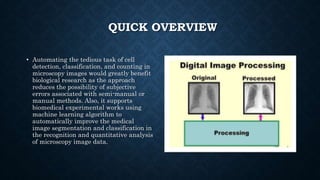
AUTOMATED ANALYSIS OF MICROSCOPY IMAGES USING
DEEP CONVOLUTIONAL NEURAL NETWORKS
Yaser M. Banadaki1*, Adetayo Okunoye2, and Safura Sharifi3,
1Department of Computer Science, Southern University, Baton Rouge, LA 70813
2Department of Computer Science, University of Georgia, Athens, GA 30602
3Department of Physics, University of Illinois Urbana Champaign, IL 61820
RESEARCH
GOALS
• To analyze deep convolutional neural network as an important tool
for the expedited analysis of high‐content microscopy image data
analysis.
• To automate interpretations of medical data which are being done
manually by medical experts including cell counting and
classifications – Processes that are time-intensive, cumbersome
and prone to human errors.
• To train and classify microscopy cellular images using TensorFlow
and achieve a result that outperforms other existing traditional
classification methods.
WHAT IS DEEP
CONVOLUTIONAL
NEURAL NETWORK?
In deep learning,
a convolutional neural
network (CNN) is a class
of deep neural networks, most
applied to analyzing visual
imagery. It simply means a
convolution neural network
with many layers.
QUICK OVERVIEW
• This work automated and analyzed
the tedious task of cell detection,
classification, and counting in
microscopy images.
• We employed DCNN to develop an
automated method for analyzing the
complex high-content microscopy
data that outperforms conventional
cell segmentation, classification, and
counting techniques.
• This would greatly benefit biological
research and the field of medicine
because of the tremendous
improvement in the detection of
complex cell morphologies.
• The notion of applying deep learning-based algorithms to biological and medical
imaging is a fascinating and growing research area. Deep Convolutional Neural
Networks (DCNN) and transfer learning approach has recently shown
remarkable success in image-based data analysis resulting in a tremendous
improvement in automated detection of complex morphologies
QUICK OVERVIEW
Deep learning technology applied to medical
imaging is the most disruptive technology
since the advent of digital imaging.
This research focuses on developing an
accurate, fast and fully automated
computational technique to analyze large-
scale high-throughput microscopy images
for fast phenotyping of functionally diverse
cell populations that outperforms
conventional cell segmentation,
classification and counting techniques.
QUICK OVERVIEW
• Automating the tedious task of cell
detection, classification, and counting in
microscopy images would greatly benefit
biological research as the approach
reduces the possibility of subjective
errors associated with semi-manual or
manual methods. Also, it supports
biomedical experimental works using
machine learning algorithm to
automatically improve the medical
image segmentation and classification in
the recognition and quantitative analysis
of microscopy image data.
THE BUILDING
BLOCKS OF DCNN
• Multi-Layer Perceptrons
(MLPs) are among the most
fundamental building blocks
in Artificial Neural Networks
(ANNs). It refers to a set of
computational models that
are loosely inspired by the
human brain. In general,
they consist of two important
elements, namely, artificial
neurons (nodes) and
synapses (weights) that
connect them. LeCun, [18]
METHODS - HOW IT WORKS
METHODS
• The microscopy image
analysis requires the use
of deep convolutional
neural network model for
thorough learning,
classification and testing
of the given images. In
this work, we have
adopted the use of tensor
flow (Google’s open source
software for machine
learning) for training and
classification
METHODS
The
research
method
procedure
are as
follows:
Preparation of samples and data sets for
analysis
Train the DCNN model
Evaluation and metrics.
METHODS
Models: Inception-v3/v4
Deep learning
techniques:
Transfer learning
(Feature
extraction, Fine
Tuning)
Deep learning
frameworks:
TensorFlow
RESULT
• This shows the result
of the simulation using
2500 datasets from each
category of the blood
samples. The classified
blood samples are
basophil, homophile,
lymphocyte, monocyte,
and neutrophil. The
graph shows that the
maximum test accuracy
of 76 percent can be
achieved using the
number of the training
samples in our dataset.
RESULT
This shows the prediction confidence of 10 randomly test images of four blood cells. We tested
the trained model with ten blood cell samples of mixed categories for identification of the type
of blood cells. It can be noticed that the model predicted neutrophils and monocyte with high
confidence margins.
CONCLUSION
• The annotation of the cells with complex morphology in the images and then the training
process of the model is time-consuming. However, the learned model would reduce the
runtime for cell classifications by orders of magnitudes. The deep convolutional neural
network and transfer learning approach used in the Inception v3 model has specifically
outperformed the binary classifier ensemble across all localization leading to an average
precision score of over 75% in classifying four white blood cells. The paper addressed the
pressing application of artificial intelligence is in the 21st century by enabling the
automated and quantitative analysis of microscopy images – bridging the gap between
existing image analysis techniques in biology and the novel data analytics techniques.
REFERENCES
• 1 Kraus, O.Z., Ba, J.L., and Frey, B.J.: ‘Classifying and segmenting microscopy images
with deep multiple instance learning’, Bioinformatics, 2016, 32, (12), pp. i52-i59
• 2 Dürr, O., and Sick, B.: ‘Single-cell phenotype classification using deep convolutional
neural networks’, Journal of biomolecular screening, 2016, 21, (9), pp. 998-1003
• 3 Pärnamaa, T., and Parts, L.: ‘Accurate classification of protein subcellular localization
from high-throughput microscopy images using deep learning’, G3: Genes, Genomes,
Genetics, 2017, 7, (5), pp. 1385-1392
• 4 Sadanandan, S.K., Ranefall, P., Le Guyader, S., and Wählby, C.: ‘Automated training
of deep convolutional neural networks for cell segmentation’, Scientific reports, 2017, 7,
(1), pp. 1-7
• 5 Xue, Y., and Ray, N.: ‘Cell Detection in microscopy images with deep convolutional
neural network and compressed sensing’, arXiv preprint arXiv:1708.03307, 2017
• 6. Abràmoff, M.D., Magalhães, P.J., and Ram, S.J.: ‘Image processing with ImageJ’,
Biophotonics international, 2004, 11, (7), pp. 36-427. Sommer, C.; Straehle, C. N.;
Koethe, U.; Hamprecht, F. A. In Ilastik: Interactive learning and segmentation toolkit,
ISBI, 2011; p 8
• 7 Dataset, B.: ‘https://github.com/Shenggan/BCCD_Dataset’
• 8 Abadi, M., Agarwal, A., Barham, P., Brevdo, E., Chen, Z., Citro, C., Corrado, G.,
Davis, A., Dean, J., and Devin, M.: ‘Tensorflow: Large-scale machine learning on
heterogeneous distributed systems’, 2015
• THANK YOU FOR
LISTENING

Automated Analysis of Microscopy Images using Deep Convolutional Neural Network

  • 1.
    AUTOMATED ANALYSIS OFMICROSCOPY IMAGES USING DEEP CONVOLUTIONAL NEURAL NETWORKS Yaser M. Banadaki1*, Adetayo Okunoye2, and Safura Sharifi3, 1Department of Computer Science, Southern University, Baton Rouge, LA 70813 2Department of Computer Science, University of Georgia, Athens, GA 30602 3Department of Physics, University of Illinois Urbana Champaign, IL 61820
  • 2.
    RESEARCH GOALS • To analyzedeep convolutional neural network as an important tool for the expedited analysis of high‐content microscopy image data analysis. • To automate interpretations of medical data which are being done manually by medical experts including cell counting and classifications – Processes that are time-intensive, cumbersome and prone to human errors. • To train and classify microscopy cellular images using TensorFlow and achieve a result that outperforms other existing traditional classification methods.
  • 3.
    WHAT IS DEEP CONVOLUTIONAL NEURALNETWORK? In deep learning, a convolutional neural network (CNN) is a class of deep neural networks, most applied to analyzing visual imagery. It simply means a convolution neural network with many layers.
  • 4.
    QUICK OVERVIEW • Thiswork automated and analyzed the tedious task of cell detection, classification, and counting in microscopy images. • We employed DCNN to develop an automated method for analyzing the complex high-content microscopy data that outperforms conventional cell segmentation, classification, and counting techniques. • This would greatly benefit biological research and the field of medicine because of the tremendous improvement in the detection of complex cell morphologies.
  • 5.
    • The notionof applying deep learning-based algorithms to biological and medical imaging is a fascinating and growing research area. Deep Convolutional Neural Networks (DCNN) and transfer learning approach has recently shown remarkable success in image-based data analysis resulting in a tremendous improvement in automated detection of complex morphologies
  • 6.
    QUICK OVERVIEW Deep learningtechnology applied to medical imaging is the most disruptive technology since the advent of digital imaging. This research focuses on developing an accurate, fast and fully automated computational technique to analyze large- scale high-throughput microscopy images for fast phenotyping of functionally diverse cell populations that outperforms conventional cell segmentation, classification and counting techniques.
  • 7.
    QUICK OVERVIEW • Automatingthe tedious task of cell detection, classification, and counting in microscopy images would greatly benefit biological research as the approach reduces the possibility of subjective errors associated with semi-manual or manual methods. Also, it supports biomedical experimental works using machine learning algorithm to automatically improve the medical image segmentation and classification in the recognition and quantitative analysis of microscopy image data.
  • 8.
    THE BUILDING BLOCKS OFDCNN • Multi-Layer Perceptrons (MLPs) are among the most fundamental building blocks in Artificial Neural Networks (ANNs). It refers to a set of computational models that are loosely inspired by the human brain. In general, they consist of two important elements, namely, artificial neurons (nodes) and synapses (weights) that connect them. LeCun, [18]
  • 9.
    METHODS - HOWIT WORKS
  • 10.
    METHODS • The microscopyimage analysis requires the use of deep convolutional neural network model for thorough learning, classification and testing of the given images. In this work, we have adopted the use of tensor flow (Google’s open source software for machine learning) for training and classification
  • 11.
    METHODS The research method procedure are as follows: Preparation ofsamples and data sets for analysis Train the DCNN model Evaluation and metrics.
  • 12.
    METHODS Models: Inception-v3/v4 Deep learning techniques: Transferlearning (Feature extraction, Fine Tuning) Deep learning frameworks: TensorFlow
  • 13.
    RESULT • This showsthe result of the simulation using 2500 datasets from each category of the blood samples. The classified blood samples are basophil, homophile, lymphocyte, monocyte, and neutrophil. The graph shows that the maximum test accuracy of 76 percent can be achieved using the number of the training samples in our dataset.
  • 14.
    RESULT This shows theprediction confidence of 10 randomly test images of four blood cells. We tested the trained model with ten blood cell samples of mixed categories for identification of the type of blood cells. It can be noticed that the model predicted neutrophils and monocyte with high confidence margins.
  • 15.
    CONCLUSION • The annotationof the cells with complex morphology in the images and then the training process of the model is time-consuming. However, the learned model would reduce the runtime for cell classifications by orders of magnitudes. The deep convolutional neural network and transfer learning approach used in the Inception v3 model has specifically outperformed the binary classifier ensemble across all localization leading to an average precision score of over 75% in classifying four white blood cells. The paper addressed the pressing application of artificial intelligence is in the 21st century by enabling the automated and quantitative analysis of microscopy images – bridging the gap between existing image analysis techniques in biology and the novel data analytics techniques.
  • 16.
    REFERENCES • 1 Kraus,O.Z., Ba, J.L., and Frey, B.J.: ‘Classifying and segmenting microscopy images with deep multiple instance learning’, Bioinformatics, 2016, 32, (12), pp. i52-i59 • 2 Dürr, O., and Sick, B.: ‘Single-cell phenotype classification using deep convolutional neural networks’, Journal of biomolecular screening, 2016, 21, (9), pp. 998-1003 • 3 Pärnamaa, T., and Parts, L.: ‘Accurate classification of protein subcellular localization from high-throughput microscopy images using deep learning’, G3: Genes, Genomes, Genetics, 2017, 7, (5), pp. 1385-1392 • 4 Sadanandan, S.K., Ranefall, P., Le Guyader, S., and Wählby, C.: ‘Automated training of deep convolutional neural networks for cell segmentation’, Scientific reports, 2017, 7, (1), pp. 1-7 • 5 Xue, Y., and Ray, N.: ‘Cell Detection in microscopy images with deep convolutional neural network and compressed sensing’, arXiv preprint arXiv:1708.03307, 2017 • 6. Abràmoff, M.D., Magalhães, P.J., and Ram, S.J.: ‘Image processing with ImageJ’, Biophotonics international, 2004, 11, (7), pp. 36-427. Sommer, C.; Straehle, C. N.; Koethe, U.; Hamprecht, F. A. In Ilastik: Interactive learning and segmentation toolkit, ISBI, 2011; p 8 • 7 Dataset, B.: ‘https://github.com/Shenggan/BCCD_Dataset’ • 8 Abadi, M., Agarwal, A., Barham, P., Brevdo, E., Chen, Z., Citro, C., Corrado, G., Davis, A., Dean, J., and Devin, M.: ‘Tensorflow: Large-scale machine learning on heterogeneous distributed systems’, 2015
  • 17.
    • THANK YOUFOR LISTENING