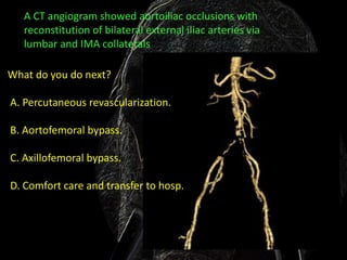
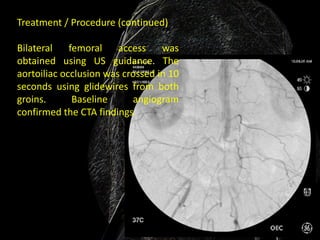
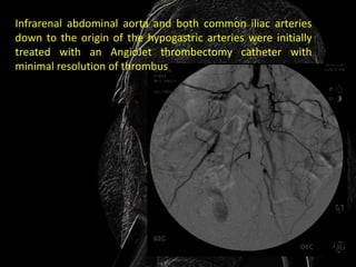
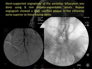
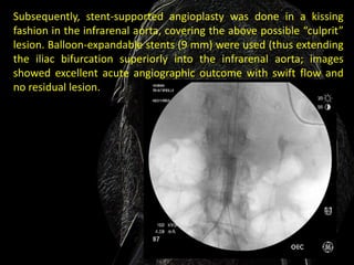
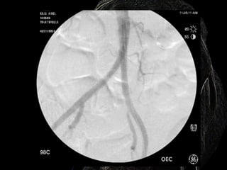
Acute Aortic Occlusion:
    Evaluation and
    Management
A 79-year-old Caucasian female presented with a 1-
month history of severe, relentless rest pain involving
both calves and feet as well as acute renal failure. She
had a major stroke 3 months prior, from which she had
been recuperating in the skilled nursing facility. Renal
function normalized with hydration in the first 48
hours.

Clinical examination showed absent femoral pulses with
soft bilateral systolic bruits and absent popliteal and
pedal pulses. There was pinkish-blue discoloration of
toes bilaterally. Her left elbow had flexion contractures
from her recent stroke. Duplex examination showed
monophasic waveforms with severely diminished peak
systolic velocities bilaterally at all levels.
What is the next step in evaluation?


 A.
Conservative management using pain
medications and Rooke’s vascular boots.
 B.
CT angiogram.

C.
MR angiogram.
D.
Surgical consultation for possible amputation.
• Why the author recommends Answer B:
  • CT angiography is a quick, inexpensive, and universally
    available method and provides excellent anatomical details
    of the aortoiliac vasculature.
• Why the author does not recommend A:
  • Obviously conservative management is not a choice for this
    patient. She has a 1-month history of rest pain with
    impalpable femoral pulses suggestive of acute or acute-on-
    chronic aortoiliac arterioocclusive disease. She is at risk of
    limb gangrene if left untreated.
• Why the author does not recommend C:
  • MR angiogram is an expensive and time-consuming method
    and may not provide high-quality imaging in patients who
    can’t stay still.
• Why the author does not recommend D:
  • Surgical consultation for amputation is unwarranted at this
    time as this patient had severely ischemic but viable limbs.
A CT angiogram showed aortoiliac occlusions with
   reconstitution of bilateral external iliac arteries via
   lumbar and IMA collaterals

What do you do next?

A. Percutaneous revascularization.

B. Aortofemoral bypass.

C. Axillofemoral bypass.

D. Comfort care and transfer to hosp.
Why the author recommends Answer A:
With advances in endovascular technology, the minimally invasive
percutaneous interventions have replaced many complex aortoiliac
lesions, which hitherto required aortofemoral or axillofemoral
bypass.
Why the author does not recommend B:
Aorto- or axillofemoral bypass surgeries are associated with major
morbidity and mortality patients such as this one, with underlying
multiple major comorbidities such as HTN, CAD, diabetes and
recent CVA.
Why the author does not recommend C:
See rationale for answer B.
Why the author does not recommend D:
This is not a choice in this patient with acute or acute-on-chronic
aortoiliac occlusions. With advances in endovascular technology,
minimally invasive percutaneous interventions can be used to treat
many complex aortoiliac lesions, which hitherto required
aortofemoral or axillofemoral bypass.
What is the best approach for percutaneous
                    revascularization?

A. Antegrade access via both brachial arteries.

B. Retrograde access via both common femoral arteries.

C. Retrograde pedal access approach under US guidance.

D. Retrograde bilateral popliteal access under US guidance.
Why the author recommends Answer B:
Antegrade access is usually preferred in aortoiliac total occlusions, since any
dissection plane extensions usually propagate distally, without compromising
the visceral or renal circulation. However, as this patient has had left arm
contractures from a recent CVA event, brachial access will be challenging.
Hence, bilateral femoral access and retrograde revascularization would be a
better choice.

Why the author does not recommend A:
See rationale for answer B.

Why the author does not recommend C:
Retrograde pedal access was not an ideal option for this patient, whose femoral
arteries are open and relatively undiseased and there was enough working
distance from a common femoral access point to the reconstituted external iliac
arteries for sheath placement.

Why the author does not recommend D:
See rationale for answer C.
Treatment / Procedure (continued)

Bilateral    femoral    access    was
obtained using US guidance. The
aortoiliac occlusion was crossed in 10
seconds using glidewires from both
groins.       Baseline     angiogram
confirmed the CTA findings
Infrarenal abdominal aorta and both common iliac arteries
down to the origin of the hypogastric arteries were initially
treated with an AngioJet thrombectomy catheter with
minimal resolution of thrombus
Stent-supported angioplasty of the aortoiliac bifurcation was
done using 8 mm balloon-expandable stents. Repeat
angiogram showed a large calcified plaque in the infrarenal
aorta superior to these kissing stents
Subsequently, stent-supported angioplasty was done in a kissing
fashion in the infrarenal aorta, covering the above possible “culprit”
lesion. Balloon-expandable stents (9 mm) were used (thus extending
the iliac bifurcation superiorly into the infrarenal aorta; images
showed excellent acute angiographic outcome with swift flow and
no residual lesion.
I am only transferred the case of
Mallik Thatipelli

Aortoiliac occlusion rx

  • 1.
    Acute Aortic Occlusion: Evaluation and Management
  • 2.
    A 79-year-old Caucasianfemale presented with a 1- month history of severe, relentless rest pain involving both calves and feet as well as acute renal failure. She had a major stroke 3 months prior, from which she had been recuperating in the skilled nursing facility. Renal function normalized with hydration in the first 48 hours. Clinical examination showed absent femoral pulses with soft bilateral systolic bruits and absent popliteal and pedal pulses. There was pinkish-blue discoloration of toes bilaterally. Her left elbow had flexion contractures from her recent stroke. Duplex examination showed monophasic waveforms with severely diminished peak systolic velocities bilaterally at all levels.
  • 3.
    What is thenext step in evaluation? A. Conservative management using pain medications and Rooke’s vascular boots. B. CT angiogram. C. MR angiogram. D. Surgical consultation for possible amputation.
  • 4.
    • Why theauthor recommends Answer B: • CT angiography is a quick, inexpensive, and universally available method and provides excellent anatomical details of the aortoiliac vasculature. • Why the author does not recommend A: • Obviously conservative management is not a choice for this patient. She has a 1-month history of rest pain with impalpable femoral pulses suggestive of acute or acute-on- chronic aortoiliac arterioocclusive disease. She is at risk of limb gangrene if left untreated. • Why the author does not recommend C: • MR angiogram is an expensive and time-consuming method and may not provide high-quality imaging in patients who can’t stay still. • Why the author does not recommend D: • Surgical consultation for amputation is unwarranted at this time as this patient had severely ischemic but viable limbs.
  • 5.
    A CT angiogramshowed aortoiliac occlusions with reconstitution of bilateral external iliac arteries via lumbar and IMA collaterals What do you do next? A. Percutaneous revascularization. B. Aortofemoral bypass. C. Axillofemoral bypass. D. Comfort care and transfer to hosp.
  • 6.
    Why the authorrecommends Answer A: With advances in endovascular technology, the minimally invasive percutaneous interventions have replaced many complex aortoiliac lesions, which hitherto required aortofemoral or axillofemoral bypass. Why the author does not recommend B: Aorto- or axillofemoral bypass surgeries are associated with major morbidity and mortality patients such as this one, with underlying multiple major comorbidities such as HTN, CAD, diabetes and recent CVA. Why the author does not recommend C: See rationale for answer B. Why the author does not recommend D: This is not a choice in this patient with acute or acute-on-chronic aortoiliac occlusions. With advances in endovascular technology, minimally invasive percutaneous interventions can be used to treat many complex aortoiliac lesions, which hitherto required aortofemoral or axillofemoral bypass.
  • 7.
    What is thebest approach for percutaneous revascularization? A. Antegrade access via both brachial arteries. B. Retrograde access via both common femoral arteries. C. Retrograde pedal access approach under US guidance. D. Retrograde bilateral popliteal access under US guidance.
  • 8.
    Why the authorrecommends Answer B: Antegrade access is usually preferred in aortoiliac total occlusions, since any dissection plane extensions usually propagate distally, without compromising the visceral or renal circulation. However, as this patient has had left arm contractures from a recent CVA event, brachial access will be challenging. Hence, bilateral femoral access and retrograde revascularization would be a better choice. Why the author does not recommend A: See rationale for answer B. Why the author does not recommend C: Retrograde pedal access was not an ideal option for this patient, whose femoral arteries are open and relatively undiseased and there was enough working distance from a common femoral access point to the reconstituted external iliac arteries for sheath placement. Why the author does not recommend D: See rationale for answer C.
  • 9.
    Treatment / Procedure(continued) Bilateral femoral access was obtained using US guidance. The aortoiliac occlusion was crossed in 10 seconds using glidewires from both groins. Baseline angiogram confirmed the CTA findings
  • 10.
    Infrarenal abdominal aortaand both common iliac arteries down to the origin of the hypogastric arteries were initially treated with an AngioJet thrombectomy catheter with minimal resolution of thrombus
  • 11.
    Stent-supported angioplasty ofthe aortoiliac bifurcation was done using 8 mm balloon-expandable stents. Repeat angiogram showed a large calcified plaque in the infrarenal aorta superior to these kissing stents
  • 12.
    Subsequently, stent-supported angioplastywas done in a kissing fashion in the infrarenal aorta, covering the above possible “culprit” lesion. Balloon-expandable stents (9 mm) were used (thus extending the iliac bifurcation superiorly into the infrarenal aorta; images showed excellent acute angiographic outcome with swift flow and no residual lesion.
  • 14.
    I am onlytransferred the case of Mallik Thatipelli