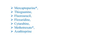
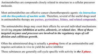
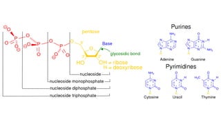
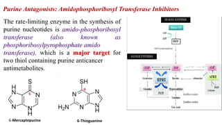
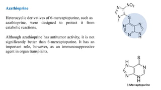
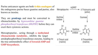
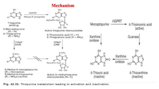
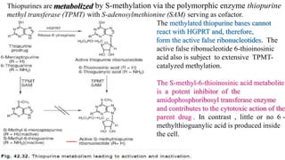
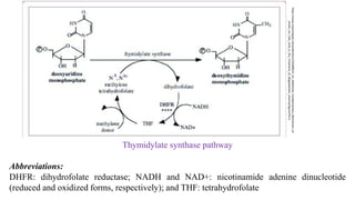
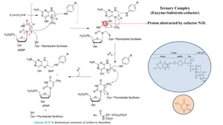
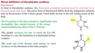
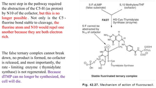
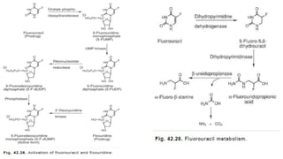
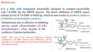
Antimetabolites
(Antineplastic Agents)
by
Prof. Jayshree Patil
Assistant Professor
AIKTC, School of Pharmacy, New Panvel
For T Y B Pharm (Sem V, PCI)
➢ Mercaptopurine*,
➢ Thioguanine,
➢ Fluorouracil,
➢ Floxuridine,
➢ Cytarabine,
➢ Methotrexate*,
➢ Azathioprine
Antimetabolites are compounds closely related in structure to a cellular precursor
molecule.
Most antimetabolites are effective cancer chemotherapeutic agents via interaction
with the biosynthesis of nucleic acids. Therefore, several of the useful drugs used
in antimetabolite therapy are purines, pyrimidines, folates, and related compounds.
The antimetabolite drugs may exert their effects by several individual mechanisms
involving enzyme inhibition at active, allosteric, or related sites. Most of these
targeted enzymes and processes are involved in the regulatory steps of cell
division and cell/tissue growth.
Often the administered drug is actually a prodrug form of an antimetabolite and
requires activation in vivo to yield the active inhibitor
These substances are generally cell cycle specific with activity in the S phase.
Purine Antagonists: Amidophosphoribosyl Transferase Inhibitors
The rate-limiting enzyme in the synthesis of
purine nucleotides is amido-phosphoribosyl
transferase (also known as
phosphoribosylpyrophosphate amido
transferase), which is a major target for
two thiol containing purine anticancer
antimetabolites.
6-Mercaptopurine
6
6-Thioguanine
6
Azathioprine
Heterocyclic derivatives of 6-mercaptopurine, such as
azathioprine, were designed to protect it from
catabolic reactions.
Although azathioprine has antitumor activity, it is not
significantly better than 6-mercaptopurine. It has an
important role, however, as an immunosuppressive
agent in organ transplants.
6-Mercaptopurine
Purine anticancer agents are both 6-thio analogues of
the endogenous purine bases guanine and purine, also
known as inosine.
They are prodrugs and must be converted to
ribonucleotides by hypoxanthine guanine
phosphoribosyl transferase (HGPRT) before they can
exert their cytotoxic actions.
Mercaptopurine , acting through a methylated
ribonucleotide metabolite , inhibits the target
amidophosphoribosyl transferase enzyme, leading to
the true antimetabolic effect of lowered AMP and
GMP biosynthesis.
Mechanism
Thiopurines are
methyl transferase (TPMT) with S-adenosylmethionine (SAM) serving as cofactor.
The methylated thiopurine bases cannot
react with HGPRT and, therefore,
form the active false ribonucleotides. The
active false ribonucleotide 6-thioinosinic
acid also is subject to extensive TPMT-
catalyzed methylation.
The S-methyl-6-thioinosinic acid metabolite
is a potent inhibitor of the
amidophosphoribosyl transferase enzyme
and contributes to the cytotoxic action of the
parent drug . In contrast , little or no 6 -
methylthioguanylic acid is produced inside
the cell.
by S-methylation via the polymorphic enzyme thiopurine
Mercaptopurine is used in the treatment of acute lymphatic and myelogenous
leukemias.
Theraupetic Uses:
Thiogunanine is administered orally in the treatment of nonlymphocytic
leukemias.
A second mechanism of antineoplastic activity for mercaptopurine (and the
predominant mechanism for thioguanine) involves the incorporation of di- and
triphosphate deoxy- and ribonucleotides generated within the tumor cell into
DNA and RNA, respectively. This illicit substitution further inhibits elongation of
the strands and promotes apoptosis.
dTMP biosynthesis
dTMP is produced via C5-methylation of deoxyuridine monophosphate (dUMP).
The rate-limiting enzyme of the dTMP synthetic pathway is the sulfhydryl-
containing thymidylate synthase, with 5,10-methylenetetrahydrofolate (5,10-
methylene-THF) serving as the methyl-donating cofactor.
All dTMP synthesis inhibitors will inhibit thymidylate synthase either directly or
indirectly, and this will result in a “ thymineless death” in actively dividing cells.
Without dTMP and its deoxythmidine triphosphate metabolite, DNA will
fragment, and the cell will die.
The synthase enzyme is very large and contains a deep pocket for the binding of
both substrate and cofactor.
Thymidylate (dTMP) biosynthesis plays an essential and
exclusive function in DNA synthesis and proper cell division, and
therefore has been an attractive therapeutic target
Thymidylate synthase pathway
Abbreviations:
DHFR: dihydrofolate reductase; NADH and NAD+: nicotinamide adenine dinucleotide
(reduced and oxidized forms, respectively); and THF: tetrahydrofolate
https://www.researchgate.net/publication/332688515_An_Additional_Complementary_Mechanism_of
_Action_for_Folic_Acid_in_the_Treatment_of_Megaloblastic_Anemia/figures?lo=1
Proton abstracted by cofactor N10.
Ternary Complex
(Enzyme-Substrate-cofactor)
1
2
3 4
5
6
Fluorouracil
To bind to thymidylate synthase, this fluorinated pyrimidine prodrug must be converted to its
deoxyribonucleotide form. The active form of fluorouracil differs from the endogenous substrate
only by the presence of the 5-fluoro group, which holds the key to the cell -killing action of this
drug.
The C6 position of the false substrate is significantly more
electrophilic than normal because of the strong
electronwithdrawing effect of the C5 fluorine.
This greatly increases the rate of attack by Cys 195 ,
resulting in a very fast formation of a fluorinated ternary
complex.
The small size of the fluorine atom assures no steric
hindrance to the formation of this false complex.
The next step in the pathway required
the abstraction of the C5-H (as proton)
by N10 of the cofactor, but this is no
longer possible . Not only is the C5 -
fluorine bond stable to cleavage, the
fluorine atom and N10 would repel one
another because they are both electron
rich.
The false ternary complex cannot break
down, no product is formed, no cofactor
is released, and most importantly, the
rate - limiting enzyme ( thymidylate
synthase) is not regenerated. Because
dTMP can no longer be synthesized, the
cell will die.
Fluorouracil is administered IV in the palliative treatment of
. Patients are treated for four consecutive
days, followed by treatment on odd-numbered days up to a maximum of 12
days.
Floxuridine
This deoxyribonucleoside prodrug is
bioconverted via 2′-deoxyuridine kinase–
mediated phosphorylation to the same active 5-
fluoro-dUMP structure generated in the
multistep biotransformation of fluorouracil.
It is given by intra-arterial infusion for the
palliative treatment of GI adenocarcinoma that
has metastasized to the liver and that cannot be
managed surgically.
5-fluorouracil
Methotrexate also is effective in inhibiting
glycine amide ribonucleotide ( G A R )
transformylase , a key enzyme in the
synthesis of purine nucleotides.
NOTE: Take note of the structural differences between
methotrexate and DHF, because these differences will be
important to an understanding of the chemical mechanism of
this anticancer agent .
Pteridine
PABA
Glutamic Acid
Methotrexate
It is a folic acid antagonist structurally designed to compete successfully
with 7,8-DHF for the DHFR enzyme. The direct inhibition of DHFR causes
cellular levels of 7,8-DHF to build up, which in turn results in feedback (indirect
) inhibition of thymidylate synthase.
Methotrexate is the classic antimetabolite of folic acid
structurally derived by N-methylation of the para-
aminobenzoic acid residue (PABA) and replacement of
a pteridine hydroxyl by the bioisosteric amino group.
The conversion of =O to -NH2 increases the basicity
of N-3 and yields greater enzyme affinity.
This drug competitively inhibits the binding of the
substrate folic acid to the enzyme DHFR, resulting
in reductions in the synthesis of nucleic acid bases,
perhaps most importantly, the conversion of
uridylate to thymidylate as catalyzed by
thymidylate synthetase.
In addition, purine synthesis is inhibited because the N-10-formyl tetrahydrofolic acid is a
formyl donor involved in purine synthesis. THFs are cofactors in the normal biosynthesis of
purines.
It has been proposed that the N5 position of DHF is
protonated by a Glu30 of DHFR and, in cationic
form, binds to DHFR Asp27 through an electrostatic
bond. N5 is the strongest base in the DHF structure,
in part because of attenuating the impact of the C4
carbonyl on electron density around N1.
Additional affinity - enhancing interactions between
enzyme and substrate also have been identified, and
once bound, the substrate 5,6- double bond is
positioned close to the NADPH cofactor so that the
transfer of hydride can proceed.
In contrast, the C4 amino substituent of methotrexate enriches
electron density at N1 through π-electron donation, increasing its
basic character between 10- and 1,000-fold and promoting
protonation by Glu30 at the expense of N5.
Because N1 and N5 are across the pteridine ring from one another ,
the interaction of N1 with the DHFR Asp27 will
effectively stand the false substrate “on its head” relative to the
orientation of 7,8-DHF
.
With the 5,6-double bond of methotrexate 180° away from the
bound NADPH cofactor and stabilized by the fully aromatic
pteridine ring, the possibility for reduction is eliminated. The
DHFR enzyme will be pseudoirreversibly bound to a molecule it
cannot reduce, which ties up the DHFR enzyme and prevents the
conversion of DHF to THF.
In turn, this halts the synthesis of the 5,10-methylene-THF
cofactor required for dTMP biosynthesis and causes feedback
inhibition of the thymidylate synthase enzyme. The cell will die a
“ thymineless death. ”
Methotrexate can be given orally in the treatment of
The sodium salt form also is marketed for IV, intramuscular, intra-arterial, or
intrathecal injection.
hydroxymetabolite (which has a three- to fivefold lower water solubility) can
precipitate in the renal tubule, causing damaging crystalluria. Methotrexate-
induced lung disease is a particularly critical problem, because it can arise at any
time and at any dose, and it can even be fatal .
Methotrexate use also can precipitate severe GI side effects, including ulcerative
stomatitis and hemorrhagic enteritis, leading to intestinal perforation. Potentially
fatal skin reactions are a risk as well .
As a Category X teratogen, this drug should not be given to women who are
pregnant or planning to become pregnant.
can occur with high doses if “ third space” fluids allow drug
to accumulate in ascites and pleural ef fusions and/or when renal excretion is
impaired by kidney disease. When used in high doses, methotrexate and its 7-
If severe methotrexate toxicity occurs, reduced folate replacement therapy with
5-formyltetrahydrofolate (leucovorin) must be initiated as soon as possible.
Leucovorin generates the folate cofactors needed by DHFR and GAR
transformylase to ensure the continued synthesis of pyrimidine and purine
nucleotides in healthy cells. “Leucovorin rescue” therapy often is given as
prophylaxis after high-dose methotrexate.
DNA Polymerase/DNA Chain Elongation
Inhibitors
Five halogenated and/or ribose-modified DNA nucleoside analogues are marketed for the treatment
of a wide variety of hematologic cancers and solid tumors.
These agents have complex and multifaceted mechanisms. All include inhibition of DNA polymerase
and/or DNA chain elongation among their actions, however , and all must be converted to
triphosphate nucleotides before activity is realized.
As nucleosides, they are actively taken up into cells via a selective nucleoside transporter protein, so
tumors deficient in this transporter system will be resistant to these anticancer agents.
Once inside the cell , specific kinases conduct the essential phosphorylation reactions. In active
triphosphate form, they can be mistakenly incorporated into the growing DNA chain, thus arresting
further elongation, and/or inhibit enzymes essential for DNA synthesis.
All drugs in this group are administered IV, are excreted via the kidneys, and induce
myelosuppression as their major use- limiting side effect.
Cytarabine
This is cytidine-based anticancer agents
undergo initial phosphorylation by
deoxycytidinekinase to the
monophosphate with subsequent
phosphorylations catalyzed by
pyrimidine monophosphate and
diphosphate kinases.
Cytaribine, an arabinoside, is catabolized
by cytidine and deoxycytidylate
(deoxycytidine monophosphate)
deaminases to inactive uracil analogues.
REFERENCES:
1. Foye’s Principles of Medicinal Chemistry, Thomas L. Lemke, David A Williams, Lippincott
Williams & Wilkins.
2. Wilson and Gisvold’s Textbook of Organic Medicinal and Pharmaceutical Chemistry, John M.
Beale, John H. Block, Lippincott Williams & Wilkins.

Antimetabolites.pdf

  • 1.
    Antimetabolites (Antineplastic Agents) by Prof. JayshreePatil Assistant Professor AIKTC, School of Pharmacy, New Panvel For T Y B Pharm (Sem V, PCI)
  • 2.
    ➢ Mercaptopurine*, ➢ Thioguanine, ➢Fluorouracil, ➢ Floxuridine, ➢ Cytarabine, ➢ Methotrexate*, ➢ Azathioprine
  • 3.
    Antimetabolites are compoundsclosely related in structure to a cellular precursor molecule. Most antimetabolites are effective cancer chemotherapeutic agents via interaction with the biosynthesis of nucleic acids. Therefore, several of the useful drugs used in antimetabolite therapy are purines, pyrimidines, folates, and related compounds. The antimetabolite drugs may exert their effects by several individual mechanisms involving enzyme inhibition at active, allosteric, or related sites. Most of these targeted enzymes and processes are involved in the regulatory steps of cell division and cell/tissue growth. Often the administered drug is actually a prodrug form of an antimetabolite and requires activation in vivo to yield the active inhibitor These substances are generally cell cycle specific with activity in the S phase.
  • 5.
    Purine Antagonists: AmidophosphoribosylTransferase Inhibitors The rate-limiting enzyme in the synthesis of purine nucleotides is amido-phosphoribosyl transferase (also known as phosphoribosylpyrophosphate amido transferase), which is a major target for two thiol containing purine anticancer antimetabolites. 6-Mercaptopurine 6 6-Thioguanine 6
  • 6.
    Azathioprine Heterocyclic derivatives of6-mercaptopurine, such as azathioprine, were designed to protect it from catabolic reactions. Although azathioprine has antitumor activity, it is not significantly better than 6-mercaptopurine. It has an important role, however, as an immunosuppressive agent in organ transplants. 6-Mercaptopurine
  • 7.
    Purine anticancer agentsare both 6-thio analogues of the endogenous purine bases guanine and purine, also known as inosine. They are prodrugs and must be converted to ribonucleotides by hypoxanthine guanine phosphoribosyl transferase (HGPRT) before they can exert their cytotoxic actions. Mercaptopurine , acting through a methylated ribonucleotide metabolite , inhibits the target amidophosphoribosyl transferase enzyme, leading to the true antimetabolic effect of lowered AMP and GMP biosynthesis.
  • 8.
  • 9.
    Thiopurines are methyl transferase(TPMT) with S-adenosylmethionine (SAM) serving as cofactor. The methylated thiopurine bases cannot react with HGPRT and, therefore, form the active false ribonucleotides. The active false ribonucleotide 6-thioinosinic acid also is subject to extensive TPMT- catalyzed methylation. The S-methyl-6-thioinosinic acid metabolite is a potent inhibitor of the amidophosphoribosyl transferase enzyme and contributes to the cytotoxic action of the parent drug . In contrast , little or no 6 - methylthioguanylic acid is produced inside the cell. by S-methylation via the polymorphic enzyme thiopurine
  • 10.
    Mercaptopurine is usedin the treatment of acute lymphatic and myelogenous leukemias. Theraupetic Uses: Thiogunanine is administered orally in the treatment of nonlymphocytic leukemias. A second mechanism of antineoplastic activity for mercaptopurine (and the predominant mechanism for thioguanine) involves the incorporation of di- and triphosphate deoxy- and ribonucleotides generated within the tumor cell into DNA and RNA, respectively. This illicit substitution further inhibits elongation of the strands and promotes apoptosis.
  • 12.
    dTMP biosynthesis dTMP isproduced via C5-methylation of deoxyuridine monophosphate (dUMP). The rate-limiting enzyme of the dTMP synthetic pathway is the sulfhydryl- containing thymidylate synthase, with 5,10-methylenetetrahydrofolate (5,10- methylene-THF) serving as the methyl-donating cofactor. All dTMP synthesis inhibitors will inhibit thymidylate synthase either directly or indirectly, and this will result in a “ thymineless death” in actively dividing cells. Without dTMP and its deoxythmidine triphosphate metabolite, DNA will fragment, and the cell will die. The synthase enzyme is very large and contains a deep pocket for the binding of both substrate and cofactor. Thymidylate (dTMP) biosynthesis plays an essential and exclusive function in DNA synthesis and proper cell division, and therefore has been an attractive therapeutic target
  • 13.
    Thymidylate synthase pathway Abbreviations: DHFR:dihydrofolate reductase; NADH and NAD+: nicotinamide adenine dinucleotide (reduced and oxidized forms, respectively); and THF: tetrahydrofolate https://www.researchgate.net/publication/332688515_An_Additional_Complementary_Mechanism_of _Action_for_Folic_Acid_in_the_Treatment_of_Megaloblastic_Anemia/figures?lo=1
  • 14.
    Proton abstracted bycofactor N10. Ternary Complex (Enzyme-Substrate-cofactor) 1 2 3 4 5 6
  • 15.
    Fluorouracil To bind tothymidylate synthase, this fluorinated pyrimidine prodrug must be converted to its deoxyribonucleotide form. The active form of fluorouracil differs from the endogenous substrate only by the presence of the 5-fluoro group, which holds the key to the cell -killing action of this drug. The C6 position of the false substrate is significantly more electrophilic than normal because of the strong electronwithdrawing effect of the C5 fluorine. This greatly increases the rate of attack by Cys 195 , resulting in a very fast formation of a fluorinated ternary complex. The small size of the fluorine atom assures no steric hindrance to the formation of this false complex.
  • 16.
    The next stepin the pathway required the abstraction of the C5-H (as proton) by N10 of the cofactor, but this is no longer possible . Not only is the C5 - fluorine bond stable to cleavage, the fluorine atom and N10 would repel one another because they are both electron rich. The false ternary complex cannot break down, no product is formed, no cofactor is released, and most importantly, the rate - limiting enzyme ( thymidylate synthase) is not regenerated. Because dTMP can no longer be synthesized, the cell will die.
  • 18.
    Fluorouracil is administeredIV in the palliative treatment of . Patients are treated for four consecutive days, followed by treatment on odd-numbered days up to a maximum of 12 days.
  • 19.
    Floxuridine This deoxyribonucleoside prodrugis bioconverted via 2′-deoxyuridine kinase– mediated phosphorylation to the same active 5- fluoro-dUMP structure generated in the multistep biotransformation of fluorouracil. It is given by intra-arterial infusion for the palliative treatment of GI adenocarcinoma that has metastasized to the liver and that cannot be managed surgically. 5-fluorouracil
  • 21.
    Methotrexate also iseffective in inhibiting glycine amide ribonucleotide ( G A R ) transformylase , a key enzyme in the synthesis of purine nucleotides. NOTE: Take note of the structural differences between methotrexate and DHF, because these differences will be important to an understanding of the chemical mechanism of this anticancer agent . Pteridine PABA Glutamic Acid Methotrexate It is a folic acid antagonist structurally designed to compete successfully with 7,8-DHF for the DHFR enzyme. The direct inhibition of DHFR causes cellular levels of 7,8-DHF to build up, which in turn results in feedback (indirect ) inhibition of thymidylate synthase.
  • 22.
    Methotrexate is theclassic antimetabolite of folic acid structurally derived by N-methylation of the para- aminobenzoic acid residue (PABA) and replacement of a pteridine hydroxyl by the bioisosteric amino group. The conversion of =O to -NH2 increases the basicity of N-3 and yields greater enzyme affinity. This drug competitively inhibits the binding of the substrate folic acid to the enzyme DHFR, resulting in reductions in the synthesis of nucleic acid bases, perhaps most importantly, the conversion of uridylate to thymidylate as catalyzed by thymidylate synthetase. In addition, purine synthesis is inhibited because the N-10-formyl tetrahydrofolic acid is a formyl donor involved in purine synthesis. THFs are cofactors in the normal biosynthesis of purines.
  • 23.
    It has beenproposed that the N5 position of DHF is protonated by a Glu30 of DHFR and, in cationic form, binds to DHFR Asp27 through an electrostatic bond. N5 is the strongest base in the DHF structure, in part because of attenuating the impact of the C4 carbonyl on electron density around N1. Additional affinity - enhancing interactions between enzyme and substrate also have been identified, and once bound, the substrate 5,6- double bond is positioned close to the NADPH cofactor so that the transfer of hydride can proceed.
  • 24.
    In contrast, theC4 amino substituent of methotrexate enriches electron density at N1 through π-electron donation, increasing its basic character between 10- and 1,000-fold and promoting protonation by Glu30 at the expense of N5. Because N1 and N5 are across the pteridine ring from one another , the interaction of N1 with the DHFR Asp27 will effectively stand the false substrate “on its head” relative to the orientation of 7,8-DHF . With the 5,6-double bond of methotrexate 180° away from the bound NADPH cofactor and stabilized by the fully aromatic pteridine ring, the possibility for reduction is eliminated. The DHFR enzyme will be pseudoirreversibly bound to a molecule it cannot reduce, which ties up the DHFR enzyme and prevents the conversion of DHF to THF. In turn, this halts the synthesis of the 5,10-methylene-THF cofactor required for dTMP biosynthesis and causes feedback inhibition of the thymidylate synthase enzyme. The cell will die a “ thymineless death. ”
  • 25.
    Methotrexate can begiven orally in the treatment of The sodium salt form also is marketed for IV, intramuscular, intra-arterial, or intrathecal injection.
  • 26.
    hydroxymetabolite (which hasa three- to fivefold lower water solubility) can precipitate in the renal tubule, causing damaging crystalluria. Methotrexate- induced lung disease is a particularly critical problem, because it can arise at any time and at any dose, and it can even be fatal . Methotrexate use also can precipitate severe GI side effects, including ulcerative stomatitis and hemorrhagic enteritis, leading to intestinal perforation. Potentially fatal skin reactions are a risk as well . As a Category X teratogen, this drug should not be given to women who are pregnant or planning to become pregnant. can occur with high doses if “ third space” fluids allow drug to accumulate in ascites and pleural ef fusions and/or when renal excretion is impaired by kidney disease. When used in high doses, methotrexate and its 7-
  • 27.
    If severe methotrexatetoxicity occurs, reduced folate replacement therapy with 5-formyltetrahydrofolate (leucovorin) must be initiated as soon as possible. Leucovorin generates the folate cofactors needed by DHFR and GAR transformylase to ensure the continued synthesis of pyrimidine and purine nucleotides in healthy cells. “Leucovorin rescue” therapy often is given as prophylaxis after high-dose methotrexate.
  • 28.
    DNA Polymerase/DNA ChainElongation Inhibitors
  • 29.
    Five halogenated and/orribose-modified DNA nucleoside analogues are marketed for the treatment of a wide variety of hematologic cancers and solid tumors. These agents have complex and multifaceted mechanisms. All include inhibition of DNA polymerase and/or DNA chain elongation among their actions, however , and all must be converted to triphosphate nucleotides before activity is realized. As nucleosides, they are actively taken up into cells via a selective nucleoside transporter protein, so tumors deficient in this transporter system will be resistant to these anticancer agents. Once inside the cell , specific kinases conduct the essential phosphorylation reactions. In active triphosphate form, they can be mistakenly incorporated into the growing DNA chain, thus arresting further elongation, and/or inhibit enzymes essential for DNA synthesis. All drugs in this group are administered IV, are excreted via the kidneys, and induce myelosuppression as their major use- limiting side effect.
  • 30.
    Cytarabine This is cytidine-basedanticancer agents undergo initial phosphorylation by deoxycytidinekinase to the monophosphate with subsequent phosphorylations catalyzed by pyrimidine monophosphate and diphosphate kinases. Cytaribine, an arabinoside, is catabolized by cytidine and deoxycytidylate (deoxycytidine monophosphate) deaminases to inactive uracil analogues.
  • 31.
    REFERENCES: 1. Foye’s Principlesof Medicinal Chemistry, Thomas L. Lemke, David A Williams, Lippincott Williams & Wilkins. 2. Wilson and Gisvold’s Textbook of Organic Medicinal and Pharmaceutical Chemistry, John M. Beale, John H. Block, Lippincott Williams & Wilkins.