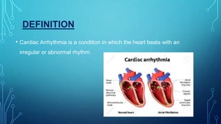
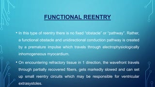
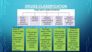
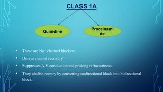
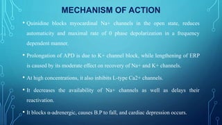
ANTI-ARRHYTHMIA
PRESENTED BY:
POOJA SWAMINATHAN
GURKIRAT KAUR
YASH SINGLA
DEFINITION
• Cardiac Arrhythmia is a condition in which the heart beats with an
irregular or abnormal rhythm.
WHY ABNORMAL RHYTHM?
 Extreme cases of abnormal rhythm
1. Bradycardia- Cardiac beats below 60 beats/min.
2. Tachycardia- Cardiac beats above 100 beats/min.
ETIOLOGY
Coronary artery disease,
Electrolyte imbalances in blood(i.e., sodium, potassium),
Changes in heart muscles,
Injury from heart attack.
CONTD…
Healing process after heart injury,
Irregular heart rhythms (can also occur in “normal, healthy” heart),
Ischemic Heart disease,
Drugs related
Other factors
SYMPTOMS
1) Palpitation 6) Sudden Cardiac death
2) Dizziness 7) Swelling
3) Chest Pain 8) Shortness of Breath
4) Dyspnea 9) Exercise Intolerance
5) Fainting
MECHANISM
• Three main mechanisms of cardiac arrhythmia:
1) Altered normal automaticity or abnormal automaticity
2) Triggered electrical activity
a) early after depolarization
b) delayed after depolarization
3) Reentry
a) Circus movement reentry
b) functional reentry
ABNORMAL AUTOMATICITY
The sinus node contains pacemaker cells that have spontaneous firing
capacity. This is called normal automaticity. Abnormal
automaticity occurs when other cells start firing spontaneously,
resulting in premature heartbeats. All cardiac cells have spontaneous
firing capacity, but at only at a very slow heart rate. Therefore, during
a normal heart rate, they will never have the chance to show off their
firing capacity. However, in pathologic conditions, such as during
extreme bradycardia, other cells can take over and cause for example
an AV-nodal heart rate.
TRIGGERED ACTIVITY
• During triggered activity heart cells contract twice, although they
only have been activated once. This is often caused by so called after
depolarization (early or delayed after depolarization EADs / DADs)
caused by electrical instability in the myocardial cell membrane.
DELAYED DEPOLARIZATION
CAUSE
S:
1) Ischemia
2) Drugs related (Potassium
Blockers)
3) Electrolyte imbalance
Effects:
1) Long QT interval
2) R on T phenomenon
3) Premature Ventricular Beat
4) Ventricular Fibrillation
REENTRY ACTIVITY
Re-entry is a common cause of arrhythmias. Ventricular
tachycardia and AV-nodal re-entry are typical examples. Re-entry can
occur when a conduction path is partly slowed down. As a result of this,
the signal is conducted by both a fast and a slow pathway. During normal
sinus rhythm this generally does not cause problems, but when an extra
systole follows rapidly upon the previous beat, the fast pathway is
sometimes still refractory and cannot conduct the signal.
Now the following sequence results in re-entry.
• The atrial signal coming from above is
conducted by the slow pathway.
• As the signal, going through the slow
pathway, reaches the end of the fast
pathway, it finds this pathway able to
conduct.
• The signal is conducted through the fast
pathway up to the beginning of the slow
pathway, which by that time is able to
conduct.
• This circle is perpetuated and a signal
generator is created. In the case of AV-
nodal re-entry this will typically generate
a signal at a frequency of 180-250 bpm.
CIRCUS MOVEMENT REENTRY
• It occurs in an anatomically defines circuit.
• A premature impulse, temporarily blocked in 1 direction by refractory
tissue, makes a one-way transit around an obstacle or though an
abnormal tract, finds the original spot in an advanced state of recovery
and reexcites it, setting up upcurrent activation of adjacent
myocardium.
• This type of reentry can be cured by radiofrequency catheter ablation
of the defined pathway.
FUNCTIONAL REENTRY
• In this type of reentry there is no fixed “obstacle” or “pathway”. Rather,
a functional obstacle and unidirectional conduction pathway is created
by a premature impulse which travels through electrophysiologically
inhomogeneous myocardium.
• On encountering refractory tissue in 1 direction, the wavefront travels
through partially recovered fibers, gets markedly slowed and can set
up small reentry circuits which may be responsible for ventricular
extrasystoles.
TREATMENT
DRUGS CLASSIFICATION
The primary
action of
drugs in class
1 is to limit
the
conductance
of Na+ and
K+ across
cell
membrane, a
local
anesthetic
The primary
action of
class 2 is to
suppress
adrenergicall
y mediated
ectopic
activity.
The main
action of this
class is
prolongation
of
repolarization
(phase 3).
Hence, AP is
widened and
ERP is
increased.
The primary
action of this
class of drugs
is to inhibit
Ca2+
mediated
slow channel
inward
current.
The main
action of
these drugs
is to act as
adenosine
receptor
agonist
(adenosine),
& to inhibit
Na+, k+, AND
ATPase
enzyme.
CLASS 1A
• These are Na+ channel blockers.
• Delays channel recovery.
• Suppresses A-V conduction and prolong refractoriness.
• They abolish reentry by converting undirectional block into bidirectional
block.
Quinidine
Procainami
de
MECHANISM OF ACTION
• Quinidine blocks myocardinal Na+ channels in the open state, reduces
automaticity and maximal rate of 0 phase depolarization in a frequency
dependent manner.
• Prolongation of APD is due to K+ channel block, while lengthening of ERP
is caused by its moderate effect on recovery of Na+ and K+ channels.
• At high concentrations, it also inhibits L-type Ca2+ channels.
• It decreases the availability of Na+ channels as well as delays their
reactivation.
• It blocks α-adrenergic, causes B.P to fall, and cardiac depression occurs.
USES
• Though quinidine is effective in many atrial and ventricular arrhythmias, it is
seldom used now, because of risk of adverse effects, including that of sudden
cardiac arrest or ventricular fibrillation.
• Given in a dose of 100-200mg TDS .
• Marketed Name: QUININGA 300mg tab, inj; NATCARDINE 100mg tab.
CLASS 1B
• It is the most commonly used local anesthetic.
• The most prominent cardiac action of lidocaine is suppression of automaticity
in ectopic foci.
• Enhanced phase-4 depolarization in partially depolarized PFs, and after
depolarization are antagonized, but SA node automaticity is not depressed.
• Lidocaine decreases APD in PF & ventricular muscle, but has practically no
effect on APD & ERP of atrial fibres.
LIDOCAINE
• Lidocaine has minimal effect on normal ECG; QT interval may decrease.
• It causes little depression of cardiac contractility or arterial BP.
Pharmacokinetics
It is inactive orally due to high first past metabolism in liver. Action of an i.v.
bolus lasts only 10-20 min because of rapid redistribution.
It is hydrolysed, deethylated and conjugated; metabolites are excreted in urine.
The t½ of early distribution phase is 8 min. While that of later elimination
phase is nearly two hours.
USES
• It is safe if given by slow i.v. injection.
• It is given only by i.v. route i.e., 50-100 mg bolus followed by 20-40 mg
every 10 to 20 mins or 1-3 mg per minute infusion.
• Marketed name: XYLOCARD, GESICARD 20 mg/ml inj.
CLASS 1C
• It is the prototype class of antiarrhythmic which markedly delays Na+
channel recovery.
• It has no consistent effect on APD and no ß adrenergic blocking property.
• It suppresses Ventricular tachycardia and PSVT.
• ↓ automaticity (↑ threshold)
• ↑ APD (K+) and ↑ refractory period, especially in rapidly depolarizing atrial
tissue.
Flecainide
USES
• Used for supraventricular arrhythmias (fibrillation)
• In premature ventricular contractions.
• Wolf-Parkinson-White syndrome.
• Marketed name : RHYTHMONORM 150 mg tab.
• Dose : 150 mg BD, 300 TDS.
CLASS 2
• These are Beta-Adrenoreceptor blocking drugs.
• Administration:
 Propranolol may injected i.v., 1mg/min. The usual oral dose is 40-20g,
2-4 times a day.
 Esmolol given i.v., only (very short acting, t½ is 9min.)
Maketed Name: MINIBLOCK 100mg/10ml.
Propranolo
l Sotalol
Esmolol
CARDIAC EFFECTS:
↑ APD & refractory period in AV node to slow AV conduction velocity.
USES:
Treating sinus and catecholamine dependent tachyarrhythmia's,
Converting reentrant arrhythmia’s in AV,
Protecting the ventricles from high atria; rates( slow AV conduction).
SIDE EFFECTS:
Bronchospasm
Hypotension
CLASS 3
1).Amiodarone: Its is incompletely and slowly absorbed from g.i.t.
It accumulates in muscle and fat from which it is slowly released and then metabolized in
liver mainly by CYP3A4. The duration of action is exceptionally long; t½ 3-8 weeks.
DOSE : 400-600mg/day for few weeks, followed by 100-200 mg.
Marketed Name: CORDARONE, ALDARONE, EURYTHMIC 100, 200 mg tabs.
Amiodaron
e
Dronedaron
e Ibutilide
Dofetilide
2.) Dronedarone-
Dose: 400mg BD oral.
Marketed name: MULTAQ 400 mg tab.
3.) Dofetilide-
These are labeled as pure class-3 antiarrhythmic.
Cardiac Effects:
↑ APD and refractory period
USES:
Maintain sinus rhythm in atrial fibrillation.
Ibutilide-
It is another new class-3rd antiarrhythmic drug, used i.v, for pharmacological
conversion of atrial flutter (AFI) and atrial fibrillation (AF) to sinus rhythm.
Cardiac Effects:
Result in ↑ APD.
USES:
Conversion OF Atrial fibrillation and flutter.
Side Effects:
Torsades de pointes(twisting of points)
CLASS 4
• VERAPAMIL
It has most prominent cardiac electrophysiological action. It blocks L-type
Ca2+ channels and delays their recovery.
The basic action is to depress Ca2+ mediated depolarization. This
suppresses automaticity and reentry dependent on slow channel response.
VERAPAMIL DILTIAZEM
• The most consistent action of verapamil is prolongation of A-V nodal ERP.
As a result A-V conduction is markedly slowed (P-R interval ↑’s) & reentry
involving A-V node is terminated.
• It ha negative inotropic action due to interference with Ca2+ mediated
excitation- contraction coupling in myocardium.
USES:
It terminates attack of PSVT.
Controls ventricles during supraventricular tachycardia.
SIDE EFFECTS:
Hypotension
↓’d Cardiac Output
Gastrointestinal problems
ADDITIONAL DRUGS
Adenosine:
It acts as a adenosine agonists by activating Ach sensitive K+ channels and
causes membrane hyperpolarization through interaction with A1 type of
adenosine GPCR’S on SA node (pacemaker depression→bradycardia), A-V
node (prolongation of ERP→ slowing of conduction) and atrium (shortening
of AP, reduced excitability).
It indirectly reduces Ca2+ current in A-V node.
Coronary dilation occurs transiently.
DIGIOXIN: (Cardiac Glycosides)
MECHANISM:
It enhances vagal activity(10th cranial nerve)
It ↑’s K+ currents, ↓’s Ca2+ currents, ↑’s refractory period
Slows A-V conduction & slow Heart Rate.
USES:
Treatment of atrial fibrillation.
ATROPINE
MECHANSIM:
Selective muscarinic antagonist.
It blocks A-V due to vagal overactivity, e.g, dititalis toxicity, some cases of
MI, it can be improved by atropine 0.6-1.2mg i.m. Atropine abbreviates
A-V node ERP and ↑’s conduction velocity in bundle of HIS.
USES:
Treat vagal bradycardia

Antiarrhythmia

  • 1.
  • 2.
    DEFINITION • Cardiac Arrhythmiais a condition in which the heart beats with an irregular or abnormal rhythm.
  • 3.
    WHY ABNORMAL RHYTHM? Extreme cases of abnormal rhythm 1. Bradycardia- Cardiac beats below 60 beats/min. 2. Tachycardia- Cardiac beats above 100 beats/min.
  • 4.
    ETIOLOGY Coronary artery disease, Electrolyteimbalances in blood(i.e., sodium, potassium), Changes in heart muscles, Injury from heart attack.
  • 5.
    CONTD… Healing process afterheart injury, Irregular heart rhythms (can also occur in “normal, healthy” heart), Ischemic Heart disease, Drugs related Other factors
  • 6.
    SYMPTOMS 1) Palpitation 6)Sudden Cardiac death 2) Dizziness 7) Swelling 3) Chest Pain 8) Shortness of Breath 4) Dyspnea 9) Exercise Intolerance 5) Fainting
  • 7.
    MECHANISM • Three mainmechanisms of cardiac arrhythmia: 1) Altered normal automaticity or abnormal automaticity 2) Triggered electrical activity a) early after depolarization b) delayed after depolarization 3) Reentry a) Circus movement reentry b) functional reentry
  • 8.
    ABNORMAL AUTOMATICITY The sinusnode contains pacemaker cells that have spontaneous firing capacity. This is called normal automaticity. Abnormal automaticity occurs when other cells start firing spontaneously, resulting in premature heartbeats. All cardiac cells have spontaneous firing capacity, but at only at a very slow heart rate. Therefore, during a normal heart rate, they will never have the chance to show off their firing capacity. However, in pathologic conditions, such as during extreme bradycardia, other cells can take over and cause for example an AV-nodal heart rate.
  • 9.
    TRIGGERED ACTIVITY • Duringtriggered activity heart cells contract twice, although they only have been activated once. This is often caused by so called after depolarization (early or delayed after depolarization EADs / DADs) caused by electrical instability in the myocardial cell membrane.
  • 10.
    DELAYED DEPOLARIZATION CAUSE S: 1) Ischemia 2)Drugs related (Potassium Blockers) 3) Electrolyte imbalance Effects: 1) Long QT interval 2) R on T phenomenon 3) Premature Ventricular Beat 4) Ventricular Fibrillation
  • 11.
    REENTRY ACTIVITY Re-entry isa common cause of arrhythmias. Ventricular tachycardia and AV-nodal re-entry are typical examples. Re-entry can occur when a conduction path is partly slowed down. As a result of this, the signal is conducted by both a fast and a slow pathway. During normal sinus rhythm this generally does not cause problems, but when an extra systole follows rapidly upon the previous beat, the fast pathway is sometimes still refractory and cannot conduct the signal. Now the following sequence results in re-entry.
  • 12.
    • The atrialsignal coming from above is conducted by the slow pathway. • As the signal, going through the slow pathway, reaches the end of the fast pathway, it finds this pathway able to conduct. • The signal is conducted through the fast pathway up to the beginning of the slow pathway, which by that time is able to conduct. • This circle is perpetuated and a signal generator is created. In the case of AV- nodal re-entry this will typically generate a signal at a frequency of 180-250 bpm.
  • 13.
    CIRCUS MOVEMENT REENTRY •It occurs in an anatomically defines circuit. • A premature impulse, temporarily blocked in 1 direction by refractory tissue, makes a one-way transit around an obstacle or though an abnormal tract, finds the original spot in an advanced state of recovery and reexcites it, setting up upcurrent activation of adjacent myocardium. • This type of reentry can be cured by radiofrequency catheter ablation of the defined pathway.
  • 14.
    FUNCTIONAL REENTRY • Inthis type of reentry there is no fixed “obstacle” or “pathway”. Rather, a functional obstacle and unidirectional conduction pathway is created by a premature impulse which travels through electrophysiologically inhomogeneous myocardium. • On encountering refractory tissue in 1 direction, the wavefront travels through partially recovered fibers, gets markedly slowed and can set up small reentry circuits which may be responsible for ventricular extrasystoles.
  • 15.
  • 16.
    DRUGS CLASSIFICATION The primary actionof drugs in class 1 is to limit the conductance of Na+ and K+ across cell membrane, a local anesthetic The primary action of class 2 is to suppress adrenergicall y mediated ectopic activity. The main action of this class is prolongation of repolarization (phase 3). Hence, AP is widened and ERP is increased. The primary action of this class of drugs is to inhibit Ca2+ mediated slow channel inward current. The main action of these drugs is to act as adenosine receptor agonist (adenosine), & to inhibit Na+, k+, AND ATPase enzyme.
  • 17.
    CLASS 1A • Theseare Na+ channel blockers. • Delays channel recovery. • Suppresses A-V conduction and prolong refractoriness. • They abolish reentry by converting undirectional block into bidirectional block. Quinidine Procainami de
  • 18.
    MECHANISM OF ACTION •Quinidine blocks myocardinal Na+ channels in the open state, reduces automaticity and maximal rate of 0 phase depolarization in a frequency dependent manner. • Prolongation of APD is due to K+ channel block, while lengthening of ERP is caused by its moderate effect on recovery of Na+ and K+ channels. • At high concentrations, it also inhibits L-type Ca2+ channels. • It decreases the availability of Na+ channels as well as delays their reactivation. • It blocks α-adrenergic, causes B.P to fall, and cardiac depression occurs.
  • 19.
    USES • Though quinidineis effective in many atrial and ventricular arrhythmias, it is seldom used now, because of risk of adverse effects, including that of sudden cardiac arrest or ventricular fibrillation. • Given in a dose of 100-200mg TDS . • Marketed Name: QUININGA 300mg tab, inj; NATCARDINE 100mg tab.
  • 20.
    CLASS 1B • Itis the most commonly used local anesthetic. • The most prominent cardiac action of lidocaine is suppression of automaticity in ectopic foci. • Enhanced phase-4 depolarization in partially depolarized PFs, and after depolarization are antagonized, but SA node automaticity is not depressed. • Lidocaine decreases APD in PF & ventricular muscle, but has practically no effect on APD & ERP of atrial fibres. LIDOCAINE
  • 21.
    • Lidocaine hasminimal effect on normal ECG; QT interval may decrease. • It causes little depression of cardiac contractility or arterial BP. Pharmacokinetics It is inactive orally due to high first past metabolism in liver. Action of an i.v. bolus lasts only 10-20 min because of rapid redistribution. It is hydrolysed, deethylated and conjugated; metabolites are excreted in urine. The t½ of early distribution phase is 8 min. While that of later elimination phase is nearly two hours.
  • 22.
    USES • It issafe if given by slow i.v. injection. • It is given only by i.v. route i.e., 50-100 mg bolus followed by 20-40 mg every 10 to 20 mins or 1-3 mg per minute infusion. • Marketed name: XYLOCARD, GESICARD 20 mg/ml inj.
  • 23.
    CLASS 1C • Itis the prototype class of antiarrhythmic which markedly delays Na+ channel recovery. • It has no consistent effect on APD and no ß adrenergic blocking property. • It suppresses Ventricular tachycardia and PSVT. • ↓ automaticity (↑ threshold) • ↑ APD (K+) and ↑ refractory period, especially in rapidly depolarizing atrial tissue. Flecainide
  • 24.
    USES • Used forsupraventricular arrhythmias (fibrillation) • In premature ventricular contractions. • Wolf-Parkinson-White syndrome. • Marketed name : RHYTHMONORM 150 mg tab. • Dose : 150 mg BD, 300 TDS.
  • 25.
    CLASS 2 • Theseare Beta-Adrenoreceptor blocking drugs. • Administration:  Propranolol may injected i.v., 1mg/min. The usual oral dose is 40-20g, 2-4 times a day.  Esmolol given i.v., only (very short acting, t½ is 9min.) Maketed Name: MINIBLOCK 100mg/10ml. Propranolo l Sotalol Esmolol
  • 26.
    CARDIAC EFFECTS: ↑ APD& refractory period in AV node to slow AV conduction velocity. USES: Treating sinus and catecholamine dependent tachyarrhythmia's, Converting reentrant arrhythmia’s in AV, Protecting the ventricles from high atria; rates( slow AV conduction). SIDE EFFECTS: Bronchospasm Hypotension
  • 27.
    CLASS 3 1).Amiodarone: Itsis incompletely and slowly absorbed from g.i.t. It accumulates in muscle and fat from which it is slowly released and then metabolized in liver mainly by CYP3A4. The duration of action is exceptionally long; t½ 3-8 weeks. DOSE : 400-600mg/day for few weeks, followed by 100-200 mg. Marketed Name: CORDARONE, ALDARONE, EURYTHMIC 100, 200 mg tabs. Amiodaron e Dronedaron e Ibutilide Dofetilide
  • 28.
    2.) Dronedarone- Dose: 400mgBD oral. Marketed name: MULTAQ 400 mg tab. 3.) Dofetilide- These are labeled as pure class-3 antiarrhythmic. Cardiac Effects: ↑ APD and refractory period USES: Maintain sinus rhythm in atrial fibrillation.
  • 29.
    Ibutilide- It is anothernew class-3rd antiarrhythmic drug, used i.v, for pharmacological conversion of atrial flutter (AFI) and atrial fibrillation (AF) to sinus rhythm. Cardiac Effects: Result in ↑ APD. USES: Conversion OF Atrial fibrillation and flutter. Side Effects: Torsades de pointes(twisting of points)
  • 30.
    CLASS 4 • VERAPAMIL Ithas most prominent cardiac electrophysiological action. It blocks L-type Ca2+ channels and delays their recovery. The basic action is to depress Ca2+ mediated depolarization. This suppresses automaticity and reentry dependent on slow channel response. VERAPAMIL DILTIAZEM
  • 31.
    • The mostconsistent action of verapamil is prolongation of A-V nodal ERP. As a result A-V conduction is markedly slowed (P-R interval ↑’s) & reentry involving A-V node is terminated. • It ha negative inotropic action due to interference with Ca2+ mediated excitation- contraction coupling in myocardium. USES: It terminates attack of PSVT. Controls ventricles during supraventricular tachycardia. SIDE EFFECTS: Hypotension ↓’d Cardiac Output Gastrointestinal problems
  • 32.
    ADDITIONAL DRUGS Adenosine: It actsas a adenosine agonists by activating Ach sensitive K+ channels and causes membrane hyperpolarization through interaction with A1 type of adenosine GPCR’S on SA node (pacemaker depression→bradycardia), A-V node (prolongation of ERP→ slowing of conduction) and atrium (shortening of AP, reduced excitability). It indirectly reduces Ca2+ current in A-V node. Coronary dilation occurs transiently.
  • 33.
    DIGIOXIN: (Cardiac Glycosides) MECHANISM: Itenhances vagal activity(10th cranial nerve) It ↑’s K+ currents, ↓’s Ca2+ currents, ↑’s refractory period Slows A-V conduction & slow Heart Rate. USES: Treatment of atrial fibrillation.
  • 34.
    ATROPINE MECHANSIM: Selective muscarinic antagonist. Itblocks A-V due to vagal overactivity, e.g, dititalis toxicity, some cases of MI, it can be improved by atropine 0.6-1.2mg i.m. Atropine abbreviates A-V node ERP and ↑’s conduction velocity in bundle of HIS. USES: Treat vagal bradycardia