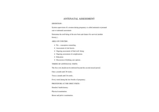
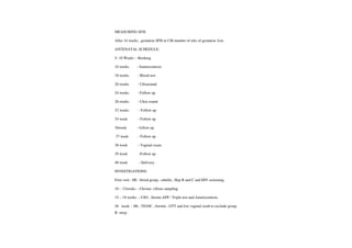
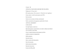
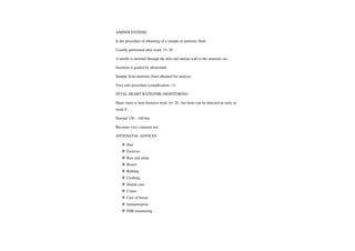
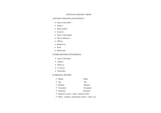
DEMONSTRATION
ON
ANTENATAL
ASSESSMENT
SUBMITTED TO SUBMITTED BY
MRS. R. ANITHA CH. HARI HARA DEEPA
LECTURER MSC NURSING 1ST YEAR
STUDENT PROFILE
NAME OF STUDENT :CH. HARI HARA DEEPA
CLASS MSC NUSING 1ST YEAR
SUBJECT :OBSTETRICAL AND GYNECOLOGICAL
NURSING
TOPIC ANTENATAL ASSESSMENT
GROUP MSC NURSING 1ST YEAR
NUMBER OF STUDENT 10
PLACE :GOVT.cOLLEGEOFNURSING
DATE :14/2/2020
DURATION :1 HOUR
AVAIDS :
BLACK BOARD , LEAF LETS, CHARTS
SUPERVISED BY MRS. R. HANSIL
OBJECTIVES
GENERAL OBJECTIVES:
At the end my demonstration student will able to in depth knowledge regarding
antenatalassessment.
SPECIFICOBJECTIVES:
At the end my demonstration student will able to in depth knowledge
regarding antenatal assessment.
.Explain definition of antenatal assessment.
Discuss aims, objectives of antenatal assessment.
.Discuss abouttime of antenatal visit.
Explain antenatal historytaking.
Elaborate antenatalphysical examination.
Discuss aboutabdominal examination
Explain aboutvaginal examination.
ANTENATAL ASSESSMENT
DEFINITION:
System supervision of a women during pregnancy is called antenatal or prenatal
care orantenatal assessment
Determine the well being of the new born and chance for survival (mother
history).
AREA OFCONCERS
Pre-conception counseling.
Assessment ofriskfactors.
Ongoing assessment of fetal well -being
Ongoing assessment ofcomplications.
Education
Discussion of birthingcare options.
TIMING OF ANTENATAL VISITS:
The first visit should not be deferred beyond the second missed period.
Once amonth until 28 weeks
Twice amonth until 36 weeks.
Every week during the last 4weeks of pregnancy
PROCEDURE AT THE FIRST VISITS
Detailed health history
Physical examination .
Breast andpelvicexamination
VITAL STASTICS
Name
Age
Ward/ unit
IP. No
Address
Religion
Occupation
Education
LMP
EDD
GA
Obstetric score
Blood group
GRAVIDA PARITY
Nulligravida Nullipara
Primigravida Primipara
Multigravida Multi para
Grand multipara
MATERNAL HISTORY:
Present obstetrical history
Diagnosis
Planned/ unplanned
Minor disorder
Immunization
Exposure to drugs / radiation.
PREGNANCYTESTS
Pregnancy kit
Urine examination
Blood examination
MATERNAL HISTORY AND RISK FACTORSS:
Comprehensive maternal history and physical examination is important to point
out risk factors.
Risk factors can be related to mother during pregnancy during labor and
delivery or afterdelivery.
Antenatal assessment starts with determination ofrisk factors.
Better knowledge about risk factors better preparation to care for the patient
ABORTION
3% ofpregnancies end in miscarriage.
Only rarely would an abortion cause problems in a subsequent pregnancy.
Increased risk of miscarriage only in women who have had multiple induced
abortions .
RISKFACTORS:
PRETERM BIRTH
What is considered preterm.
The second greatest causes of morbidity and mortality in neonates.
Previous preterm birth increases the subsequent preterm birth.
IPrior: 15% subsequentpreterm visit .
2prior: 32% subsequentpreterm birth
INCOMPTENTCERVIX:
Caused by cervical trauma, previous surgery ormaybe congenital.
Usually leads tomembrane rupture and premature delivery.
lf severe , a suture a round the cervical canal is performed
MATERNAL SMOKING AND ALCOHOL INTAKE
In the US, about 10% pregnant mothers smoke , drink alcohol or use drugs .
Maternal intake ofalcohol leads to fetal growth problems.
Smoking HBCO decrease availability of oxygen of placenta and fetus.
MATERNAL HYPERTENSION
Complicates 6-8 % ofpregnancies
Hypertensions during pregnancy ( afterW24 ) is termed Preeclampsia.
Pre eclampsia ( High BP. proteinuria , edema)
Can lead to placental abruption and preterm delivery.
DIABETES
Increase the risk for CV and CNS malformations and metabolic disturbances.
When appearduring pregnancy ( gestational diabetes, GDM)
Treatment :glycemic control
INFECTIONS DISEASE
Infections can be transmitted to fetus.
Early screening and detection ofthe infection is important.
Complicated by the rupture of the membrane.
PROBLEMS INPILACENTA,UC AND FETAIL MEMBRANE
Premature rupture: causes 50 % of preterm births.
UC( ulcerative colitis): prolapse
,short, single artery (3% )
Placental problems .
ANTENATAL ASSESSMENT:
Height
Weight
Pallor
Jaundice
Vital signs.
BREAST EXAMINATION:
Flat nipple does not protrude with stimulation.
Retracted (nipple bulls back slightly )
Inverted (nipple pulls in ward when compressed).
ABDOMIAL EXAMINATION:
INSPECTION
Size
Shape
Counter
Flank
Skin
Bladder
Fetal movements
MEASURING SFH:
After 14 weeks , gestation SFH in CM number of wks of gestation 3cm.
ANTENATAL SCHEDULE:
5-10 Weeks -
Booking
l6 weeks Amniocentesis
18 weeks -
Blood test
20 weeks -
Ultrasound
24 weeks -
Follow up
28 weeks -
Ultra sound
32 weeks -
Follow up
34 week -
Follow up
36week -
follow up
37 week -
Follow up
38 week -
Vaginal exam
39 week -Follow up
40 week -Delivery.
INVESTIGATIONS:
First visit: Hb. blood group, rubella, Hep B andC and HIV screening.
10- 12weeks -
Chronic villous sampling
15- 18 weeks -USG, Serum AFP/Tripletest and Amniocentesis.
28 week Hb, TD/DC ferritin, GTTand low vaginal swab to exclude group
B strep
36 weeks -
Hb
ANTENATAL CHART SHOULD RECORD THE FOLLOWING
Weight gain (12-15 kgs in total )
BP ( diastolic pressure >90 or increase of>20 from first visit is significant)
Urinary analysis ( watch for protein glucose and UTIS )
Fetal movementS.
Uterine size in accordance with dates and ultrasound
Fetal lie, presentation and engagementespecially after 36 weeks.
ANTNATAL ASSESSMENT:
ULTRA SOUND
Uses high frequency sound waves.
Hand- held transducer is placed directly over mother's abdomen and reflected
waves recorded on screen image
Can give valuable information about pregnancy and fetus.
CLINICAL USES OFULTRASOUND
Identify pregnancy
Determine fetal age.
Observe amnioticfluid abnormalities.
Detect fetal anomalies .
Identify placental abnormalities
Determine fetal position.
Examine fetal HR and RR.
AMINOCENTESIS:
Is the procedure of obtaining ofasample of amniotic fluid.
Usually performed afterweek 15-20.
A needle is inserted through the skin and uterine wall to the amniotic sac.
Insertion is guided by ultrasound
Sample from amniotic fluid obtained for analysis.
Very safe procedure (complication <1)
FETAL HEARTRATE(FHR) MONITORING
Heart starts to beat between week 16- 20, but beats can be detected as early as
week 8.
Normal 120- 160 bm
Becomes very commontest.
ANTENATAL ADVICES
Diet
Exercise
Rest and sleep
Bowel
Bathing
Clothing
Dental care
Coitus
Care of breast
Immunization
FHR monitoring
ANTENATAL HISTORY TAKING
I.MOTHER'S IDENTIFICATIONPROFILE
Name of the mother
Address:
Phone number
E-mail id:
Name of the hospital:
Date of admission
OPD no
Inpatientno:
Ward:
Obstetrician:
FATHER IDENTIFICATION PROFILE:
Name of the father
Address:
Phone no:
.E-mailid:
Nationality:
II. PERSONAL HISTORY :
Mother Father
Age Age
Religion Religion
Occupation Occupation
Education Education
Supportive system :family /husband /others
Habits :smoking /alcohol/drug/ tobacco/ coffee /tea.
Life style
Hobbies
Hygiene :good / fair /poor
Elimination ( bowel) :Good/ fair/ poor
(Bladder): Good /fair /poor
Rest and sleep :
III. FAMILY MEDICAL HISTORY
a. Health status of parents / sibling (if decreased, mention cause of
death
b. History ofthefollowing disease in parents /sibling /close relative's
Diabetes mellitus Tuberculosis
Vascular disease Congenital disease
Hypertension Cancer
Renal disease Complication of pregnancy
Cardiopulmonary disease Multiple pregnancy
Neuromuscular condition
Psychiatric disease
IV. PERSONAL MEDIACL HISTORY
Child hood disease.
Immunization
Hospitalization
Surgery
Blood transfusion
Allergy
HSTORY OF ANY OF THE FOLLOWING DISEASE:
Diabetic mellitus tuberculosis
Rheumatic fever Cancer
Cardiopulmonary condition Mal nutrition
Asthma Hypertension
Endocrinopathy
Blood dyscarasias
Sexually transmitted disease
Renal/urinary tractinfections
Psychiatric disorder
Injury to pelvic organ
V. MENSTRUAL HISTORY :
Age of menarche
Duration of cycle
Duration
Amount of flow
Regular/ irregular
Clot
VI. SEXUALHISTORY:
Attitude toward sexually, particular affected pregnancy.
Contraceptive history and practice.
Sexual transmitted disease ( if any ) type
Treatment taken
PREGANNACY HISTORY -PASTOBSTRITICAL HISTORY:
Year Length of gestation Abortion Type of delivery Complication Baby
Of labor
VIII PRENATAL OBSTETRIC HISTORY:
Gravida
Parity
Expected date of birth
Signs and symptoms of pregnancy
Rest and sleep
Activity and employment plan
Sexual activity
IX. PSYCHOSOCIAL HISTORY:
Psychiatric and mental health history
Self concept or self esteenm issues
Support system available to contact at the time of need
Stressor -
occupational or personal that may affect pregnancy of the fetus
Past history
Emotional/ changes and adjustment to pregnancy
ANTENATAL PHYSICAL EXAMINATION
PART EXAMINED NORMAL FINDINGS ABNORMAL
EXAMINATION FINDINGS
TECHNIQUE
HEAD AND NECK
Inspection with otoscope Hyperemia of nasal enlarged lymph
And ophthalmoscope and and buccal mucosa nodes,thyroid
Visual inspection palpation membrane slight tenderness nodules
Of nodes thyroid . Diffuse enlargement orirregular,
Thyroid. enlargement lesions
Of eyes or mouth caries
And abscesses ofteeth,
Earinfections.
CHEST AND HEAT
Auscultation with lungs clear, heart Adventitious lungs
Stethoscope
,percussion in regular rhythm . sound, irregular
And inspection cardiac rhythm
Non physiologic
Membrane.
BREAST
Palpation and inspection Enlargement of breast Masses or nodules
Ofnipple, breast and with increased vascular bloody or serious
Axilla. pattern, darkened areola nipple
With prominent tubercles, discharge ,lesions
Thin
,yellow fluid secreted erythema.
From nipple in late
Pregnancy .
SKIN
Inspection and Pigmentation changes Pallor. jaundice
Palpation rash, skinlesions .
EXTRIMITIES
Visual inspection and Mild pre tibal and Limitation of motion
Palpation, percussion ankle edema in third varicosity more than
Auscultation reflex trimester slight edema slight, hand or ankle
Hammeer of hands in hot weather edema hyper reflex
ABDOMEN
Palpation, inspection Enlarged uterus palpation uterus too large
Auscultation palpation fetal outline in later or too small for
Percussion pregnancy, fetal heart sound dates absence fetal
Contraction last trimester heart sound beyond
10 weeks
PELIVS
Speculum examination speculum examination speculum examination
Bimanual examination bluish discolorations yellow, purulentfrothy
With inspection and ofmucosa ofvagina , cherry white, foul
Palpation
,collection of and cervix , congenital smelling
Specimen cervix multigravida
Bimanual examination Bimanual
Cervix soft
,admits a finger examination
Or two, uterus soft and cervix is dilated and
Enlarged fetal head or parts affected cervical or
May be felt in lower uterine vaginal masses
Segment gynecoid pelvic amnioticfluid..
Configuration masses or fullness
Rectal masses
Hemorrhoids.
Pap smear: squamous , pap smear ; inflammation
Metaplasia negative or presence of trichomonas of
Normal, adequate or fungi diminished or absent
Increasedestrogen estrogen
THE MAIN OBJECTIVES OF ABDOMINAL EXAMINATION:
.To observe the signs ofpregnancy
.To assess fetal size and growth
To auscultate the fetal heart
.To locatefetal parts
To identify any abnormalities.
PREAPRATION OF THE MOTHER FOR ABDOMINAL EXAMINATION:
Prior to starting abdominal examination, obstetrical nurse/midwife.mustwash
her hands and keep the dry and warm
She should conduct the examination standing to the right side of the mother.
Explain the procedure to her.
Provide privacy
Askherto empty her bladder.
Lay her in supine position with knee flexed.
Ensure that she is lying comfortable with her arms by her side to relax to
abdominalmuscles.
Drape her in such a way that only the abdomen is exposed for examination.
Keep the articles try for abdominal examination.
STEPS ABDOMINAL EXAMINATION:
INSPECTON
ASSESSMENT OF FUNDAL HEIGHT
ABDOMINAL PALPATION
AUSULTATION OF FETAL HEART.
INSPECTION:
During inspection, the obstetrical nurse /mid wife should observe the following
features:
Striae gravidarum
Linea nigra
Skin rashes
Sores on the skin
Injury
Surgical scar
SIZE OT THE ABDOMEN:
Fundal height is measured to assess the size of the uterus to see whether, it is
growing according to the period of gestation as well as to document the size and
to compare with the next assessment. the size of the abdomen is determined by
measuring the fundal height.
Shape of the abdomen: the general counters of the abdomen is noted
Oval shaped -
abdomen indicates a longitudinal lie of the fetus and that
fetus and that it is well flexed.
A round shaped - abdomen indicates polyhydramnios or multiple
gestation.
A heart shaped -indicates bicornuate uterus an abdomen widerthan it is
long suggests transverse lie
A flabby shaped -
indicates grand multiparity
A saucer shaped-depression in the center suggests fetal back lying
posteriorly in mother's abdomen.
FETAL MOVEMENT:
From the 20h week of pregnancy fetal movement should be present. It indicates
that the fetus is alive
UTERINE CONTRACTION:
The mother may show signs of discomfort or pain during contraction.
ASSESSMENT OF FUNDAL HEIGHT:
Fundal height is measured to evaluate the fetus gestation age. it is measured
from the top of the fundus to the top of the symphysis pubis using a non-
stretchable tape. it is documented in centimeters.
The uterus also grows and it is assessed by measuring the fundal height.
Height of the fundus is measured every 4 weeks. during pregnancy starting from
12 weeks to 36 weeks.
Because the fundus of the uterus is not palpable till 12 weeks.
This assessment is very important to know the intrauterine fetal development,
when the fundal height is less the period of gestation if suggests intra uterine
growth restriction
PROCEDURE:
Take consent from the mother
Always advice the women to empty her bladder before symphysis fundal height
measurement is taken because fundal height 17th to 20th week of pregnancy has
been found to measure 3cm higher in the presence of a full bladder.
Help the mother lie down in a comfortable semi recumbent position
Place the non-stretchable tape touching the skin down ward from the top of
uterus to the upper part of the symphysis pubis.
Documented the findings in centimeters.
When the SFH measure shorter than the period of gestation, if suggests
The baby may be small and unhealthy
Engagement ofthe fetal presenting part into maternal pelvis
Inaccurate expected date ofEDD
Baby is small for gestation
Transverse lie.
Oligohydramnios.
When the SFH measure longer than the period of gestation, if suggests
The baby may be large
Multiple pregnancy
Inaccurate EDD
Baby is large forgestationalage
Breech presentation when the presenting part is not engaged.
Polyhydramnios.
ABDOMINAL PALPATION:
The fundal height the midwife/ obstetrical nurse are palpation to identify the
lie, position attitude and presenting part of the fetus (Leopold maneuver)
There are four steps in abdominal palpation
Fundal palpation
Lateral palpation
Pawlick palpation
Pelvic palpation
THE AIM IS TO:
Look for signs ofpregnancy
Assess fetal growth
Locate fetal parts
Auscultate the fetal heart
Note abnormalities.
The women is asked to empty her bladder as a full bladder can cause discomfort
while examining and shows an increased height of the uterus while palpating.
Help the mother lie down in comfortable semi recumbent position.
Place the non-stretchable tape touching the skin down ward from the top of
uterus to the upper part of the symphysis pubis.
Document the findings in centimeters.
When the SFH measure shorter than the period of gestation, if
suggests
The baby may be small and un healthy
Engagement of fetal presenting part into maternal pelvis
Inaccurate expected date of delivery
Baby is small for gestation age
Transverse lie.
When the SFH measure longer than the period of gestation, if
suggests
The baby may be large
Multiple pregnancy
LEOPOLD MANEUVERS
Overall objectives: to determinate fetal position by systemic abdominal
palpation.
NURSING ACTION:
Explain procedure to women and rationale what is being done.
Instruct women to empty bladder before beginning procedure. if she has not
recently voided.
Instruct women to lie flat on her back, with her knee flexed slightly, place a
small pillow or rolled towel under one side.
Wash hands with warm water.
FIRST MANEUVER:
Objectives: To determine in fetal head breech is in fundus.
Observe women s abdomen before beginning palpation.
Stand at the foot of the examining table, facing the women and gently place
both hands flat on the abdomen.
Palpate upper abdomen with both hands.
Determine if the mass palpated is the hands or butocks by observing the
relative consistency, shape and mobility.
Assess ballottement the ability of the head to be moved back and forth against
the examining fingers.
SECOND MANEUVER:
OBJECTIVE: To locate the back of the fetus in relation to the right and left side
of the abdomen.
Facing the women, place the palmer surfaces of the hands on either side of the
abdomen and apply gentle but deep pressure.
Hold one hand still while using the flat surface to the fingers on the other hand
to gradually palpate the opposite side from the top to the lower segment of the
uterus show.
Palpate the fetal out line.
Reassure actions to the hands to palpate the outside.
THIRD MANEUVER:
OBJECTIVES: To determine if the fetal head is at the pelvic inlet and
determined its mobility.
Gently grasp the lower portion of the abdomen, just above the symphysis pubis
between the them band the fingers of the hand.
Press thunmb and fingers together in an attempt to grasp the presenting part.
FOUTH MANEUVER:
OBJECIVES: To determine if the fetal head is well flexed and how far it is
distended in the pelvic, to confirm location of fetal back.
Stand facing women s feet.
Place hips of first fingers on both sides of the mid line about 2 inches above
pouparts igament
Exert pressure downward and in the direction of the birth canal, moving skin of
the abdomen down ward a long with the fingers.
Allow fingers of one hand to be carried down ward well under the pouparts
ligament.
Slide fingers of the other hand as far as possible.
AUSULTATION OF FETAL HEART:
The fetal heart is auscultated, but putting the stethoscope or fetal doppler on the
fetal back at the position of the fetal heart. it is moved about till the maximum
intensity of sound is obtained.
The fetal heart is counted and to determine whether it is not maternal, the pulse
is counted a long side the normal range of the fetal heart rate is 110-160 bpm.
PELVIC EXAMINATION:
A complete pelvic examination is done during the initial antepartum
examination.
This include speculum examination, bimanual examination and retro vaginal
examination as well as evaluation of the bony pelvis for assessment pelvic
capacity.
SUMMARY:
At the end of my topic student will able to knowledge regarding definition of
antenatal assessment, aims and objectives of antenatal assessment, time of
antenatal visits, antenatal history and physical examination, abdominal
examination and vaginal examination.
CONCULISION:
At the end of my topic student will able to knowledge regarding, definition of
antenatal assessment, aims and objectives of antenatal assessment, time of
antenatal visits. antenatal history collection, physical examination, abdominal
examination and vaginal examination.
BIBLIOGRAPHY:
Dutta DC 2015 "A text book of obstetrics ", Hiralal konar 8th edition
,new
Delhi, health sciencepublishers.
Annamma Jacob, "A text book of midwifery and gynecological nursing "*:
5th
edition,Jaypeepublications.
Myles, "
A text book ofmidwifes " 16th edition. published by Elsevier
publications.
www.Nursing journals .com
www.Obsgyn. com
ANTENATAL ASSESSMENT1.pdf

ANTENATAL ASSESSMENT1.pdf

  • 1.
    DEMONSTRATION ON ANTENATAL ASSESSMENT SUBMITTED TO SUBMITTEDBY MRS. R. ANITHA CH. HARI HARA DEEPA LECTURER MSC NURSING 1ST YEAR
  • 2.
    STUDENT PROFILE NAME OFSTUDENT :CH. HARI HARA DEEPA CLASS MSC NUSING 1ST YEAR SUBJECT :OBSTETRICAL AND GYNECOLOGICAL NURSING TOPIC ANTENATAL ASSESSMENT GROUP MSC NURSING 1ST YEAR NUMBER OF STUDENT 10 PLACE :GOVT.cOLLEGEOFNURSING DATE :14/2/2020 DURATION :1 HOUR AVAIDS : BLACK BOARD , LEAF LETS, CHARTS SUPERVISED BY MRS. R. HANSIL
  • 3.
    OBJECTIVES GENERAL OBJECTIVES: At theend my demonstration student will able to in depth knowledge regarding antenatalassessment. SPECIFICOBJECTIVES: At the end my demonstration student will able to in depth knowledge regarding antenatal assessment. .Explain definition of antenatal assessment. Discuss aims, objectives of antenatal assessment. .Discuss abouttime of antenatal visit. Explain antenatal historytaking. Elaborate antenatalphysical examination. Discuss aboutabdominal examination Explain aboutvaginal examination.
  • 4.
    ANTENATAL ASSESSMENT DEFINITION: System supervisionof a women during pregnancy is called antenatal or prenatal care orantenatal assessment Determine the well being of the new born and chance for survival (mother history). AREA OFCONCERS Pre-conception counseling. Assessment ofriskfactors. Ongoing assessment of fetal well -being Ongoing assessment ofcomplications. Education Discussion of birthingcare options. TIMING OF ANTENATAL VISITS: The first visit should not be deferred beyond the second missed period. Once amonth until 28 weeks Twice amonth until 36 weeks. Every week during the last 4weeks of pregnancy PROCEDURE AT THE FIRST VISITS Detailed health history Physical examination . Breast andpelvicexamination
  • 5.
    VITAL STASTICS Name Age Ward/ unit IP.No Address Religion Occupation Education LMP EDD GA Obstetric score Blood group GRAVIDA PARITY Nulligravida Nullipara Primigravida Primipara Multigravida Multi para Grand multipara MATERNAL HISTORY: Present obstetrical history Diagnosis Planned/ unplanned Minor disorder
  • 6.
    Immunization Exposure to drugs/ radiation. PREGNANCYTESTS Pregnancy kit Urine examination Blood examination MATERNAL HISTORY AND RISK FACTORSS: Comprehensive maternal history and physical examination is important to point out risk factors. Risk factors can be related to mother during pregnancy during labor and delivery or afterdelivery. Antenatal assessment starts with determination ofrisk factors. Better knowledge about risk factors better preparation to care for the patient ABORTION 3% ofpregnancies end in miscarriage. Only rarely would an abortion cause problems in a subsequent pregnancy. Increased risk of miscarriage only in women who have had multiple induced abortions . RISKFACTORS: PRETERM BIRTH What is considered preterm. The second greatest causes of morbidity and mortality in neonates. Previous preterm birth increases the subsequent preterm birth.
  • 7.
    IPrior: 15% subsequentpretermvisit . 2prior: 32% subsequentpreterm birth INCOMPTENTCERVIX: Caused by cervical trauma, previous surgery ormaybe congenital. Usually leads tomembrane rupture and premature delivery. lf severe , a suture a round the cervical canal is performed MATERNAL SMOKING AND ALCOHOL INTAKE In the US, about 10% pregnant mothers smoke , drink alcohol or use drugs . Maternal intake ofalcohol leads to fetal growth problems. Smoking HBCO decrease availability of oxygen of placenta and fetus. MATERNAL HYPERTENSION Complicates 6-8 % ofpregnancies Hypertensions during pregnancy ( afterW24 ) is termed Preeclampsia. Pre eclampsia ( High BP. proteinuria , edema) Can lead to placental abruption and preterm delivery. DIABETES Increase the risk for CV and CNS malformations and metabolic disturbances. When appearduring pregnancy ( gestational diabetes, GDM) Treatment :glycemic control INFECTIONS DISEASE Infections can be transmitted to fetus. Early screening and detection ofthe infection is important.
  • 8.
    Complicated by therupture of the membrane. PROBLEMS INPILACENTA,UC AND FETAIL MEMBRANE Premature rupture: causes 50 % of preterm births. UC( ulcerative colitis): prolapse ,short, single artery (3% ) Placental problems . ANTENATAL ASSESSMENT: Height Weight Pallor Jaundice Vital signs. BREAST EXAMINATION: Flat nipple does not protrude with stimulation. Retracted (nipple bulls back slightly ) Inverted (nipple pulls in ward when compressed). ABDOMIAL EXAMINATION: INSPECTION Size Shape Counter Flank Skin Bladder Fetal movements
  • 9.
    MEASURING SFH: After 14weeks , gestation SFH in CM number of wks of gestation 3cm. ANTENATAL SCHEDULE: 5-10 Weeks - Booking l6 weeks Amniocentesis 18 weeks - Blood test 20 weeks - Ultrasound 24 weeks - Follow up 28 weeks - Ultra sound 32 weeks - Follow up 34 week - Follow up 36week - follow up 37 week - Follow up 38 week - Vaginal exam 39 week -Follow up 40 week -Delivery. INVESTIGATIONS: First visit: Hb. blood group, rubella, Hep B andC and HIV screening. 10- 12weeks - Chronic villous sampling 15- 18 weeks -USG, Serum AFP/Tripletest and Amniocentesis. 28 week Hb, TD/DC ferritin, GTTand low vaginal swab to exclude group B strep
  • 10.
    36 weeks - Hb ANTENATALCHART SHOULD RECORD THE FOLLOWING Weight gain (12-15 kgs in total ) BP ( diastolic pressure >90 or increase of>20 from first visit is significant) Urinary analysis ( watch for protein glucose and UTIS ) Fetal movementS. Uterine size in accordance with dates and ultrasound Fetal lie, presentation and engagementespecially after 36 weeks. ANTNATAL ASSESSMENT: ULTRA SOUND Uses high frequency sound waves. Hand- held transducer is placed directly over mother's abdomen and reflected waves recorded on screen image Can give valuable information about pregnancy and fetus. CLINICAL USES OFULTRASOUND Identify pregnancy Determine fetal age. Observe amnioticfluid abnormalities. Detect fetal anomalies . Identify placental abnormalities Determine fetal position. Examine fetal HR and RR.
  • 11.
    AMINOCENTESIS: Is the procedureof obtaining ofasample of amniotic fluid. Usually performed afterweek 15-20. A needle is inserted through the skin and uterine wall to the amniotic sac. Insertion is guided by ultrasound Sample from amniotic fluid obtained for analysis. Very safe procedure (complication <1) FETAL HEARTRATE(FHR) MONITORING Heart starts to beat between week 16- 20, but beats can be detected as early as week 8. Normal 120- 160 bm Becomes very commontest. ANTENATAL ADVICES Diet Exercise Rest and sleep Bowel Bathing Clothing Dental care Coitus Care of breast Immunization FHR monitoring
  • 12.
    ANTENATAL HISTORY TAKING I.MOTHER'SIDENTIFICATIONPROFILE Name of the mother Address: Phone number E-mail id: Name of the hospital: Date of admission OPD no Inpatientno: Ward: Obstetrician: FATHER IDENTIFICATION PROFILE: Name of the father Address: Phone no: .E-mailid: Nationality: II. PERSONAL HISTORY : Mother Father Age Age Religion Religion Occupation Occupation Education Education Supportive system :family /husband /others Habits :smoking /alcohol/drug/ tobacco/ coffee /tea.
  • 13.
    Life style Hobbies Hygiene :good/ fair /poor Elimination ( bowel) :Good/ fair/ poor (Bladder): Good /fair /poor Rest and sleep : III. FAMILY MEDICAL HISTORY a. Health status of parents / sibling (if decreased, mention cause of death b. History ofthefollowing disease in parents /sibling /close relative's Diabetes mellitus Tuberculosis Vascular disease Congenital disease Hypertension Cancer Renal disease Complication of pregnancy Cardiopulmonary disease Multiple pregnancy Neuromuscular condition Psychiatric disease IV. PERSONAL MEDIACL HISTORY Child hood disease. Immunization Hospitalization Surgery Blood transfusion
  • 14.
    Allergy HSTORY OF ANYOF THE FOLLOWING DISEASE: Diabetic mellitus tuberculosis Rheumatic fever Cancer Cardiopulmonary condition Mal nutrition Asthma Hypertension Endocrinopathy Blood dyscarasias Sexually transmitted disease Renal/urinary tractinfections Psychiatric disorder Injury to pelvic organ V. MENSTRUAL HISTORY : Age of menarche Duration of cycle Duration Amount of flow Regular/ irregular Clot VI. SEXUALHISTORY: Attitude toward sexually, particular affected pregnancy. Contraceptive history and practice.
  • 15.
    Sexual transmitted disease( if any ) type Treatment taken PREGANNACY HISTORY -PASTOBSTRITICAL HISTORY: Year Length of gestation Abortion Type of delivery Complication Baby Of labor VIII PRENATAL OBSTETRIC HISTORY: Gravida Parity Expected date of birth Signs and symptoms of pregnancy Rest and sleep Activity and employment plan Sexual activity IX. PSYCHOSOCIAL HISTORY: Psychiatric and mental health history Self concept or self esteenm issues Support system available to contact at the time of need Stressor - occupational or personal that may affect pregnancy of the fetus Past history Emotional/ changes and adjustment to pregnancy
  • 16.
    ANTENATAL PHYSICAL EXAMINATION PARTEXAMINED NORMAL FINDINGS ABNORMAL EXAMINATION FINDINGS TECHNIQUE HEAD AND NECK Inspection with otoscope Hyperemia of nasal enlarged lymph And ophthalmoscope and and buccal mucosa nodes,thyroid Visual inspection palpation membrane slight tenderness nodules Of nodes thyroid . Diffuse enlargement orirregular, Thyroid. enlargement lesions Of eyes or mouth caries And abscesses ofteeth, Earinfections. CHEST AND HEAT Auscultation with lungs clear, heart Adventitious lungs Stethoscope ,percussion in regular rhythm . sound, irregular And inspection cardiac rhythm Non physiologic Membrane. BREAST Palpation and inspection Enlargement of breast Masses or nodules Ofnipple, breast and with increased vascular bloody or serious
  • 17.
    Axilla. pattern, darkenedareola nipple With prominent tubercles, discharge ,lesions Thin ,yellow fluid secreted erythema. From nipple in late Pregnancy . SKIN Inspection and Pigmentation changes Pallor. jaundice Palpation rash, skinlesions . EXTRIMITIES Visual inspection and Mild pre tibal and Limitation of motion Palpation, percussion ankle edema in third varicosity more than Auscultation reflex trimester slight edema slight, hand or ankle Hammeer of hands in hot weather edema hyper reflex ABDOMEN Palpation, inspection Enlarged uterus palpation uterus too large Auscultation palpation fetal outline in later or too small for Percussion pregnancy, fetal heart sound dates absence fetal Contraction last trimester heart sound beyond 10 weeks PELIVS Speculum examination speculum examination speculum examination Bimanual examination bluish discolorations yellow, purulentfrothy
  • 18.
    With inspection andofmucosa ofvagina , cherry white, foul Palpation ,collection of and cervix , congenital smelling Specimen cervix multigravida Bimanual examination Bimanual Cervix soft ,admits a finger examination Or two, uterus soft and cervix is dilated and Enlarged fetal head or parts affected cervical or May be felt in lower uterine vaginal masses Segment gynecoid pelvic amnioticfluid.. Configuration masses or fullness Rectal masses Hemorrhoids. Pap smear: squamous , pap smear ; inflammation Metaplasia negative or presence of trichomonas of Normal, adequate or fungi diminished or absent Increasedestrogen estrogen THE MAIN OBJECTIVES OF ABDOMINAL EXAMINATION: .To observe the signs ofpregnancy .To assess fetal size and growth To auscultate the fetal heart .To locatefetal parts To identify any abnormalities.
  • 19.
    PREAPRATION OF THEMOTHER FOR ABDOMINAL EXAMINATION: Prior to starting abdominal examination, obstetrical nurse/midwife.mustwash her hands and keep the dry and warm She should conduct the examination standing to the right side of the mother. Explain the procedure to her. Provide privacy Askherto empty her bladder. Lay her in supine position with knee flexed. Ensure that she is lying comfortable with her arms by her side to relax to abdominalmuscles. Drape her in such a way that only the abdomen is exposed for examination. Keep the articles try for abdominal examination. STEPS ABDOMINAL EXAMINATION: INSPECTON ASSESSMENT OF FUNDAL HEIGHT ABDOMINAL PALPATION AUSULTATION OF FETAL HEART. INSPECTION: During inspection, the obstetrical nurse /mid wife should observe the following features: Striae gravidarum Linea nigra Skin rashes
  • 20.
    Sores on theskin Injury Surgical scar SIZE OT THE ABDOMEN: Fundal height is measured to assess the size of the uterus to see whether, it is growing according to the period of gestation as well as to document the size and to compare with the next assessment. the size of the abdomen is determined by measuring the fundal height. Shape of the abdomen: the general counters of the abdomen is noted Oval shaped - abdomen indicates a longitudinal lie of the fetus and that fetus and that it is well flexed. A round shaped - abdomen indicates polyhydramnios or multiple gestation. A heart shaped -indicates bicornuate uterus an abdomen widerthan it is long suggests transverse lie A flabby shaped - indicates grand multiparity A saucer shaped-depression in the center suggests fetal back lying posteriorly in mother's abdomen. FETAL MOVEMENT: From the 20h week of pregnancy fetal movement should be present. It indicates that the fetus is alive UTERINE CONTRACTION: The mother may show signs of discomfort or pain during contraction.
  • 21.
    ASSESSMENT OF FUNDALHEIGHT: Fundal height is measured to evaluate the fetus gestation age. it is measured from the top of the fundus to the top of the symphysis pubis using a non- stretchable tape. it is documented in centimeters. The uterus also grows and it is assessed by measuring the fundal height. Height of the fundus is measured every 4 weeks. during pregnancy starting from 12 weeks to 36 weeks. Because the fundus of the uterus is not palpable till 12 weeks. This assessment is very important to know the intrauterine fetal development, when the fundal height is less the period of gestation if suggests intra uterine growth restriction PROCEDURE: Take consent from the mother Always advice the women to empty her bladder before symphysis fundal height measurement is taken because fundal height 17th to 20th week of pregnancy has been found to measure 3cm higher in the presence of a full bladder. Help the mother lie down in a comfortable semi recumbent position Place the non-stretchable tape touching the skin down ward from the top of uterus to the upper part of the symphysis pubis. Documented the findings in centimeters. When the SFH measure shorter than the period of gestation, if suggests The baby may be small and unhealthy Engagement ofthe fetal presenting part into maternal pelvis Inaccurate expected date ofEDD
  • 22.
    Baby is smallfor gestation Transverse lie. Oligohydramnios. When the SFH measure longer than the period of gestation, if suggests The baby may be large Multiple pregnancy Inaccurate EDD Baby is large forgestationalage Breech presentation when the presenting part is not engaged. Polyhydramnios. ABDOMINAL PALPATION: The fundal height the midwife/ obstetrical nurse are palpation to identify the lie, position attitude and presenting part of the fetus (Leopold maneuver) There are four steps in abdominal palpation Fundal palpation Lateral palpation Pawlick palpation Pelvic palpation THE AIM IS TO: Look for signs ofpregnancy Assess fetal growth Locate fetal parts Auscultate the fetal heart Note abnormalities.
  • 23.
    The women isasked to empty her bladder as a full bladder can cause discomfort while examining and shows an increased height of the uterus while palpating. Help the mother lie down in comfortable semi recumbent position. Place the non-stretchable tape touching the skin down ward from the top of uterus to the upper part of the symphysis pubis. Document the findings in centimeters. When the SFH measure shorter than the period of gestation, if suggests The baby may be small and un healthy Engagement of fetal presenting part into maternal pelvis Inaccurate expected date of delivery Baby is small for gestation age Transverse lie. When the SFH measure longer than the period of gestation, if suggests The baby may be large Multiple pregnancy LEOPOLD MANEUVERS Overall objectives: to determinate fetal position by systemic abdominal palpation. NURSING ACTION: Explain procedure to women and rationale what is being done.
  • 24.
    Instruct women toempty bladder before beginning procedure. if she has not recently voided. Instruct women to lie flat on her back, with her knee flexed slightly, place a small pillow or rolled towel under one side. Wash hands with warm water. FIRST MANEUVER: Objectives: To determine in fetal head breech is in fundus. Observe women s abdomen before beginning palpation. Stand at the foot of the examining table, facing the women and gently place both hands flat on the abdomen. Palpate upper abdomen with both hands. Determine if the mass palpated is the hands or butocks by observing the relative consistency, shape and mobility. Assess ballottement the ability of the head to be moved back and forth against the examining fingers. SECOND MANEUVER: OBJECTIVE: To locate the back of the fetus in relation to the right and left side of the abdomen. Facing the women, place the palmer surfaces of the hands on either side of the abdomen and apply gentle but deep pressure. Hold one hand still while using the flat surface to the fingers on the other hand to gradually palpate the opposite side from the top to the lower segment of the uterus show. Palpate the fetal out line.
  • 25.
    Reassure actions tothe hands to palpate the outside. THIRD MANEUVER: OBJECTIVES: To determine if the fetal head is at the pelvic inlet and determined its mobility. Gently grasp the lower portion of the abdomen, just above the symphysis pubis between the them band the fingers of the hand. Press thunmb and fingers together in an attempt to grasp the presenting part. FOUTH MANEUVER: OBJECIVES: To determine if the fetal head is well flexed and how far it is distended in the pelvic, to confirm location of fetal back. Stand facing women s feet. Place hips of first fingers on both sides of the mid line about 2 inches above pouparts igament Exert pressure downward and in the direction of the birth canal, moving skin of the abdomen down ward a long with the fingers. Allow fingers of one hand to be carried down ward well under the pouparts ligament. Slide fingers of the other hand as far as possible. AUSULTATION OF FETAL HEART: The fetal heart is auscultated, but putting the stethoscope or fetal doppler on the fetal back at the position of the fetal heart. it is moved about till the maximum intensity of sound is obtained. The fetal heart is counted and to determine whether it is not maternal, the pulse is counted a long side the normal range of the fetal heart rate is 110-160 bpm.
  • 26.
    PELVIC EXAMINATION: A completepelvic examination is done during the initial antepartum examination. This include speculum examination, bimanual examination and retro vaginal examination as well as evaluation of the bony pelvis for assessment pelvic capacity. SUMMARY: At the end of my topic student will able to knowledge regarding definition of antenatal assessment, aims and objectives of antenatal assessment, time of antenatal visits, antenatal history and physical examination, abdominal examination and vaginal examination. CONCULISION: At the end of my topic student will able to knowledge regarding, definition of antenatal assessment, aims and objectives of antenatal assessment, time of antenatal visits. antenatal history collection, physical examination, abdominal examination and vaginal examination. BIBLIOGRAPHY: Dutta DC 2015 "A text book of obstetrics ", Hiralal konar 8th edition ,new Delhi, health sciencepublishers. Annamma Jacob, "A text book of midwifery and gynecological nursing "*: 5th edition,Jaypeepublications. Myles, " A text book ofmidwifes " 16th edition. published by Elsevier publications. www.Nursing journals .com www.Obsgyn. com