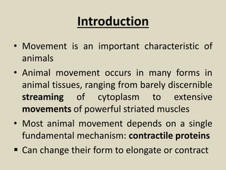
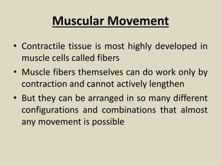
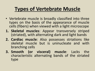
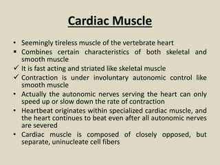
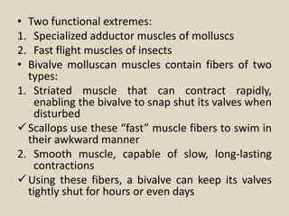
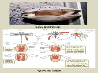
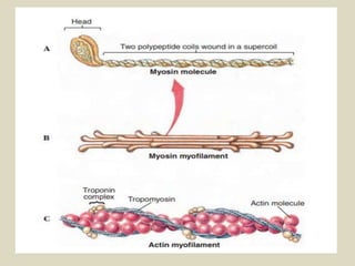
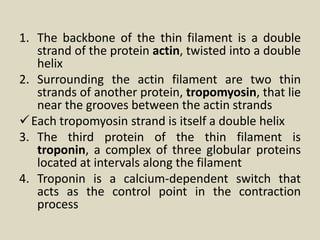
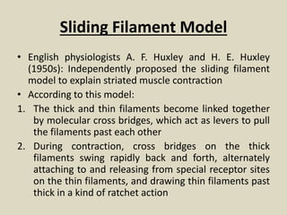
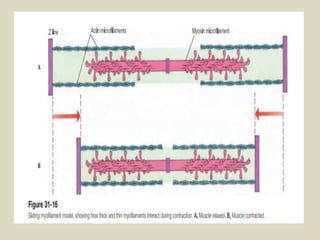
This slide contains the data regarding various mechanisms of movements adopted by invertebrates and vertebrate organisms. It also contains the types of muscles involved in movement of these animals. The data has been retrieved from Hickman's Integrated Principles of Zoology.
2. Introduction
• Movement is an important characteristic of
animals
• Animal movement occurs in many forms in
animal tissues, ranging from barely discernible
streaming of cytoplasm to extensive
movements of powerful striated muscles
• Most animal movement depends on a single
fundamental mechanism: contractile proteins
Can change their form to elongate or contract
3. This contractile machinery is always composed of
ultrafine fibrils arranged to contract when
powered by ATP
Ultrafine fibrils: fine filaments, striated fibrils, or
tubular fibrils (microtubules)
• By far the most important protein contractile
system is the actomyosin system
Composed of two proteins, actin and myosin
This is an almost universal biomechanical system
found from protozoa to vertebrates
Performs a long list of diverse functional roles
Cilia and flagella: Composed of different proteins
4. Ameboid Movement
• Ameboid movement is a form of movement especially
characteristic of amebas and other unicellular forms
It is also found in many wandering cells of metazoans, such as
white blood cells, embryonic mesenchyme, and numerous
other mobile cells that move through the tissue spaces
Ameboid cells change their shape by sending out and
withdrawing pseudopodia (false feet) from any point on the
cell surface
Beneath the plasmalemma lies a nongranular layer, the gel-
like ectoplasm, which encloses the more liquid endoplasm
Research with a variety of ameboid cells, including the
pathogen-fighting phagocytes present in blood, has produced
a consensus model to explain pseudopodial extension and
ameboid crawling
5.
6. • Optical studies of an ameba in movement suggest the
outer layer of ectoplasm surrounds a rather fluid core
of endoplasm
• Movement depends on actin and other regulatory
proteins
• According to one hypothesis, as the pseudopod
extends, hydrostatic pressure forces actin subunits into
the pseudopod where they assemble into a network to
form a gel state
• At the trailing edge of the gel, where the network
disassembles, freed actin interacts with myosin to
create a contractile force that pulls the cell along
behind the extending pseudopod
• Locomotion is assisted by membrane-adhesion
proteins that attach temporarily to the substrate to
provide traction, enabling the cell to crawl steadily
forward
7.
8. Ciliary Movement
• Cilia: Minute, hairlike, motile processes that extend from the
surfaces of the cells of many animals
They are a particularly distinctive feature of ciliate protistans,
Except for nematodes in which motile cilia are absent and
arthropods in which they are rare, cilia are found in all major
groups of animals
• Perform many roles:
Moves small organisms such as unicellular ciliates and
ctenophores through their aquatic environment
Propels fluids and materials across epithelial surfaces of larger
animals
• Cilia are of remarkably uniform diameter (0.2 to 0.5 m)
wherever they are found
9.
10. Electron microscope: Each cilium contains a peripheral
circle of nine double microtubules arranged around
two single microtubules in the center
Each microtubule is composed of a spiral array of
protein subunits called tubulin
The microtubule doublets around the periphery are
connected to each other and to the central pair of
microtubules by a complex system of connective
elements
Also extending from each doublet is a pair of arms
composed of the protein dynein
The dynein arms, which act as cross bridges between
the doublets, operate to produce a sliding force
between the microtubules
11.
12. Flagella
• A flagellum is a whiplike structure longer than a cilium and
usually present singly or in small numbers at one end of a
cell
They are found in members of flagellate protistans, in
animal spermatozoa, and in sponges
The main difference between a cilium and a flagellum is in
their beating pattern rather than in their structure, since
both look alike internally
A flagellum beats symmetrically with snakelike undulations
so that water is propelled parallel to the long axis of the
flagellum
A cilium beats asymmetrically with a fast power stroke in
one direction followed by a slow recovery during which the
cilium bends as it returns to its original position
Water is propelled parallel to the ciliated surface
13. • The mechanism of ciliary movement is not completely
understood
• It is known that microtubules behave as “sliding
filaments” that move past one another much like the
sliding filaments of vertebrate skeletal muscle
• During ciliary flexion:
1. Dynein arms link to adjacent microtubules
2. They swivel and release in repeated cycles, causing
microtubules on the concave side to slide outward
past microtubules on the convex side
This process increases curvature of the cilium
During the recovery stroke microtubules on the
opposite side slide outward to bring the cilium back to
its starting position
14. Muscular Movement
• Contractile tissue is most highly developed in
muscle cells called fibers
• Muscle fibers themselves can do work only by
contraction and cannot actively lengthen
• But they can be arranged in so many different
configurations and combinations that almost
any movement is possible
15.
16. Types of Vertebrate Muscle
• Vertebrate muscle is broadly classified into three
types on the basis of the appearance of muscle
cells (fibers) when viewed with a light microscope
1. Skeletal muscles: Appear transversely striped
(striated), with alternating dark and light bands
2. Cardiac muscle: Also possesses striations like
skeletal muscle but is uninucleate and with
branching cells
3. Smooth (or visceral) muscle: Lacks the
characteristic alternating bands of the striated
type
17.
18. Skeletal Muscle
• Typically organized into sturdy, compact bundles or bands
• It is called skeletal muscle because it is attached to skeletal
elements and is responsible for movements of the trunk,
appendages, respiratory organs, eyes, mouthparts, and other
structure
• Skeletal muscle fibers are extremely long cylindrical, multinucleate
cells that may reach from one end of the muscle to the other
They are packed into bundles called fascicles (L. fasciculus, small
bundle)
Enclosed by tough connective tissue
• The fascicles are in turn grouped into a discrete muscle surrounded
by a thick connective tissue layer
• Most skeletal muscles taper at their ends, where they connect to
bones by tendons
19.
20. • Other muscles, such as the ventral abdominal muscles,
are flattened sheets
• In most fishes, amphibians, and to some extent lizards
and snakes, there is a segmented organization of
muscles alternating with the vertebrae
• The skeletal muscles of other vertebrates, by splitting,
fusion, and shifting, have developed into specialized
muscles best suited for manipulating jointed
appendages that have evolved for locomotion on land
• Skeletal muscle contracts powerfully and quickly but
fatigues more rapidly than does smooth muscle
• Skeletal muscle is sometimes called voluntary muscle
because it is stimulated by motor fibers and is under
conscious cerebral control
21. Smooth Muscle
• Lacks the striations typical of skeletal muscle
• Cells are long, tapering strands, each containing a single
nucleus
• Smooth muscle cells are organized into sheets of muscle
circling the walls of the alimentary canal, blood vessels,
respiratory passages, and urinary and genital ducts
• Smooth muscle is typically slow acting and can maintain
prolonged contractions with very little energy expenditure
• It is under the control of the autonomic nervous system
• The principal functions of smooth muscles are to push
material in a tube, such as the intestine, along its way by
active contractions or to regulate the diameter of a tube,
such as a blood vessel, by sustained contraction
22. Cardiac Muscle
• Seemingly tireless muscle of the vertebrate heart
Combines certain characteristics of both skeletal and
smooth muscle
It is fast acting and striated like skeletal muscle
Contraction is under involuntary autonomic control like
smooth muscle
• Actually the autonomic nerves serving the heart can only
speed up or slow down the rate of contraction
• Heartbeat originates within specialized cardiac muscle, and
the heart continues to beat even after all autonomic nerves
are severed
• Cardiac muscle is composed of closely opposed, but
separate, uninucleate cell fibers
23. Types of Invertebrate Muscle
• Smooth and striated muscles are also characteristic of
invertebrate animals
• There are many variations of both types and even
instances in which structural and functional features of
vertebrate smooth and striated muscle are combined
• Striated muscle appears in invertebrate groups as
diverse as cnidarians and arthropods
• The thickest muscle fibers known, approximately 3 mm
in diameter and 6 cm long, are those of giant barnacles
and of Alaska king crabs living along the Pacific coast of
North America
24. • Two functional extremes:
1. Specialized adductor muscles of molluscs
2. Fast flight muscles of insects
• Bivalve molluscan muscles contain fibers of two
types:
1. Striated muscle that can contract rapidly,
enabling the bivalve to snap shut its valves when
disturbed
Scallops use these “fast” muscle fibers to swim in
their awkward manner
2. Smooth muscle, capable of slow, long-lasting
contractions
Using these fibers, a bivalve can keep its valves
tightly shut for hours or even days
26. • Such adductor muscles use little metabolic energy
and receive remarkably few nerve impulses to
maintain the activated state
• The contracted state has been likened to a “catch
mechanism” involving some kind of stable cross-
linkage between contractile proteins within the fiber
Despite considerable research, there is still much
uncertainty about how this adductor mechanism
works
• Insect flight muscles are virtually the functional
antithesis of the slow, holding muscles of bivalves
• The wings of some small flies operate at frequencies
greater than 1000 beats per second
27. • The so-called fibrillar muscle, which contracts at these
frequencies— far greater than even the most active of
vertebrate muscles—shows unique characteristics
It has very limited extensibility
Wing leverage system is arranged so that the muscles
shorten only slightly during each downbeat of the
wings
Muscles and wings operate as a rapidly oscillating
system in an elastic thorax
Muscles rebound elastically and are activated by
stretch during flight
They receive impulses only periodically rather than one
impulse per contraction
One reinforcement impulse for every 20 or 30
contractions is enough to keep the system active
28. Structure of Striated Muscle
• Striated muscle is so named because of periodic bands that pass
across the widths of muscle cells
• Each cell, or fiber, is a multinucleated tube containing numerous
myofibrils, packed together and invested by the cell membrane, the
sarcolemma
• The myofibril contains two types of myofilaments:
Thick filaments composed of the protein myosin
Thin filaments, composed of the protein actin
These are the actual contractile proteins of the muscle
• Thin filaments are held together by a dense structure called the Z
line
• The functional unit of the myofibril, the sarcomere, extends
between successive Z lines
• Each thick filament is made up of myosin molecules packed
together in an elongate bundle
29.
30. • Each myosin molecule is composed of two
polypeptide chains, each having a club-shaped
head
• Lined up as they are in a bundle to form a
thick filament, the double heads of each
myosin molecule face outward from the
center of the filament
• These heads act as molecular cross bridges
that interact with the thin filaments during
contraction
• Thin filaments are more complex because they
are composed of three different proteins
31.
32. 1. The backbone of the thin filament is a double
strand of the protein actin, twisted into a double
helix
2. Surrounding the actin filament are two thin
strands of another protein, tropomyosin, that lie
near the grooves between the actin strands
Each tropomyosin strand is itself a double helix
3. The third protein of the thin filament is
troponin, a complex of three globular proteins
located at intervals along the filament
4. Troponin is a calcium-dependent switch that
acts as the control point in the contraction
process
33. Sliding Filament Model
• English physiologists A. F. Huxley and H. E. Huxley
(1950s): Independently proposed the sliding filament
model to explain striated muscle contraction
• According to this model:
1. The thick and thin filaments become linked together
by molecular cross bridges, which act as levers to pull
the filaments past each other
2. During contraction, cross bridges on the thick
filaments swing rapidly back and forth, alternately
attaching to and releasing from special receptor sites
on the thin filaments, and drawing thin filaments past
thick in a kind of ratchet action
34. 3. As contraction continues, the Z lines are pulled
closer together
4. The sarcomere shortens
5. As all sarcomere units shorten together, the
muscle contracts
6. Relaxation is a passive process
7. When cross bridges between the thick and thin
filaments release, the sarcomeres are free to
lengthen
8. This requires some force, which is usually
supplied by antagonistic muscles or the force of
gravity
35.
36. Control of Contraction
• Muscle contracts in response to nerve stimulation
• If the nerve supply to a muscle is severed, the
muscle atrophies, or wastes away
• Skeletal muscle fibers are innervated by motor
neurons whose cell bodies are located in the
spinal cord
• Each cell body gives rise to a motor axon that
leaves the spinal cord to travel by way of a
peripheral nerve trunk to a muscle where it
branches repeatedly into many terminal branches
37. • Each terminal branch innervates a single muscle
fiber
• Depending on the type of muscle, a single motor
axon may innervate as few as three or four
muscle fibers (where very precise control is
needed)
• The motor neuron and all muscle fibers it
innervates is called a motor unit
The motor unit is the functional unit of skeletal
muscle
When a motor neuron fires, the action potential
passes to all fibers of the motor unit and each is
stimulated to contract simultaneously
38.
39. • Total force exerted by a muscle depends on the
number of motor units activated
• Precise control of movement is achieved by
varying the number of motor units activated at
any one time
• Motor unit recruitment: A smooth and steady
increase in muscle tension is produced by
increasing the number of motor units brought
into play
• Myoneural Junction: The place where a motor
axon terminates on a muscle fiber is called the
myoneural junction
At the junction is a tiny gap, or synaptic cleft, that
thinly separates a nerve fiber and muscle fiber
40. In the vicinity of the junction, the neuron stores a
chemical, acetylcholine, in minute vesicles known as
synaptic vesicles
Acetylcholine is released when a nerve impulse
reaches a synapse
This substance is a chemical mediator that diffuses
across the narrow junction and acts on the muscle
fiber membrane to generate an electrical
depolarization
The depolarization spreads rapidly through the muscle
fiber, causing it to contract
Synapse: A special chemical bridge that couples
together the electrical activities of nerve and muscle
fibers
41.
42. • Built into vertebrate skeletal muscle is an
elaborate conduction system that serves to carry
the depolarization from the myoneural junction
to the densely packed filaments within the fiber
• Along the surface of the sarcolemma are
numerous invaginations that project as a system
of tubules into the muscle fiber
This is called the T-system
It is continuous with the sarcoplasmic reticulum,
a system of fluid-filled channels that runs parallel
to the myofilaments
• The system is ideally arranged for speeding the
electrical depolarization from the myoneural
junction to the myofilaments within the fiber
43. Excitation-Contraction Coupling
• In resting, unstimulated muscle, shortening does not occur
because thin tropomyosin strands surrounding the actin
myofilaments lie in a position that prevents the myosin
heads from attaching to actin
1. When muscle is stimulated and the electrical
depolarization arrives at the sarcoplasmic reticulum
surrounding the fibrils, calcium ions are released
2. Some calcium binds to the control protein troponin
3. Troponin immediately undergoes changes in shape that
allow tropomyosin to move out of its blocking position,
exposing active sites on the actin myofilaments
4. The myosin heads then bind to these sites, forming cross
bridges between adjacent thick and thin myofilaments
5. This sets in motion an attach-pull-release cycle that occurs
in a series of steps
44. 1. Release of bond energy from ATP activates the
myosin head, which swings 45 degrees, at the
same time releasing a molecule of ADP
2. This is the power stroke that pulls the actin
filament a distance of about 10 nm
3. It comes to an end when another ATP molecule
binds to the myosin head, inactivating the site
• Thus each cycle requires expenditure of energy in
the form of ATP
• Shortening will continue as long as nerve
impulses arrive at the myoneural junction and
free calcium remains available around the
myofilaments
45. • The attach-pull-release cycle can repeat again and
again, 50 to 100 times per second, pulling thick and
thin filaments past each other
• While the distance each sarcomere can shorten is very
small, this distance is multiplied by the thousands of
sarcomeres lying end to end in a muscle fiber
• Consequently, a strongly contracting muscle may
shorten by as much as one-third its resting length
• When stimulation stops, calcium is quickly pumped
back into the sarcoplasmic reticulum
• Troponin resumes its original configuration
• Tropomyosin moves back into its blocking position on
actin, and the muscle relaxes
46. Energy for Contraction
• Muscle contraction requires large amounts of energy
• ATP is the immediate source of energy, but the amount
present will sustain contraction for only a second or two
• Muscle cells immediately call on the second level of energy
reserve, creatine phosphate
Creatine phosphate is a high-energy phosphate compound
that stores bond energy during periods of rest
• As ADP is produced during contraction, creatine phosphate
releases its stored bond energy to convert ADP to ATP
This reaction can be summarized as:
47. • Within a few seconds (30 seconds), depending on the
rapidity of muscle contraction, the reserves of creatine
phosphate are depleted
• The contracting muscle now must be fueled from its third
and largest store of energy, glycogen
Glycogen is a polysaccharide chain of glucose molecules
stored in both liver and muscle
Muscle has by far the larger store—some 3/4th of all the
glycogen in the body is stored in muscle
• As a supply of energy for contraction, glycogen has three
important advantages:
1. It is relatively abundant
2. It can be mobilized quickly
3. It can provide energy under anoxic conditions
• As soon as the muscle’s store of creatine phosphate
declines, enzymes break down glycogen, through glycolysis
that leads to the generation of ATP
Editor's Notes
Discernible: Noticeable
Metazoan: Those animals whose body is arranged into tissues, organs and organ systems. They are multicellular organisms.
Mesenchyme is a type of loosely organized animal embryonic connective tissue of undifferentiated cells that give rise to most tissues, such as skin, blood or bone
Trailing: Dragging Traction: Pull
Synchronous are muscles in which the frequency of nervous stimulation and contraction are consistent
Asynchronous is muscle that contracts more than once per nerve impulse
Ratchet: Linear or rotary motion in only one direction. Common examples of ratchets are clocks, jacks, etc.
Atrophy: Degeneration of the cell
The sarcolemma is the plasma membrane of the muscle cell
Reticulum: a fine network or netlike structure