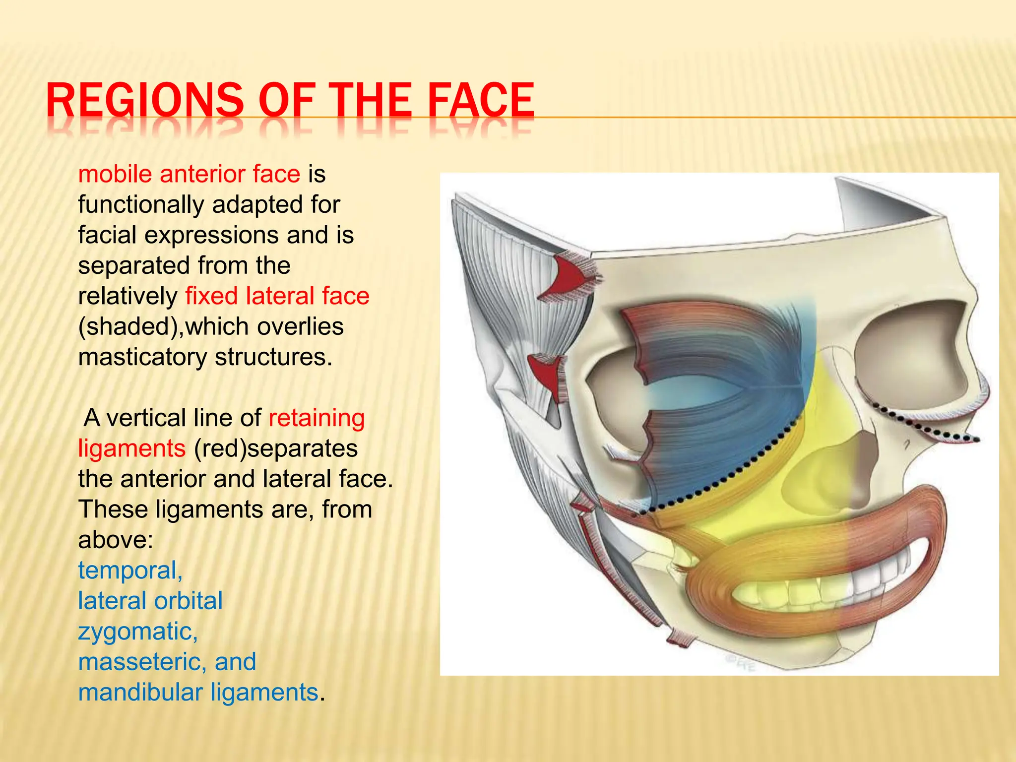
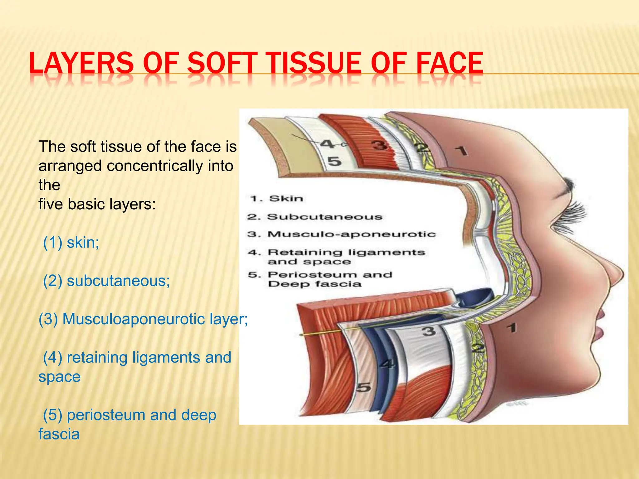
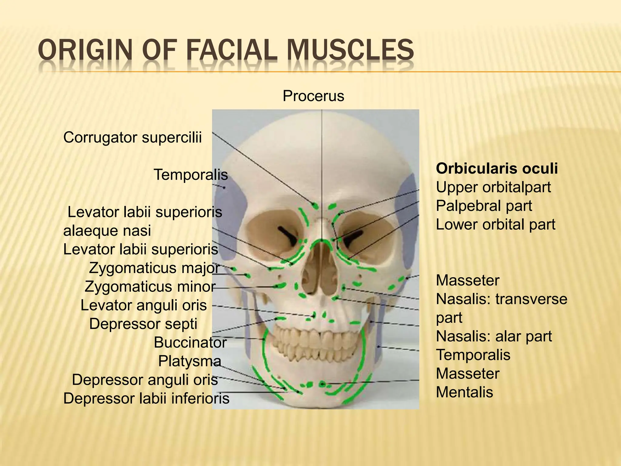
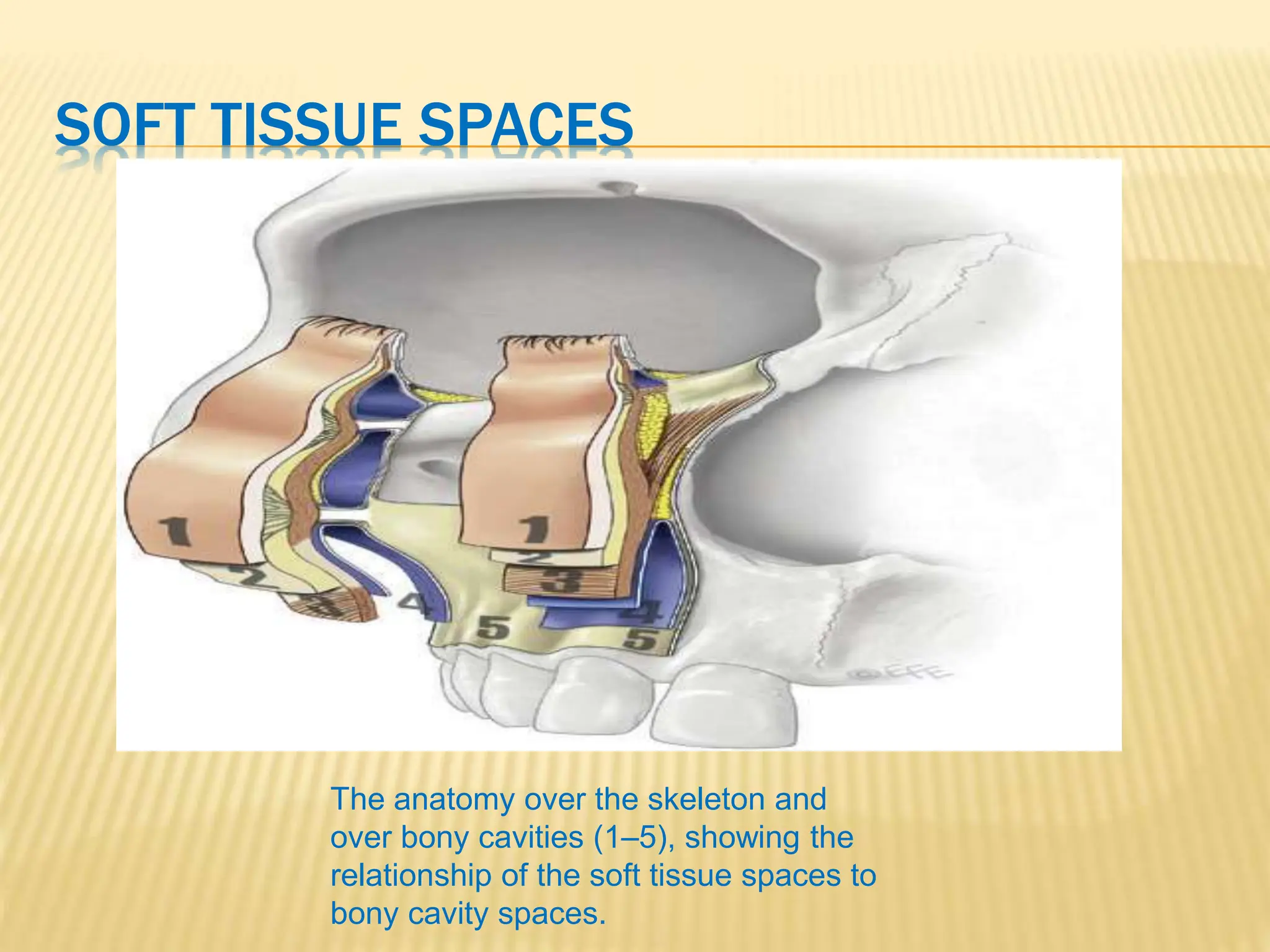
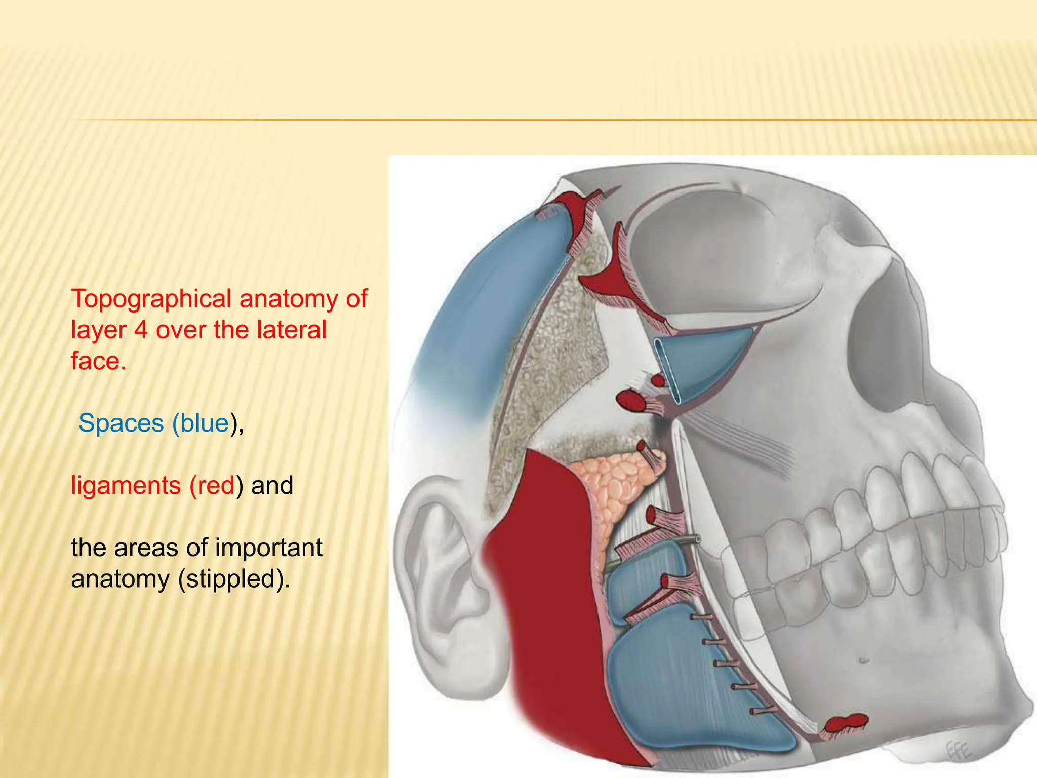
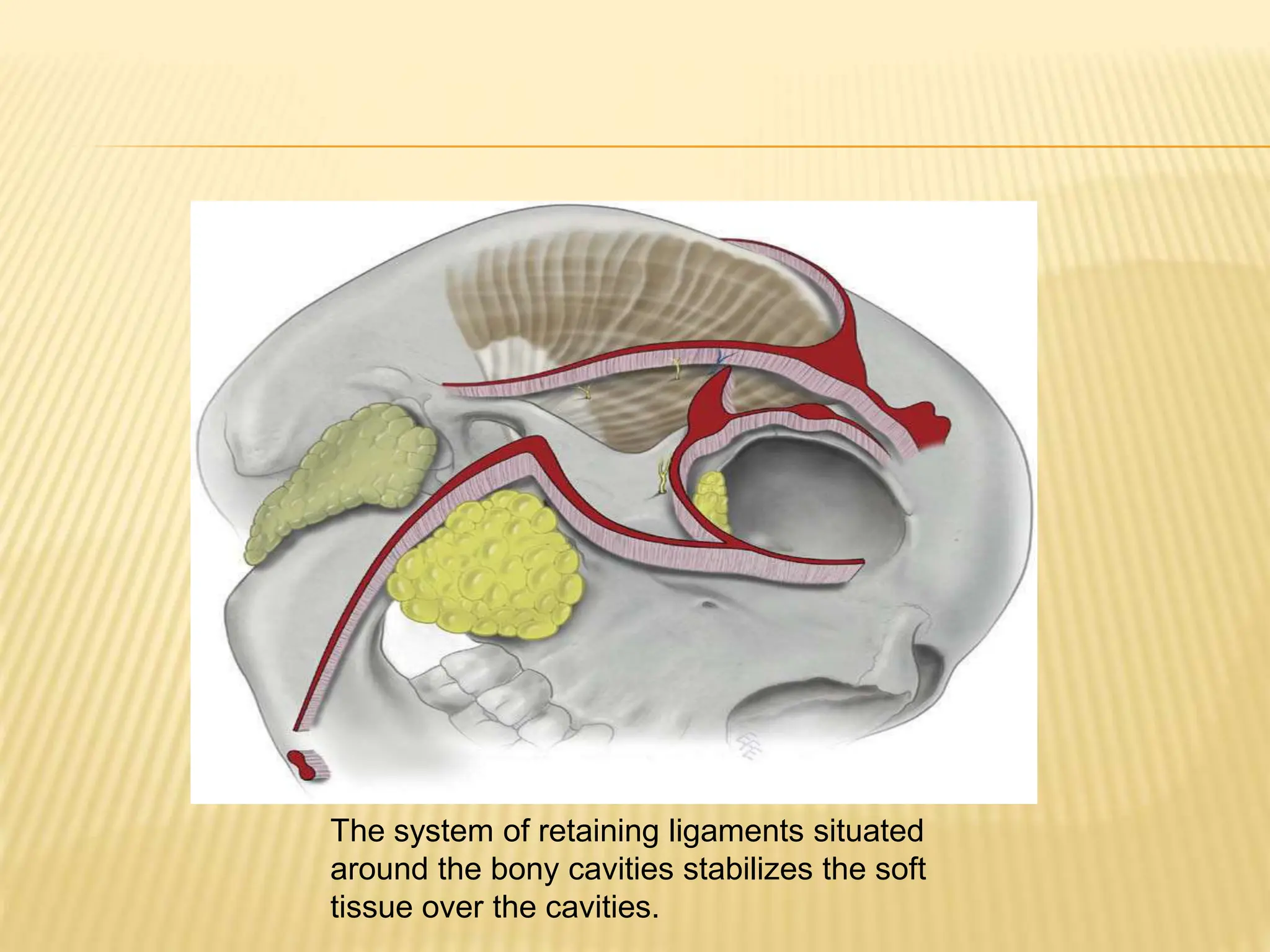
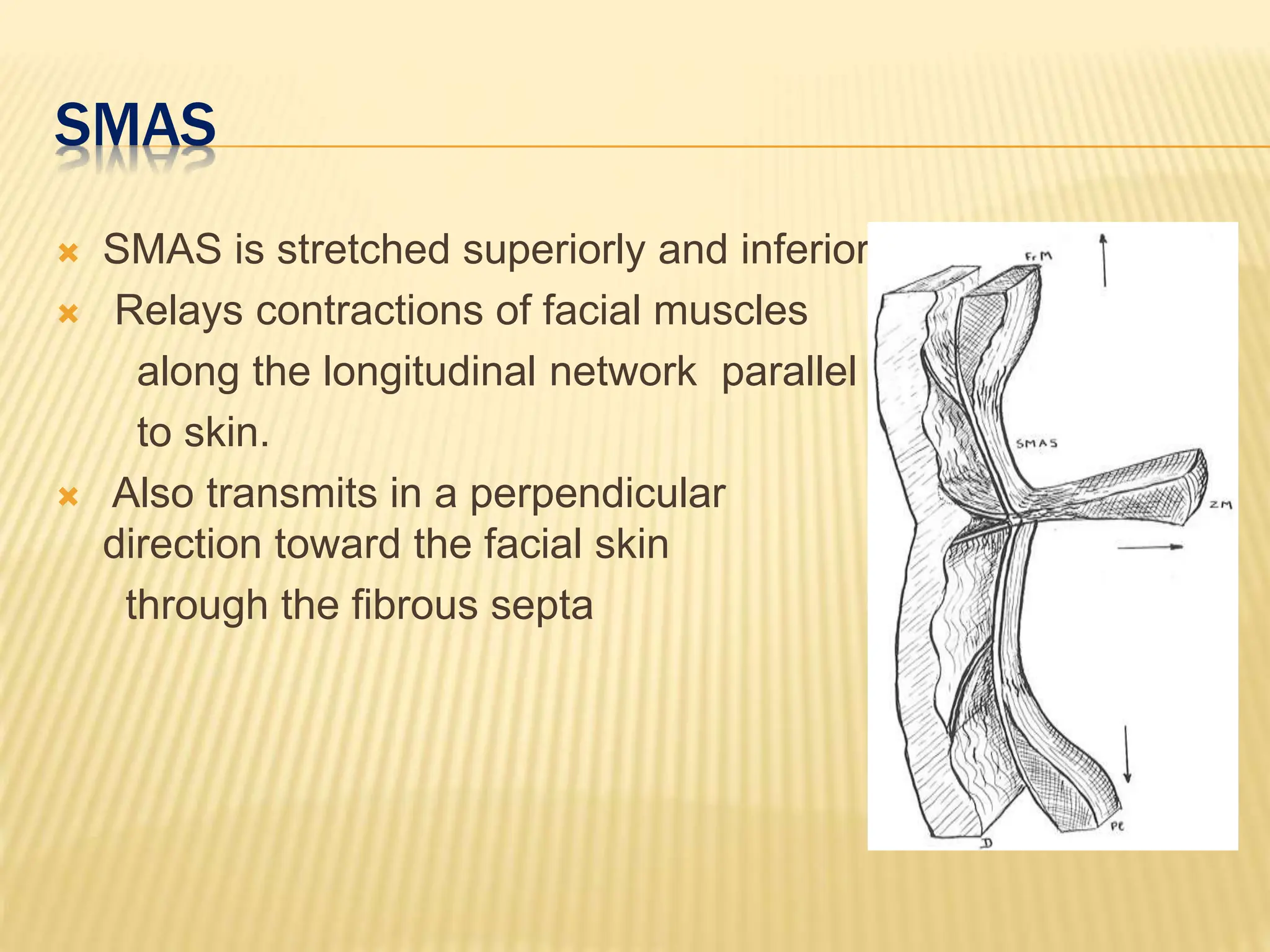
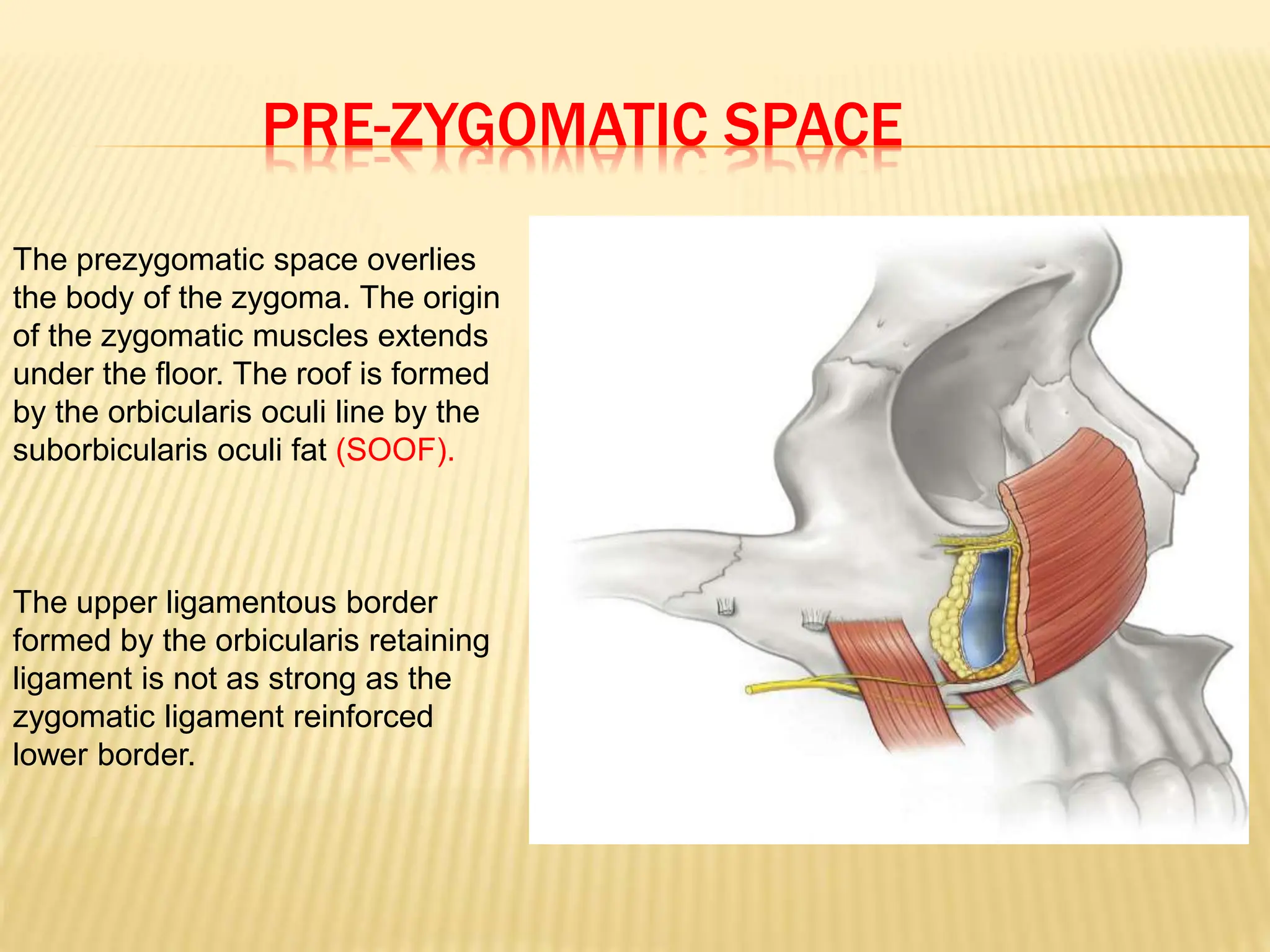
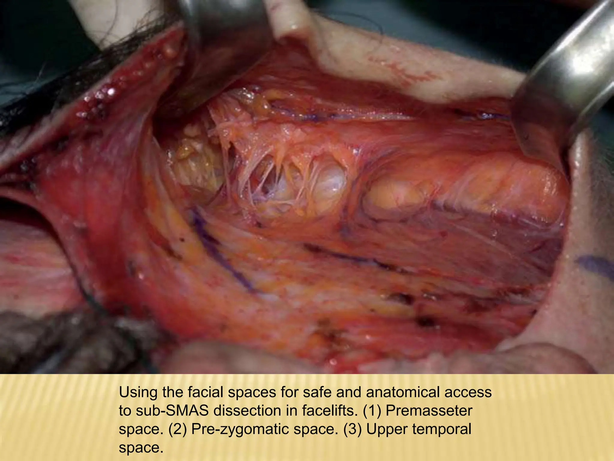
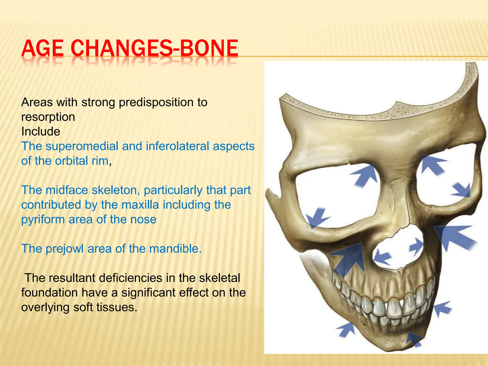
ANATOMY OF AGING FACE-THE RELEVANCE OF
RETAINING LIGAMENTS
INTRODUCTION
 Aging of the face- a multifactorial process
 - can be explained on an
anatomical basis.
 The face - constructed of five basic
layers
 - bound together by a system
of facial retaining ligaments.
 To facilitate the mobility needed for facial
expression, a series of soft tissue spaces are
incorporated into the architecture of the face.
 This presentation will describe, in detail, the
five-layered construct of the face, including
the spaces and retaining ligaments, and will
highlight the relevance of these structures in
the aging face.
DIVISION OF FACE
 The traditional approach
 upper,
 middle, and
 lower thirds
From a functional perspective, the face has an
anterior aspect and
a lateral aspect—
(A vertical line descending from the lateral orbital rim is
the approximate division between the anterior and
lateral zones of the face)
REGIONS OF THE FACE
mobile anterior face is
functionally adapted for
facial expressions and is
separated from the
relatively fixed lateral face
(shaded),which overlies
masticatory structures.
A vertical line of retaining
ligaments (red)separates
the anterior and lateral face.
These ligaments are, from
above:
temporal,
lateral orbital
zygomatic,
masseteric, and
mandibular ligaments.
LAYERS OF SOFT TISSUE OF FACE
The soft tissue of the face is
arranged concentrically into
the
five basic layers:
(1) skin;
(2) subcutaneous;
(3) Musculoaponeurotic layer;
(4) retaining ligaments and
space
(5) periosteum and deep
fascia
SKIN
Epidermis-cell rich layer composed mainly
of keratinocytes
-also melanocytes & langerhans
cells
Dermis -predominantly extra cellular matrix
secreted by fibroblasts
-thinnest in eyelid
-thickest over forehead& nasal tip.
LAYER2-SUBCUTANEOUS LAYER
 The subcutaneous layer has two
components
 subcutaneous fat- provides volume
 fibrous retinacular cutis- binds the dermis to
the underlying SMAS.
LAYER2-SUBCUTANEOUS FAT
DISTRIBUTION
Superior orbital
Inferior orbital
Nasolabial
Jowl
Lateral Temporal cheek Middle
(forehead)
Central
Lateral orbital
Medial
Middle
Lateral
temporal-cheek
Pre-platysma
fat
LAYER2-RETINACULAR CUTIS
 The density and strength-varies in
different areas of the face.
 Over retaining ligaments,
 fibers are denser and oriented more
vertically.
 sharp release is usually necessary to
raise a subcutaneous flap.
 in areas overlying a space,
 the fibers are less dense and oriented
more horizontally.
 it is relatively easy to elevate a
subcutaneous facelift flap overlying a
space.
RETINACULAR CUTIS
RETINACULAR CUTIS
 Within the subcutaneous tissue-
attachment to the overlying
dermis is stronger and denser than to the
underlying SMAS.
 easier to perform subcutaneous dissection
in the deeper subcutaneous level (just on the
surface of the underlying SMAS) than more
superficially nearer the dermis, as there are
fewer retinacular cutis fibers .
LAYER 3: MUSCULO-APONEUROTIC LAYER
 muscles of facial expression
unique,
situated within the superficial fascia
move the soft tissues.
 different from skeletal muscles- beneath the
deep fascia,
 All muscles of facial expression have either
all or the majority of their course within layer
3
LAYER 3
 Layer 3 is continuous over the entire face,
although for descriptive purposes, different
names are given to certain parts according to
the superficial muscle within. It is called ,
 the galea-- over the
scalp,
 the temporo parietal- fascia over the
temple
ORIGIN OF FACIAL MUSCLES
Orbicularis oculi
Upper orbitalpart
Palpebral part
Lower orbital part
Masseter
Nasalis: transverse
part
Nasalis: alar part
Temporalis
Masseter
Mentalis
Corrugator supercilii
Temporalis
Levator labii superioris
alaeque nasi
Levator labii superioris
Zygomaticus major
Zygomaticus minor
Levator anguli oris
Depressor septi
Buccinator
Platysma
Depressor anguli oris
Depressor labii inferioris
Procerus
LAYER4-SOFT TISSUE SPACES &
RETAINING LIGAMENTS
 It is an area of significant complexity and
 contains the following structures:
 (1) soft tissue spaces;
 (2) retaining ligaments;
 (3) deep layers of the intrinsic muscles
passing from their bone attachment to their
more superficial soft tissue origin; and
 (4) facial nerve branches, passing from deep
to superficial.
SOFT TISSUE SPACES
 Functionally, a series of soft tissue spaces
exist in layer 4 to allow independent
movement of the periorbital and perioral
muscle of facial expressions over the deep
fascia responsible for mastication directly
beneath the muscles of facial expression.
SOFT TISSUE SPACES
The anatomy over the skeleton and
over bony cavities (1–5), showing the
relationship of the soft tissue spaces to
bony cavity spaces.
Topographical anatomy of
layer 4 over the lateral
face.
Spaces (blue),
ligaments (red) and
the areas of important
anatomy (stippled).
RETAINING LIGAMENTS
 The retaining ligaments of
the face are strategically
placed within the
boundaries between
the soft tissue spaces,
and functions to reinforce
the boundaries.
Orbicularis retaining
ligament
Zygomatic ligament
(McGregor’s patch)
Masseteric
ligament
t
Mandibular ligament
Platysma-auricular
ligament
RETAINING LIGAMENTS
 True ligaments
 the zygomatic ligament -connects the inferior
border of the zygomatic arch to the dermis
 Mandibular ligament- connects the
periosteum of the mandible just medial to the
origin of depressor anguli oris to the
overlying dermis.
 lateral orbital thickening- on the superolateral
orbital rim that arises as a thickening of the
orbicularis retaining ligament,
The system of retaining ligaments situated
around the bony cavities stabilizes the soft
tissue over the cavities.
THE RELATIONSHIP BETWEEN FACIAL
NERVE BRANCHES, SPACES& RETAINING
LIGAMENTS.
The nerves stay deep to and outside of
the spaces at all times in the lateral
face. In the boundary between the
lateral and anterior face, the facial nerve
branches transition from under layer 5
to enter layer 3, always in close
association with the retaining ligaments
of the face.
Extra care should be taken when
releasing ligaments as important facial
nerve branches are intimately related to
ligaments, such as the zygomatic and
mandibular retaining ligaments.
 Layer 4 is the plane in which dissection is
performed in subSMAS facelifts.
 Platysma auricular fascia-
-in the lateral face,
-immediately in front of the ear,
-extending 25–30 mm forward of the
ear cartilage to the posterior border of the
platysma,
-As no facial expressions occur here,
LAYER 5-DEEP FASCIA
 The deepest soft tissue layer of the face,
-- is formed by the periosteum where it
overlies bone.
 -- Over the lateral face, where the muscle
of mastication (temporalis and masseter)
overlie the bone, the deep fascia is instead
the fascial covering of the muscles,
 -the deep temporal fascia above the
zygomatic arch, and
SMAS -SUPERFICIAL MUSCULO
APONEUROTIC SYSTEM
-- system or network of collagen fibers,
elastic fibers, and fat cells
--connects the mimetic muscles to the
overlying dermis and
-- plays an important functional role in facial
expression.
In simple terms, the
 SMAS can be considered as a sheet of
SMAS
 SMAS is stretched superiorly and inferiorly
 Relays contractions of facial muscles
along the longitudinal network parallel
to skin.
 Also transmits in a perpendicular
direction toward the facial skin
through the fibrous septa
SMAS-SUPERFICIAL MUSCULO
APONEUROTIC SYSTEM
LAYERS OF FACIAL SKIN
FACIAL NERVE
 The facial nerve branches exit the parotid
gland and remain deep to layer 5 in the
lateral face.
 As they approach the anterior face, the
branches traverse layer 4 to reach the
underside of mimetic muscles of the face. It
is at these transition points across layer 4
that the nerves are at greatest risk of injury.
FACIAL NERVE
 Facial motor function is provided by
 facial nerve &
 oculomotor nerve ( eyelid elevation)
 CNVII divides into its major trunks within the
parotid gland.
In the lower face, the branches of the facial
nerve are
deep to the SMAS and
FACIAL NERVE
Facial nerve
––Protected by superficial lobe
of the parotid gland
––travels beneath the parotido-
masseteric fascia
––Innervates superficial facial
mimetic muscles from deeper
surface
FACIAL NERVE
 The temporal and mandibular branches are
the most significant in terms of surgical risks
because of the lack of cross innervation of
their target muscle.
FACIAL NERVE-SAFETY ZONE
FACIAL SPACES
 A large part of the subSMAS layer 4 consists
of soft tissue “spaces.”
 These spaces have defined boundaries that
are strategically reinforced by retaining
ligaments.
 anatomically “safe spaces”, as no structures
crosses within spaces.
 As the roof of each space is the least
supported part, it is more prone to
developing laxity with aging, compared with
UPPER TEMPORAL SPACE
 separates the superficial temporal fascia
from the deep temporal fascia
 No structures cross the temporal space
 safe surgical access to the lateral brow and
upper mid-cheek.
PRE-ZYGOMATIC SPACE
 This triangular-shaped space overlies the
body of the zygoma,its floor covering the
origins of the zygomatic muscles.
 With the laxity of aging, the roof of the
space rests at a lower level than in youth.
 This aging of the prezygomatic space,
with bulging over its roof accentuated by
its well-supported boundaries, is the
anatomical basis for the clinical entity
variously described as malar mounds,
bags or malar crescent.
MALAR MOUNDS
PRE-ZYGOMATIC SPACE
The prezygomatic space overlies
the body of the zygoma. The origin
of the zygomatic muscles extends
under the floor. The roof is formed
by the orbicularis oculi line by the
suborbicularis oculi fat (SOOF).
The upper ligamentous border
formed by the orbicularis retaining
ligament is not as strong as the
zygomatic ligament reinforced
lower border.
PREMASSETER SPACE
 overlies the lower half of the masseter
 allows opening of the jaw without restriction
 The roof is formed by the platysma.
 it is the anatomical basis for the development
of jowls with aging.
 Laxity in the roof of the space, particularly
where it has a weakened attachment to the
anterior masseter by the masseteric
ligaments and its inferior boundary where
there is no ligament, manifests as the
BUCCAL SPACE
 one of the deep facial spaces,
 located in the anterior face, medial to the
anterior border of the masseter above the
level of the oral commissure in youth.
 Aging and attrition of the boundaries,
particularly of the masseteric ligaments
inferiorly result in the platysma being less
firmly bound to the masseter. This allows the
space to enlarge and also allows the buccal
fat to prolapse inferiorly,
In youth, the spaces are tight. The retaining ligaments are stout and
the transition between spaces is not discernible. With aging, spaces
expand to a greater extent than the laxity that develops in ligaments
within their boundaries, resulting in bulges between areas of relative
fixations. These spaces open with relative ease with blunt dissection.
Using the facial spaces for safe and anatomical access
to sub-SMAS dissection in facelifts. (1) Premasseter
space. (2) Pre-zygomatic space. (3) Upper temporal
space.
AGE CHANGES IN SKIN
 With aging, the supple skin of youth
becomes thinned and flattened, with loss of
elasticity and architectural regularity.
 Atrophy of the extracellular matrix is reflected
by the decreased fibroblasts
decreased collagen ( I and III) and
decreased elastin in the dermis.
 As the underlying collagen weakens and the
skin thins, the dermis loses its capacity to
resist the constant force of the muscles and
AGE CHANGES-SUBCUTANEOUS TISSUE
 The fibrous and fat components in the
subcutaneous tissue are not a uniform but
arranged in discrete compartments. specific
names such as the malar fat pad and naso
labial fat are given.
 The boundary-correspond to the location of
the retaining ligaments. Distinct
compartmentalization by the retaining
ligaments holds the fat in its relative
positions.
MUSCLE AGING
 Skeletal muscles- atrophy up to 50% with
age-applicable to temporalis and masseter
 the mimetic muscle of the face, in contrast to
skeletal muscles, may not undergo the same
degree of degeneration with aging because
of their constant use with facial expression.
 The orbicularis oculi- has been noted to
remain histologically unchanged with no loss
of muscle fibers aging.
AGE CHANGES IN SPACES
 The spaces expand with aging as well, to a
greater extent than the laxity that develop in
the ligaments within their boundaries,
resulting in bulges between areas of relative
fixations.
older patients- dissect easily .
 In young people, the spaces are more
potential that real and do not open quite so
easily with blunt dissection
AGE CHANGES-BONE
Areas with strong predisposition to
resorption
Include
The superomedial and inferolateral aspects
of the orbital rim,
The midface skeleton, particularly that part
contributed by the maxilla including the
pyriform area of the nose
The prejowl area of the mandible.
The resultant deficiencies in the skeletal
foundation have a significant effect on the
overlying soft tissues.
 The retrusion of the facial skeleton causes
the origin of the multi-linked fibrous retaining
ligaments to be displaced posteriorly. This
pulls the skin inwards, exaggerating the
concavity between the areas of relative
convexity that develop with aging.
LOWER FACE
 The jowl and the labiomandibular fold in the
lower face are not present in youth, and
develop with aging.
 Distension of the weaker masseteric
ligaments at the anterior border of the lower
pre-masseter space (below the key
masseteric ligament) and inferior
displacement of the buccal fat (within the
buccal space) is the anatomical basis for the
development of the labiomandibular fold.
JOWL
LABIOMANDIBULAR FOLD
 The mandibular ligament demarcates the
transition from the labiomandibular fold
above and the jowl below.
 The jowl develops as a result of distension of
the roof of the lower premasseter space with
resultant descent of the tissues below the
body of the mandible.
CONCLUSION
 There are both surgical and nonsurgical
treatment options available for correction of
age related changes of face.A thorough
understanding of anatomical basis of aging
of face will help the surgeon in choosing the
appropriate treatment plan.
BIBLIOGRAPHY
 Aesthetic Surgery of the Face(chapter-
anatomy of the aging face)
 Authors:Bryan Mendelson and Chin-Ho
Wong

anatomy face.pptx

  • 1.
    ANATOMY OF AGINGFACE-THE RELEVANCE OF RETAINING LIGAMENTS
  • 2.
    INTRODUCTION  Aging ofthe face- a multifactorial process  - can be explained on an anatomical basis.  The face - constructed of five basic layers  - bound together by a system of facial retaining ligaments.  To facilitate the mobility needed for facial expression, a series of soft tissue spaces are incorporated into the architecture of the face.
  • 3.
     This presentationwill describe, in detail, the five-layered construct of the face, including the spaces and retaining ligaments, and will highlight the relevance of these structures in the aging face.
  • 4.
    DIVISION OF FACE The traditional approach  upper,  middle, and  lower thirds From a functional perspective, the face has an anterior aspect and a lateral aspect— (A vertical line descending from the lateral orbital rim is the approximate division between the anterior and lateral zones of the face)
  • 5.
    REGIONS OF THEFACE mobile anterior face is functionally adapted for facial expressions and is separated from the relatively fixed lateral face (shaded),which overlies masticatory structures. A vertical line of retaining ligaments (red)separates the anterior and lateral face. These ligaments are, from above: temporal, lateral orbital zygomatic, masseteric, and mandibular ligaments.
  • 6.
    LAYERS OF SOFTTISSUE OF FACE The soft tissue of the face is arranged concentrically into the five basic layers: (1) skin; (2) subcutaneous; (3) Musculoaponeurotic layer; (4) retaining ligaments and space (5) periosteum and deep fascia
  • 7.
    SKIN Epidermis-cell rich layercomposed mainly of keratinocytes -also melanocytes & langerhans cells Dermis -predominantly extra cellular matrix secreted by fibroblasts -thinnest in eyelid -thickest over forehead& nasal tip.
  • 8.
    LAYER2-SUBCUTANEOUS LAYER  Thesubcutaneous layer has two components  subcutaneous fat- provides volume  fibrous retinacular cutis- binds the dermis to the underlying SMAS.
  • 9.
    LAYER2-SUBCUTANEOUS FAT DISTRIBUTION Superior orbital Inferiororbital Nasolabial Jowl Lateral Temporal cheek Middle (forehead) Central Lateral orbital Medial Middle Lateral temporal-cheek Pre-platysma fat
  • 10.
    LAYER2-RETINACULAR CUTIS  Thedensity and strength-varies in different areas of the face.  Over retaining ligaments,  fibers are denser and oriented more vertically.  sharp release is usually necessary to raise a subcutaneous flap.  in areas overlying a space,  the fibers are less dense and oriented more horizontally.  it is relatively easy to elevate a subcutaneous facelift flap overlying a space.
  • 11.
  • 12.
    RETINACULAR CUTIS  Withinthe subcutaneous tissue- attachment to the overlying dermis is stronger and denser than to the underlying SMAS.  easier to perform subcutaneous dissection in the deeper subcutaneous level (just on the surface of the underlying SMAS) than more superficially nearer the dermis, as there are fewer retinacular cutis fibers .
  • 13.
    LAYER 3: MUSCULO-APONEUROTICLAYER  muscles of facial expression unique, situated within the superficial fascia move the soft tissues.  different from skeletal muscles- beneath the deep fascia,  All muscles of facial expression have either all or the majority of their course within layer 3
  • 14.
    LAYER 3  Layer3 is continuous over the entire face, although for descriptive purposes, different names are given to certain parts according to the superficial muscle within. It is called ,  the galea-- over the scalp,  the temporo parietal- fascia over the temple
  • 15.
    ORIGIN OF FACIALMUSCLES Orbicularis oculi Upper orbitalpart Palpebral part Lower orbital part Masseter Nasalis: transverse part Nasalis: alar part Temporalis Masseter Mentalis Corrugator supercilii Temporalis Levator labii superioris alaeque nasi Levator labii superioris Zygomaticus major Zygomaticus minor Levator anguli oris Depressor septi Buccinator Platysma Depressor anguli oris Depressor labii inferioris Procerus
  • 16.
    LAYER4-SOFT TISSUE SPACES& RETAINING LIGAMENTS  It is an area of significant complexity and  contains the following structures:  (1) soft tissue spaces;  (2) retaining ligaments;  (3) deep layers of the intrinsic muscles passing from their bone attachment to their more superficial soft tissue origin; and  (4) facial nerve branches, passing from deep to superficial.
  • 17.
    SOFT TISSUE SPACES Functionally, a series of soft tissue spaces exist in layer 4 to allow independent movement of the periorbital and perioral muscle of facial expressions over the deep fascia responsible for mastication directly beneath the muscles of facial expression.
  • 18.
    SOFT TISSUE SPACES Theanatomy over the skeleton and over bony cavities (1–5), showing the relationship of the soft tissue spaces to bony cavity spaces.
  • 19.
    Topographical anatomy of layer4 over the lateral face. Spaces (blue), ligaments (red) and the areas of important anatomy (stippled).
  • 20.
    RETAINING LIGAMENTS  Theretaining ligaments of the face are strategically placed within the boundaries between the soft tissue spaces, and functions to reinforce the boundaries. Orbicularis retaining ligament Zygomatic ligament (McGregor’s patch) Masseteric ligament t Mandibular ligament Platysma-auricular ligament
  • 21.
    RETAINING LIGAMENTS  Trueligaments  the zygomatic ligament -connects the inferior border of the zygomatic arch to the dermis  Mandibular ligament- connects the periosteum of the mandible just medial to the origin of depressor anguli oris to the overlying dermis.  lateral orbital thickening- on the superolateral orbital rim that arises as a thickening of the orbicularis retaining ligament,
  • 22.
    The system ofretaining ligaments situated around the bony cavities stabilizes the soft tissue over the cavities.
  • 23.
    THE RELATIONSHIP BETWEENFACIAL NERVE BRANCHES, SPACES& RETAINING LIGAMENTS. The nerves stay deep to and outside of the spaces at all times in the lateral face. In the boundary between the lateral and anterior face, the facial nerve branches transition from under layer 5 to enter layer 3, always in close association with the retaining ligaments of the face. Extra care should be taken when releasing ligaments as important facial nerve branches are intimately related to ligaments, such as the zygomatic and mandibular retaining ligaments.
  • 24.
     Layer 4is the plane in which dissection is performed in subSMAS facelifts.  Platysma auricular fascia- -in the lateral face, -immediately in front of the ear, -extending 25–30 mm forward of the ear cartilage to the posterior border of the platysma, -As no facial expressions occur here,
  • 25.
    LAYER 5-DEEP FASCIA The deepest soft tissue layer of the face, -- is formed by the periosteum where it overlies bone.  -- Over the lateral face, where the muscle of mastication (temporalis and masseter) overlie the bone, the deep fascia is instead the fascial covering of the muscles,  -the deep temporal fascia above the zygomatic arch, and
  • 26.
    SMAS -SUPERFICIAL MUSCULO APONEUROTICSYSTEM -- system or network of collagen fibers, elastic fibers, and fat cells --connects the mimetic muscles to the overlying dermis and -- plays an important functional role in facial expression. In simple terms, the  SMAS can be considered as a sheet of
  • 27.
    SMAS  SMAS isstretched superiorly and inferiorly  Relays contractions of facial muscles along the longitudinal network parallel to skin.  Also transmits in a perpendicular direction toward the facial skin through the fibrous septa
  • 28.
  • 29.
  • 30.
    FACIAL NERVE  Thefacial nerve branches exit the parotid gland and remain deep to layer 5 in the lateral face.  As they approach the anterior face, the branches traverse layer 4 to reach the underside of mimetic muscles of the face. It is at these transition points across layer 4 that the nerves are at greatest risk of injury.
  • 31.
    FACIAL NERVE  Facialmotor function is provided by  facial nerve &  oculomotor nerve ( eyelid elevation)  CNVII divides into its major trunks within the parotid gland. In the lower face, the branches of the facial nerve are deep to the SMAS and
  • 32.
    FACIAL NERVE Facial nerve ––Protectedby superficial lobe of the parotid gland ––travels beneath the parotido- masseteric fascia ––Innervates superficial facial mimetic muscles from deeper surface
  • 33.
    FACIAL NERVE  Thetemporal and mandibular branches are the most significant in terms of surgical risks because of the lack of cross innervation of their target muscle.
  • 34.
  • 35.
    FACIAL SPACES  Alarge part of the subSMAS layer 4 consists of soft tissue “spaces.”  These spaces have defined boundaries that are strategically reinforced by retaining ligaments.  anatomically “safe spaces”, as no structures crosses within spaces.  As the roof of each space is the least supported part, it is more prone to developing laxity with aging, compared with
  • 36.
    UPPER TEMPORAL SPACE separates the superficial temporal fascia from the deep temporal fascia  No structures cross the temporal space  safe surgical access to the lateral brow and upper mid-cheek.
  • 37.
    PRE-ZYGOMATIC SPACE  Thistriangular-shaped space overlies the body of the zygoma,its floor covering the origins of the zygomatic muscles.  With the laxity of aging, the roof of the space rests at a lower level than in youth.  This aging of the prezygomatic space, with bulging over its roof accentuated by its well-supported boundaries, is the anatomical basis for the clinical entity variously described as malar mounds, bags or malar crescent.
  • 38.
  • 39.
    PRE-ZYGOMATIC SPACE The prezygomaticspace overlies the body of the zygoma. The origin of the zygomatic muscles extends under the floor. The roof is formed by the orbicularis oculi line by the suborbicularis oculi fat (SOOF). The upper ligamentous border formed by the orbicularis retaining ligament is not as strong as the zygomatic ligament reinforced lower border.
  • 40.
    PREMASSETER SPACE  overliesthe lower half of the masseter  allows opening of the jaw without restriction  The roof is formed by the platysma.  it is the anatomical basis for the development of jowls with aging.  Laxity in the roof of the space, particularly where it has a weakened attachment to the anterior masseter by the masseteric ligaments and its inferior boundary where there is no ligament, manifests as the
  • 42.
    BUCCAL SPACE  oneof the deep facial spaces,  located in the anterior face, medial to the anterior border of the masseter above the level of the oral commissure in youth.  Aging and attrition of the boundaries, particularly of the masseteric ligaments inferiorly result in the platysma being less firmly bound to the masseter. This allows the space to enlarge and also allows the buccal fat to prolapse inferiorly,
  • 43.
    In youth, thespaces are tight. The retaining ligaments are stout and the transition between spaces is not discernible. With aging, spaces expand to a greater extent than the laxity that develops in ligaments within their boundaries, resulting in bulges between areas of relative fixations. These spaces open with relative ease with blunt dissection.
  • 44.
    Using the facialspaces for safe and anatomical access to sub-SMAS dissection in facelifts. (1) Premasseter space. (2) Pre-zygomatic space. (3) Upper temporal space.
  • 45.
    AGE CHANGES INSKIN  With aging, the supple skin of youth becomes thinned and flattened, with loss of elasticity and architectural regularity.  Atrophy of the extracellular matrix is reflected by the decreased fibroblasts decreased collagen ( I and III) and decreased elastin in the dermis.  As the underlying collagen weakens and the skin thins, the dermis loses its capacity to resist the constant force of the muscles and
  • 46.
    AGE CHANGES-SUBCUTANEOUS TISSUE The fibrous and fat components in the subcutaneous tissue are not a uniform but arranged in discrete compartments. specific names such as the malar fat pad and naso labial fat are given.  The boundary-correspond to the location of the retaining ligaments. Distinct compartmentalization by the retaining ligaments holds the fat in its relative positions.
  • 47.
    MUSCLE AGING  Skeletalmuscles- atrophy up to 50% with age-applicable to temporalis and masseter  the mimetic muscle of the face, in contrast to skeletal muscles, may not undergo the same degree of degeneration with aging because of their constant use with facial expression.  The orbicularis oculi- has been noted to remain histologically unchanged with no loss of muscle fibers aging.
  • 48.
    AGE CHANGES INSPACES  The spaces expand with aging as well, to a greater extent than the laxity that develop in the ligaments within their boundaries, resulting in bulges between areas of relative fixations. older patients- dissect easily .  In young people, the spaces are more potential that real and do not open quite so easily with blunt dissection
  • 49.
    AGE CHANGES-BONE Areas withstrong predisposition to resorption Include The superomedial and inferolateral aspects of the orbital rim, The midface skeleton, particularly that part contributed by the maxilla including the pyriform area of the nose The prejowl area of the mandible. The resultant deficiencies in the skeletal foundation have a significant effect on the overlying soft tissues.
  • 50.
     The retrusionof the facial skeleton causes the origin of the multi-linked fibrous retaining ligaments to be displaced posteriorly. This pulls the skin inwards, exaggerating the concavity between the areas of relative convexity that develop with aging.
  • 52.
    LOWER FACE  Thejowl and the labiomandibular fold in the lower face are not present in youth, and develop with aging.  Distension of the weaker masseteric ligaments at the anterior border of the lower pre-masseter space (below the key masseteric ligament) and inferior displacement of the buccal fat (within the buccal space) is the anatomical basis for the development of the labiomandibular fold.
  • 53.
  • 54.
  • 55.
     The mandibularligament demarcates the transition from the labiomandibular fold above and the jowl below.  The jowl develops as a result of distension of the roof of the lower premasseter space with resultant descent of the tissues below the body of the mandible.
  • 56.
    CONCLUSION  There areboth surgical and nonsurgical treatment options available for correction of age related changes of face.A thorough understanding of anatomical basis of aging of face will help the surgeon in choosing the appropriate treatment plan.
  • 57.
    BIBLIOGRAPHY  Aesthetic Surgeryof the Face(chapter- anatomy of the aging face)  Authors:Bryan Mendelson and Chin-Ho Wong