
Recommended
More Related Content
Similar to Skin Basics UICAM.pptx
Similar to Skin Basics UICAM.pptx (20)
Recently uploaded
Recently uploaded (20)
Skin Basics UICAM.pptx
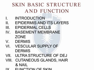
- 1. SKIN BASIC STRUCTURE AND FUNCTION I. INTRODUCTION II. EPIDERMIS AND ITS LAYERS III. EPIDERMAL CELLS IV. BASEMENT MEMBRANE ZONE V. DERMIS VI. VESCULAR SUPPLY OF DERMIS VII. ULTRA STRUCTURE OF DEJ VIII. CUTANEOUS GLANDS, HAIR & NAIL
- 2. • Skin is the largest organ in the body. • A 70 kg individual the Skin weighs over 5 kg & surface area 2 meter square. • The skin is composed of two basic layers 1. outermost layer-Epidermis 2. underlying connective tissue-Dermis BELOW THE DERMIS Subcutaneous fatty layer is Hypodermis
- 4. Layers of epidermis • Stratum basale (the deepest layer) • Stratum spinosum • Stratum granulosum • Stratum lucidum (only in thick skin) • Stratum corneum (most superficial layer of epidermis).
- 5. Layers of epidermis SPINOUS LAYER Upper most STRATUM CORNEUM GRANULAR LAYER Lowest BASAL LAYER Epidermis formed by the division of cells in the basal layer. which give rise to the spinous layer. This layer contains cells that move outwards and progressively differentiate, forming the granular layer and the stratum corneum.
- 6. EPIDERMI S :- • Epidermis derived from ectoderm • The epidermis is mainly composed of keratinocytes , • Thickness is 0.05–0.1 mm . • Thickest on plams and soles (approx- 1.5mm) • Thinnest on eyelids & scrotum (approx- 0.1mm) • The cellular progression from the basal layer to the skin surface takes about 30
- 8. Stratum Basale • The stratum germinativum (basal layer) - single layer of basophilic columnar or cuboidal cells • All cells contain intermediate keratin filaments. • Number of keratin filaments increases as cells progress upward.
- 9. Stratum Spinosum • Also contain the dividing cells as in basale. Cells contain bundles of intermediate filament (tonofilaments) projecting into the processses of cells which give attachment to the desmosomes, so giving spined appearance hence called Prickle Layer • Tonofilaments provide resistant to the abrasion so this layer is thicker in the areas prone to abrasion (thick skin) . • Keratinization begins in the stratum spinosum
- 10. stratum granulosum • Consists of polygonal cells , cytoplasm of which is filled with the basophilic granule ,keratohyaline granules. • It is rich in phosphorylated histidine and cystine. Cells contain, lamellated bodies, made up of lipid. • It fuses with the cell membrane and it come out of cells and function as a intercellular cement or sealing agent.
- 11. Stratum Lucidum • More prominent in thick skin . • Cellular organells and nuclei are not prominent. • It is composed of clear non-nucleated cells. • In the palms and soles, the stratum lucidum is present. • The tan colored protein blocks the underlying melanocytes from view
- 12. Stratum corneum • The main difference between thick skin and thin skin relates to the thickness of the Stratum corneum. • These are the dead cells, flaking off. The cells lose their nucleus and fuse to form squamous sheets, which are eventually shed from the surface (desquamation). • The mean turnover or renewal time of epidermis is 28-30 Days i.e. time for a cell to move from the stratum basale to the distal edge of the stratum corneum and shed.
- 13. CELLS OF EPIDERMIS • A. Keratinocyte • B. Melanocyte • C. Langerhans Cells • D. Merkel Cells ( Haascheiben cells)
- 14. A. Keratinocyte • The cells are named after its intermediate filament called keratin which provides mechanical strength to this cells ( strength is required for resisting daily trauma and mechanical pressure on the epidermis ). • Keratinocytes devide and generate the upper layers. Transit time / turnover time is the time it takes for basal cells to reach the skin surface it is 28 days. • More the need for strength, more the need for keratin
- 16. • Since SC require the maximum strength maximum keratin is in SC and least amount of keratin is in SB. • This process of increasing the keartin in the cells from SB upward to SC is called keratinization of epidermis • As function of SC is protection and it does not divide, it sheds off its nucleus .
- 17. Keratin filamentsjoin together in the upper epidermis by Filaggrin. Since upper layers require more strength than lower layer, Filaggrin synthesis is in upper layer
- 18. Disease with abnormal keratinization • A. Parakeratosis:- in psoriasis the basal layer divides too fast
- 19. • In psoriasis the basal layer divides too fast. Hence the basal layer reach the SC faster and donot shed the nucleus • Hence as nuclei are retained in psoriasis, nucleated stratum corneum is seen on histopathology this is called parakeratosis • In the granular layer the keratohyaline granules contain these filaggrin molecules (Hence granular layer is called granular)
- 20. B. Melanocytes • Present in basal layer • Derived from the neural crest • Produce Melanin • Package it in organells (Melanosomes) and Transfer it via finger-like processes called Dendrites to Keratinocyes • Each melanocyte transfer melanin to 36 keratinocytes • Forming the epidermal-melanin unit [1:36]
- 21. Through these Dendrites melanin transfer to keratinocytes Keratinocyt es Dendriti c process Melanocyte in basal layer
- 22. • Melanin protects cells form Uv light. • They differ from keratinocytes by possessi ng no desmosomes. • Colour of skin depends on :- 1. Amount of melanin inside melanocytes 2. Number and size of melanosomes 3. Degree of transfer into keratinocytes
- 23. • It does not depend on the number of melanocyte (all humans have the same number of melanocytes) Types of melanin :- 1. Eumelanin:- brown or black pigment found in dark colored races. 2. Pheomelanin:- yellow-red in caucasian skin
- 24. C. Langerhans Cells • Derived from bone marrow • Dendritic cells expressing :-CD45, HLA-DR, CD 1C • Role in adaptive immune responses in the skin since they are antigen-presenting cells (APC) Processing antigens and transporting them to local lymph nodes • Like melanocytes they are not connected to adjacent keratinocyte by the desmosomes. • Contain tennis racket shaped “Birbeck
- 25. D. Merkel Cells ( Haascheiben cells) • Seen amongst the basal keratinocytes. contain desmosomes • Derivation of this cell is controversial- evidence supports both differentiation from epidermal keratinocytes as well as migration from the neural crest. • Slow adapting touch receptors.
- 26. Dermi s • It is connective tissue that support the epidermis and attaches the epidermis to the hypodermis. • Dermis is 15-40 times thicker than the epidermis • Its surface consists of many ridges (dermal papillae) which interdigitate with epidermal ridges. • The dermis is also the area where all the glands of the body are located. • Has 2 layers/compartments 1.A thin zone immediately beneath the epidermis the papillary dermis 2. A thick zone of Reticular dermis that extends from the base of the papillary dermis to the surface of the subcutaneous fat
- 27. Papillary dermis • Papillary layer –The papillary dermis is the uppermost layer of the dermis,composed of thin haphazardly arranged collagen bundles,delicate branching elastic fibers,numerous fibrocytes,abundant ground substance. • A highly developed microcirculation composed of arterioles,capillaries and venules Its superior surface is uneven (fingerlike projections) which forms the characteristic fingerprint of the finger. • This layer provides the epidermis with nutrients. Pain and touch receptors are found here
- 28. Reticular dermis • Dense irregular Connective Tissue Has thick bundles of Collagen and coarse Elastic fibers. • Proportionally, there are fewer fibrocytes and blood vessels and less ground substance compared to papillary dermis • Arrangement of bundle in the direction of mechanical force give rise to the cleavage lines of Langer. • Strongest layer of the Dermis. Gives the area strength.Contains sweat,sebaceous glands and pressure receptors • Leather is made of this layer.
- 29. HYPODER MIS • Consists of loose connective tissue which helps in sliding the skin over the deep structure. • Consists of layer of fat according to the nutritional status of the person. • Also called as superficial fascia or panniculus adiposus
- 30. VESSELS IN SKIN • Arteries form the 2 plexuses One at the junction of papillary and reticular layer( sub- papillary plexus) and another at junction of dermis and hypodermis (cutaneous plexus). • Veins form the 3 plexuses – 2 in same position as for arterial and another in the middle of the dermis
- 31. BASEMENT MEMBRANE ZONE • THE JUNCTION BETWEEN EPIDERMIS AND DERMIS IS CALLED THE BASEMENT MEMBRANE ZONE • ULTRASTRUCTURALLY THIS ZONE IS COMPOSED OF THREE COMPONENTS FROM TOP TO BOTTOM
- 32. 1. BASAL KERATINOCYTE 2. DEJ 3. DERMAL CONNECTIVE TISSUE
- 33. ULTRA STRUCTURE OF DEJ HEMIDESMOSO MES LAMINA DENSA LAMINA LUCIDA DE J TYPE 4 COLLAGEN FIBER ANCHORING FIBER DERMAL CONNETIVE TISSUE
- 34. • PLASMA MEMBRANE OF BASAL KERATINOCYTE WITH THE SPECIALIZED ATTACHMENT PLATES = HEMIDESMOSOMES • DE JUNCTION:- IT IS DEVIDED INTO THE UPPER – LAMINA LUCIDA LOWER – LAMINA DENSA TYPE –IV COLLAGEN IS THE MAJOR COMPONENT OF LAMINA DENSA • ANCHORING FIBERS:-ATTACHES THE LAMINA DENSA ABOVE TO THE UNDERLYING CONNECTIVE TISSUES TYPE VII COLLAGEN IS THE MAJOR PROTEIN IN THE ANCHORING FIBER
- 35. BASAL KERATINOCYTE TYPE -4 COLL.FIBER BP- 1 BP- 2 LAMINI N DERMAL CONNECTIVE TISSUE ANCHORING FIBER BP (anti body against BP-1) BP,CP,LAD (anti-b against BP-2) EBA (antibody against collagen 7) EBD (absent collagen 7 since birth) EBJ (Absent laminin since birth)
- 36. Cutaneous Glands • 1. Sebaceous (oil) glands- Sebaceous glands are microscopic glands in the skin which secrete an oily matter, called sebum, In the hair follicles to lubricate the hair. • In humans, they are found in greatest abundance on the face and scalp, though they are distributed throughout all skin sites except the palms and soles. • infection causes acne
- 37. 2. Sweat glands – • Sweat glands are exocrine glands, found in the skin , that are used for body temperature regulation. a)Eccrine glands -Eccrine glands (or merocrine glands) are found at virtually all sites on the human body. They produce clear liquid (perspiration), consisting of water, salts, and urea. b)Apocrine glands- Apocrine glands are found in axillary and genital areas, secrete a milky protein and fat substance. This mixture is an excellent source of nutrients for bacteria which produce body odour.
- 38. HAI R • Follicle- A hair follicle is a part of the skin that grows hair by packing old cells together
- 39. • Arrector pili -Arrectores pilorum (singular Arrector pili) are tiny smooth muscle fibers attached to each hair follicle, which contract to make the hairs stand on end, causing goose bumps.
- 40. Nail s • Fingernails and toenails are made of a tough protein called keratin. Along with hair and teeth they are an appendage of the skin.
- 41. • Free edge- The part of the nail that extends past the finger, beyond the nail plate. There should always be a free edge present to prevent infections. • Nail folds (cuticle)- A fold of hard skin overlapping the base and sides of a fingernail or toenail. • Nail Matrix- This is the only living part of the nail. It is situated behind and underneath the Nail Fold and produces protein keratin which makes up the Nail Plate
- 43. • The skin of the embryo begins to form during the first 20 to 30 days of embryonic life, the period of active organogenesis in human development • The skin arises by the juxtaposition of two major embryological elements 1. The prospective epidermis, originates from a surface area of the early gastrula; ectoderm. 2. The prospective mesoderm, which is brought into contact with the inner surface of the epidermis. • The neural crest also makes contribution to the skin
- 44. Development of epidermis EMBRYONIC GESTATIONAL AGE EVENTS 3 Weeks Single Layer Of Flattened Epithelial Cells 4 Weeks Basal Germinative Layer & Periderm 3 Months Intermediate Cells Tonofilaments- desmosomes 5 Months Keratohyaline Granules, signs Of Cornification Starts 6 Months Cornification Completed Term Increase In Thickness Of Cornified Layers
- 45. Physiological function of skin Epidermis is a barrier to water loss and also barrier to prevent entry of harmful substance like microbes and UV light into the body . Loss of this barrier will lead to increase of infection and more loss of fluids from skin (in burn patient) This barrier is created by polygonal shape of keratinocytes and better sticking together by glue like lipid secreted by keratinocytes. It is compared to a brick and cement model where bricks are synonymous with keratinocytes and cement is the glue.
- 46. BRICK=KERATINOCY TE CEMENT = INTER CELLULAR LIPID LAMELLAR BODY IN KERATINOCYTE SYNTHESIZES LIPID AND EXPELS IT INTO LIPID INTO INTERCELLULAR SPACE
- 47. BARRIER LOSS • Barrier loss can either be due to problem in the keratinocytes (brick damage) or problem in the glue (cement collapse). A. Problem in the keratinocytes (brick damage) :- 1.Damage to intercellular junction desmosomes 2. Primary destruction of the keratinocyte itself (cytolysis) 3. Problem in calcium pump
- 48. • B. Problem in the Glue (cement damage) :- – Intercellular cement contain ceramide, Squalene, fatty acids. – Loss of intercellular glue cause eczema. The small gap creates a barrier function loss. This Gap then fill up with fluid this is called intercellular edema or spongiosis. – Fluid can ooze out of skin clinically. These microscopic gap also allows entry of organism in to skin [ Pt. with atopic eczema have more infectious especially staphylococcal ]. – In eczema to replace this loss of ceramide moisturising containing ceramide are often given to replace this intercellular cement.
- 49. • Langerhans cells which serve as sentinel cells whose prime function is to survey the epidermal environment and to initiate an immune response against microbial threats. • Melanin, which is mostly found in basal keratinocytes, also provides some protection against DNA damage from ultraviolet radiation. • Thermoregulation:-Vasodilatation or vasoconstriction of the blood vessels in the deep or superficial plexuses helps regulate heat loss. • Skin lubrication and waterproofing is provided by sebum secreted from sebaceous glands. • Subcutaneous fat has important roles in cushioning trauma aswell as providing insulation and a calorie reserve. • Endocrine function:- releasing the hormone leptin, which acts on the hypothalamus to regulate hunger and energy metabolism • Vitamin D synthesis from UV-B