More Related Content Similar to Anatomia y fisiopatologia de la injuria asociada con la anestesia regional y la medicina del dolor
Similar to Anatomia y fisiopatologia de la injuria asociada con la anestesia regional y la medicina del dolor (20) More from Silvestre Degreéf
More from Silvestre Degreéf (12) 1. Anatomy and Pathophysiology of Spinal Cord Injury
Associated With Regional Anesthesia and Pain Medicine
2015 Update
Joseph M. Neal, MD,* Sandra L. Kopp, MD,† Jeffrey J. Pasternak, MD,†
William L. Lanier, MD,† and James P. Rathmell, MD‡
Background and Objectives: In March 2012, the American Society
of Regional Anesthesia and Pain Medicine convened its second Practice
Advisory on Neurological Complications in Regional Anesthesia and
Pain Medicine. This update is based on the proceedings of that confer-
ence and relevant information published since its conclusion. This arti-
cle updates previously described information on the pathophysiology
of spinal cord injury and adds new material on spinal stenosis, blood
pressure control during neuraxial blockade, neuraxial injury subse-
quent to transforaminal procedures, cauda equina syndrome/local anes-
thetic neurotoxicity/arachnoiditis, and performing regional anesthetic
or pain medicine procedures in patients concomitantly receiving gen-
eral anesthesia or deep sedation.
Methods: Recommendations are based on extensive review of research
on humans or employing animal models, case reports, pathophysiology re-
search, and expert opinion.
Results: The pathophysiology of spinal cord injury associated with re-
gional anesthetic techniques is reviewed in depth, including that related
to mechanical trauma from direct needle/catheter injury or mass lesions,
spinal cord ischemia or vascular injury from direct needle/catheter trauma,
and neurotoxicity from local anesthetics, adjuvants, or antiseptics. Specific
recommendations are offered that may reduce the likelihood of spinal
cord injury associated with regional anesthetic or interventional pain
medicine techniques.
Conclusions: The practice advisory’s recommendations may, in select
cases, reduce the likelihood of injury. However, many of the described in-
juries are neither predictable nor preventable based on our current state
of knowledge.
What’s New: Since publication of initial recommendations in 2008, new
information has enhanced our understanding of 5 specific entities: spinal
stenosis, blood pressure control during neuraxial anesthesia, neuraxial injury
subsequent to transforaminal techniques, cauda equina syndrome/local an-
esthetic neurotoxicity/arachnoiditis, and performing regional anesthetic or
pain procedures in patients concomitantly receiving general anesthesia or
deep sedation.
(Reg Anesth Pain Med 2015;40: 506–525)
Injury to the neuraxis associated with regional anesthesia or pain
medicine procedures is ultimately linked to anatomic and/or
physiologic damage to the spinal cord, the spinal nerve roots,
or their blood supply. Mechanisms of injury are sometimes iden-
tifiable, as in the case of epidural hematoma or abscess, but can
also be exceedingly difficult to pinpoint, as exemplified by most
cases of presumed spinal vascular injury. The goal of our origi-
nal advisory on this topic and the updated material contained
herein is to provide an anatomic and pathophysiologic basis
from which to build an understanding of neuraxial complica-
tions associated with regional anesthesia and pain medicine.
Consistent with a recent editorial call to focus practice advi-
sory and consensus conference updates on new material,1
we have
crafted this review in 2 sections. First, to provide perspective, we
will briefly review those topics and associated recommendations
for which no substantially new knowledge has emerged. To pro-
vide consistency over time or when appropriate, the current review
will present text and especially recommendations that are essen-
tially verbatim from those of our original work. Interested readers
can find the detailed explanations and their specific literature-
based citations by revisiting those 2008 articles.2,3
The second
section will focus on 5 topics that either have significantly new in-
formation to add to our previous understanding and/or we believe
deserve more extensive discussion than was provided in the first
iteration of this practice advisory.
METHODS
Standard search engines and cross-referenced citations iden-
tified the literature basis for the updated material contained within
this review. PubMed and Ovid were searched from 2006 for-
ward to identify new material by using MeSH terms as individ-
ual headings or in relevant combinations: “spinal cord injury,”
“hypotension,” “neurotoxicity,” “transforaminal,” “cauda equina syn-
drome,” “anterior spinal artery syndrome,” “needle injury,” “spinal
stenosis,” “spinal cord ischemia,” and “spinal cord infarction.”
As specifically noted in our 2008 review, “The strength of
scientific evidence that is used to arrive at these recommendations
is not easily measured by traditional stratification methodologies
such as the United States Agency for Health Care Policy and Re-
search structure for ranking Statements of Evidence and Grades of
Recommendation.4
Because of the extreme rarity of the specific
complications that are addressed in this article, traditional method-
ologies such as randomized controlled trials, meta-analysis, or large
human case series rarely exist and are unlikely to exist in the future.
Our recommendations are therefore based on methodologies that
are necessarily less robust, such as anatomic or pathophysiologic
studies of human cadavers or animals, nonrandomized trials, ret-
rospective series, case reports, or expert opinion. The grading of
recommendations offered by this practice advisory has been mod-
ified from an American College of Cardiology/American Heart
Association construct that classifies the strength of guidelines
for perioperative cardiac evaluation.”2
Readers of this article are reminded that practice advisories
are created when data on a subject are limited or nonexistent.
From the *Department of Anesthesiology, Virginia Mason Medical Center,
Seattle, WA; †Mayo Clinic College of Medicine, Rochester, MN; and
‡Harvard Medical School, Boston, MA.
Accepted for publication June 3, 2015.
Address correspondence to: Joseph M. Neal, MD, 1100 9th Ave (B2-AN),
Seattle, WA 98101 (e‐mail: Joseph.Neal@virginiamason.org).
Portions of this article were presented as a part of the American Society of
Regional Anesthesia and Pain Medicine’s second Practice Advisory on
Neurological Complications in Regional Anesthesia and Pain Medicine in
San Diego, California, on March 16, 2012.
This study has no funding sources.
The authors declare no conflict of interest.
Copyright © 2015 by American Society of Regional Anesthesia and Pain
Medicine
ISSN: 1098-7339
DOI: 10.1097/AAP.0000000000000297
SPECIAL ARTICLE
506 Regional Anesthesia and Pain Medicine • Volume 40, Number 5, September-October 2015
Copyright © 2015 American Society of Regional Anesthesia and Pain Medicine. Unauthorized reproduction of this article is prohibited.
2. Advisories rely on limited clinical and animal data, and as such,
the synthesis and interpretation of data by 1 group of experts
may differ from conclusions by another set of equally qualified
experts. Thus, practice advisories represent a level of recom-
mendation that is less than that offered by standards or clinical
practice guidelines.5
The recommendations contained herein do
not define standard of care. They are not intended to replace clin-
ical judgment as applied to a specific patient scenario. “These
recommendations are intended to encourage optimal patient care,
but cannot ensure the avoidance of adverse outcomes. As with
any practice advisory recommendation, these are subject to revi-
sion as knowledge of specific complications advances.”6
REVIEW OF PREVIOUS RECOMMENDATIONS
Mechanical Injury
Some neuraxial anesthetic complications are secondary to
mechanical injury of the spinal cord, spinal nerve roots, or
the spinal nerves as they exit the intervertebral foramina. Injury
to these structures may involve direct needle or catheter trauma
or lesions within the vertebral canal that compress neural struc-
tures and thereby cause ischemic injury. These various mecha-
nisms ultimately lead to loss of anatomic and/or physiologic
neural integrity and can result in permanent injury.7
We have
not changed the majority of recommendations related to mechan-
ical injury (Table 1).
Iatrogenic Spinal Cord Trauma
The incidence of spinal cord–related needle/catheter trauma
is unknown, but decidedly rare. Anesthesiologist-reported or quality-
assurance databases may well underreport this complication,
whereas medicolegal databases are likely to skew data in the oppo-
site direction. For instance, direct spinal cord injury was noted in
6 (0.73%) of 821 neuraxial claims from the American Society of
Anesthesiologists (ASA) Closed Claims database, which does not
provide a denominator of total cases.7
Conversely, direct needle
trauma was detected in only 9 of 1.7 million neuraxial anes-
thetics (0.0005%) over a 10-year period in Sweden,12
and only
1 case was reported in a 2000 survey from French spinal cord re-
habilitation centers (from an estimated 1 million neuraxial anes-
thetics performed that year in their catchment area).13
Three anatomic characteristics of the human neuraxis con-
tribute to its potential for sustaining needle or catheter injury. First,
although the conus medullaris is typically described as terminat-
ing at the L1–2 vertebral interspace in adults (and terminates more
caudad in the first few months of life), its terminus varies widely
from T12 to L4. When this variation is coupled with practitioners’
inaccurate determination of which spinal interspace they are pal-
pating,14
it is not surprising that needle injury to the spinal cord
has been reported in instances where the conus medullaris termi-
nated more caudad than expected.12
Indeed, a recent study demon-
strated that in 40% of term parturients, the perceived vertebral
level identified by palpation at the intercristal line was in reality
at the L3 interspace or higher.15
Neuraxial ultrasonography may
improve estimation of the vertebral level because it is more accu-
rate than palpation for identifying surface landmarks, especially in
challenging anatomic scenarios,16
such as obesity, scoliosis, or
previous spinal surgery.17
Second, the customary expectation of
encountering resistance prior to entering the epidural space is
not always fulfilled in those individuals in whom the ligamentum
flavum has failed to fuse in the midline, a condition that is more
prominent in the upper thoracic (4%–21% midline gaps at T3–4
and above) and cervical neuraxis (51%–74% midline gaps).18
Un-
anticipated congenital dysraphisms can also contribute to acciden-
tal entry into the spinal cord.19
Third, the margin for error during
needle advancement is significantly diminished as one proceeds
from the lumbar posterior epidural space with its 5- to 13-mm
dorsal-to-ventral dimensions, to the 2- to 4-mm thoracic posterior
epidural space, to the average 0.4-mm cervical posterior epidural
TABLE 1. Recommendations: Factors That May Limit Neuraxial Injury
These recommendations are intended to encourage optimal patient care, but cannot ensure the avoidance of adverse outcomes.
As with any practice advisory recommendation, these are subject to revision as knowledge advances regarding specific complications.
Anatomic factors
• Misidentification of vertebral level, unrecognized lateral needle placement or deviation, abnormal caudad termination of the spinal cord, or
failure of the ligamentum flavum to fuse in the midline may contribute to direct needle injury of the spinal cord. Clinicians are advised to be
aware of these anatomic conditions, particularly in patients with challenging surface anatomy (eg, as may occur with obesity, kyphoscoliosis,
and other conditions). Ultrasonography or fluoroscopy could be considered as an adjunct for accurate determination of vertebral level in these
challenging patients. (Class I)
• Surgical positioning, severe spinal stenosis, and specific space-occupying extradural lesions (eg, epidural lipomatosis, ligamentum flavum
hypertrophy, synovial cysts, or ependymoma) have been associated with temporary or permanent spinal cord injury in conjunction with
neuraxial regional anesthetic techniques. These conditions are particularly relevant when they coexist with an epidural hematoma or
abscess. Awareness of these conditions should prompt consideration of risk-versus-benefit when contemplating neuraxial regional anesthetic
techniques. (Class I)
• Patients with known tumor in the epidural space should undergo neuraxial imaging studies to define the extent of tumor mass. If the tumor
is close to the planned site of epidural solution injection, alternative methods of anesthesia or analgesia should be considered. (Class II)
• For patients receiving neuraxial injection for treatment of pain (eg, cervical epidural injection of steroids via an interlaminar route), radiologic
imaging studies such as computed tomography (CT) or magnetic resonance imaging should be used to assess the dimensions of the spinal
canal, and this information should be considered in the overall risk-to-benefit analysis as well as guiding the selection of the safest level
for entry. (Class II)
Physiologic factors
• Clinicians are advised to be aware of and to avoid conditions that have been linked to the formation of epidural hematoma or epidural abscess,
as noted in previous American Society of Regional Anesthesia and Pain Medicine Practice Advisories. Such conditions include concurrent
or imminent anticoagulation, the use of multiple anticoagulants, improper aseptic technique, and needle placement during untreated
active infection.8–11
(Class I)
Recommendations contained within Table 1 have been modified minimally from the authors’ 2008 advisory.2
Significant changes are in italics.
Regional Anesthesia and Pain Medicine • Volume 40, Number 5, September-October 2015 Pathophysiology of Spinal Cord Injury
© 2015 American Society of Regional Anesthesia and Pain Medicine 507
Copyright © 2015 American Society of Regional Anesthesia and Pain Medicine. Unauthorized reproduction of this article is prohibited.
3. space. Indeed, because the epidural space is a potential space, the
cervical posterior epidural space may be nonexistent, particularly
at higher vertebral levels.20–22
Mass lesions can also lead to spinal cord injury. Intradural or
extradural lesions compete for cross-sectional space within the
spinal canal and in so doing potentially decrease spinal cord per-
fusion pressure (SCPP) by inhibiting arterial inflow, inhibiting
venous outflow, or elevating cerebrospinal fluid (CSF) pressure
([SCPP = mean arterial pressure − spinal cord CSF pressure]23
;
in rare circumstances, direct venous outflow pressure may also
impact regional SCPP.) If SCPP is sufficiently diminished, it can
lead to spinal cord ischemia that in more severe instances can pro-
duce infarction. Epidural hematoma and abscess are commonly
recognized complications of neuraxial anesthetic or pain medicine
techniques, and they can lead to consequential mass lesions.8,24
Less well appreciated is the potential for transient pressure eleva-
tions secondarytoexcessivevolumes oflocalanesthetic25
(espe-
ciallyin infants),26
compromised egress of local anesthetic or blood
through stenotic intervertebral foramina,27
or unusual mass lesions
such as tumors, granulomas from chronic intrathecal morphine
administration,28 epidural lipomatosis,29,30
synovial cysts,13
or
ependymoma. Many of these conditions are occult to patient and
practitioner and become relevant only when blood, pus, or local
anesthetic competes for limited cross-sectional area within the
vertebral canal. The presence of increased volume within the ver-
tebral canal, whether by fluids or mass lesions, can have a “Star-
ling resistor” effect on blood vessels, limiting blood ingress into
and egress from the affected tissues. Moreover, patients with
severe spinal stenosis or other mass effects may be at additional
risk of compromised tissue blood flow when surgical field expo-
sure requires certain positions such as extreme lordosis, lithotomy,
or the flexed lateral position.2,31
Spinal Nerve Root and Spinal Nerve Trauma
The spinal nerves are protected during midline or para-
median approaches to the neuraxis because of their lateral posi-
tion and the partial protection afforded by the vertebral laminae,
transverse processes, and facet joints. Therefore, midline procedure-
related injury to the spinal nerve, or to the anterior or posterior
ramus outside the intervertebral foramen, occurs only when
needles deviate laterally. Spinal nerves can also be contacted un-
intentionally during procedures such as paravertebral or psoas
compartment block when the needle is directed too medially.
Needlesmedialtothefacet withinthelateralrecess mayuninten-
tionally contact the dorsal nerve roots2
(Fig. 1).
As concluded in our 2008 review, “mechanical injury to the
neuraxis can arise consequent to direct needle trauma or to space-
occupying lesions whose mass effect compromises spinal cord
blood flow (SCBF). Evidence to support contribution to injury
varies with the mechanism of injury. In the case of epidural
hematoma or abscess, extensive literature supports causation.
Conversely, neuraxis injury in the setting of rare extraspinal mass
lesions or relatively common surgical positions only establishes
association or chance occurrence.”2
Limited additional informa-
tion since our 2008 publication has not altered this conclusion
(Table 1), except for new information on the association of spinal
stenosis with neuraxial injury, as will be presented subsequently.
Spinal Cord Ischemia and Vascular Injury
Disruption of SCBF can occur from a variety of mechanisms,
including needle trauma affecting the spinal vasculature (Fig. 2),
compressive mass lesions (Fig. 3), or vascular spasm (Fig. 4). Spi-
nal cord blood flow may also be compromised from low-flow
states, such as might occur from significant and prolonged
systemic hypotension, embolic phenomena, or vascular stenosis.
The frequency of spinal cord ischemia is distinctly rare and our
understanding limited. Our previous article2
extensively reviewed
the human spinal cord blood supply. The spinal cord and cauda
equina receive two-thirds of their blood supply from the ante-
rior spinal artery, which cannot be injured directly by midline
or paramedian needles without first traversing the spinal cord
(Fig. 2). Damage to the spinal cord’s posterior blood supply
is largely mitigated by the redundancy of the posterior spinal
artery system. However, the segmental or spinal branch arteries
are exposed to needle-related trauma when the needle deviates
far laterally or is intentionally placed near a segmental artery (such
as during perispinal techniques or celiac plexus block).33
Radic-
ular arteries can sustain needle injury during a transforaminal
approach, whichcanbeimportantifthatarteryis1ofthefewradic-
ular arteries that continue on to become a medullary artery supply-
ing the spinal cord (Fig. 5). Disruption of SCBF might also occur
from procedure-induced hematoma or drug-induced vasospasm
associated with neurolytic procedures such as celiac plexus block
(Fig. 4), although clear proof of these mechanisms is lacking.2
Within the category of vascular injury to the neuraxis, the
majority of our previous recommendations remain intact (Table 1).
However, we will henceforth discuss 2 topics for which new infor-
mation justifies strengthening of previous advice: blood pressure
control during neuraxial anesthesia and central nervous system
(CNS) injury during transforaminal pain medicine procedures.
Neurotoxicity
Our previous review concluded, “Neuraxial local anesthetics,
opioids, adjuvants, and preservatives in clinically recommended
doses are remarkably safe in the vast majority of patients. Never-
theless, a patient may rarely be vulnerable to local anesthetic and
adjuvant neurotoxicity even in normal clinical situations. Clin-
ical evidence comes from case reports of patients who sustained
neuraxis injury that was presumed secondary to a neurotoxic mech-
anism, even though they received standard doses of spinal or epi-
dural local anesthetic with or without adjuvant. Neurotoxicity is
more likely to occur in conjunction with physical disruption of
the blood–spinal cord barrier by needle or catheter trauma, or
from iatrogenic conditions leading to maldistribution and/or over-
dosing of neuraxial local anesthetics.”2
While our previous rec-
ommendations have not substantially changed (Table 1), we will
henceforth discuss newer information concerning cauda equina
syndrome (CES), arachnoiditis, and clinical experience with intra-
thecal 2-chloroprocaine (2-CP).
NEW RECOMMENDATIONS FROM THE SECOND
PRACTICE ADVISORY
Spinal Stenosis
Spinal hematoma, abscess, tumor spread, epidural lipoma-
tosis, spinal arachnoid cysts, ankylosing spondylitis,12,34 or ex-
treme surgical positioning such as hyperlordosis or extreme
lateral flexion31,35
are mechanical causes that can contribute to
the development of spinal cord ischemia or infarction in the
perioperative period. Recent literature has focused on degener-
ative spinal stenosis and its association with various manifesta-
tions of neuraxial injury in the setting of regional anesthesia.
Degenerative spinal stenosis is caused by osteoporosis and/or
hypertrophy of the ligamentum flavum and bony elements of
the spinal canal that effectively reduces spinal canal cross-
sectional area and competes with the spinal cord and nerve
roots for space. Similar mechanisms might contribute to local
anesthetic neurotoxicity by causing maldistribution and/or
Neal et al Regional Anesthesia and Pain Medicine • Volume 40, Number 5, September-October 2015
508 © 2015 American Society of Regional Anesthesia and Pain Medicine
Copyright © 2015 American Society of Regional Anesthesia and Pain Medicine. Unauthorized reproduction of this article is prohibited.
4. reduced clearance of relatively undiluted drug.36,37
Degenera-
tive changes may also cause narrowing of the vertebral foram-
ina, which compromises egress of fluids22,38
and consequently
results in an increase in vertebral canal pressure27
and tran-
siently diminished neural blood flow.39
Spinal stenosis repre-
sents a continuum of severity, from mild and inconsequential to
severe; 19% of patients in their 60s will have absolute spinal ste-
nosis (<1-cm anteroposterior diameter of the spinal canal).40
Al-
though commonly discovered during imaging for the diagnosis
of back pain, there are subsets of patients with undiagnosed spi-
nal stenosis that is discovered only during workup of injury af-
ter a neuraxial anesthetic.12
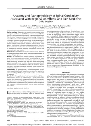
FIGURE 1. Midline or paramedian approaches (needles A and B) may directly traumatize the spinal cord, whereas unintentional lateral
deviation of the needle (C) may contact the spinal nerve or the anterior or posterior primary ramus outside the foramen. Intentional lateral
approaches, for example, transforaminal approach (needle D), have the potential to come in close proximity to the spinal nerve or a spinal
artery. Note that transforaminal approaches are typically at the cervical or lumbar levels, not the T6 level as illustrated. Illustration by Gary J.
Nelson. Reproduced with permission from Neal and Rathmell, Complications in Regional Anesthesia and Pain Medicine.32
Regional Anesthesia and Pain Medicine • Volume 40, Number 5, September-October 2015 Pathophysiology of Spinal Cord Injury
© 2015 American Society of Regional Anesthesia and Pain Medicine 509
Copyright © 2015 American Society of Regional Anesthesia and Pain Medicine. Unauthorized reproduction of this article is prohibited.
5. The potential for spinal stenosis to cause or worsen neuraxial
injury in the setting of a regional anesthetic has been the object of
speculation for decades.41
However, the first strong signal of the
relationship between spinal pathology and increased risk of
neuraxial injury was identified in Moen and colleagues’12
report
of the association between spinal stenosis and neuraxial injury
in 1.7 million neuraxial anesthetics conducted in Sweden between
1990 and 1999. Of the 33 cases of spinal hematoma, one-third
were associated with coagulopathy or thromboprophylaxis (in one-
third of those cases, thromboprophylaxis was administered in accor-
dance with published guidelines). This report contrasted the
extreme rarity of spinal hematoma in young women who re-
ceived neuraxial anesthesia for childbirth (1:200,000) versus
the 1:22,000 incidence of spinal hematoma in elderly women
undergoing hip fracture repair or 1:3600 incidence in those elderly
women receiving total joint arthroplasty. During diagnostic imag-
ing, 6 of the 33 cases were noted to have previously undiagnosed
spinal stenosis or ankylosing spondylitis. These conditions may
have compromised spinal cord circulation to a greater extent in
the mostly elderly women who constituted the majority of this
cohort, as compared with younger obstetric patients with an
uncompromised spinal canal who likely would have experi-
enced a lesser degree of circulatory impairment from a simi-
larly sized hematoma.12,42
Since the Swedish publication, confirmatory reports have
emerged.13,43
For example, a retrospective analysis of neuraxial
anesthetics performed in patients with known spinal canal pathol-
ogy (spinal stenosis or lumbar disc disease) observed a 1.1% inci-
dence of neuraxial complications, which was higher than expected
for patients without spinal canal pathology who underwent
similar surgeries at the same institution.44
While this study cor-
roborates observations from previous investigations and points
to a higher incidence in those patients with known spinal steno-
sis compared with those with unsuspected disease, it also points
to the difficulties in firmly establishing spinal canal pathology
and subsequent neuraxial injury as cause and effect, rather than
association. Case reports and large registries do not provide a
general anesthetic control group and cannot distinguish whether
FIGURE 2. Midline or paramedian approaches may directly traumatize the posterior spinal arteries, whereas unintentional lateral deviation of
the needle may contact the spinal branch artery. Direct injury to the anterior spinal artery would require placement of a needle or catheter
through the spinal cord. Illustration by Gary J. Nelson. Reproduced with permission from Neal and Rathmell, Complications in Regional
Anesthesia and Pain Medicine.32
Neal et al Regional Anesthesia and Pain Medicine • Volume 40, Number 5, September-October 2015
510 © 2015 American Society of Regional Anesthesia and Pain Medicine
Copyright © 2015 American Society of Regional Anesthesia and Pain Medicine. Unauthorized reproduction of this article is prohibited.
6. FIGURE 3. Extradural mass lesions. Note how various conditions can reduce spinal canal cross-sectional area and either directly compress the
spinal cord or the cauda equina (arrows) or increase epidural space or CSF pressures through their mass effect. Illustration by Gary J. Nelson.
Reproduced with permission from Neal and Rathmell, Complications in Regional Anesthesia and Pain Medicine.32
Regional Anesthesia and Pain Medicine • Volume 40, Number 5, September-October 2015 Pathophysiology of Spinal Cord Injury
© 2015 American Society of Regional Anesthesia and Pain Medicine 511
Copyright © 2015 American Society of Regional Anesthesia and Pain Medicine. Unauthorized reproduction of this article is prohibited.
7. the observed injury results from underlying spinal canal pathol-
ogy, disease progression, surgical factors, patient positioning, or
a combination thereof.
The advisory panel therefore acknowledges growing evi-
dence of an association between spinal stenosis or other spinal
canal pathology and a higher incidence of complications after
neuraxial blockade.12,13,41,44
However, causation cannot be estab-
lished definitively. To this point, the panel also acknowledges
that a multitude of neuraxial regional anesthetics and interven-
tional pain medicine procedures are performed daily on patients
with varying degrees of spinal stenosis. In some of these cases,
spinal stenosis has been diagnosed and may indeed be the indi-
cation for intervention. Furthermore, spinal stenosis can con-
tribute to neurologic injury from surgery or positioning even
in the absence of neuraxial anesthetic techniques.45,46
Based
on details gleaned from the limited literature on this topic and in
accordance with the double-crush theory,47
we believe it reason-
able to speculate that patients with moderate to severe spinal ste-
nosis might be more vulnerable to injury if there are coexisting
conditions such as neuraxial surgery, preexisting neurologic dis-
ease, mucopolysaccharidosis,48
nonneutral patient positioning, or
conditions known or unknown that compete for limited cross-
sectional area within the spinal canal. Although the preponderance
of spinal stenosis has been associated with epidural and combined
spinal-epidural techniques,12
association with spinal anesthesia
has also been reported.35,44
As noted previously, the presence of
spinal stenosis may be unknown to the clinician and the patient.
However, those patients who report neurogenic claudication with
symptoms that progress with ambulation are likely to have severe
stenosis, even if not formally diagnosed. Recommendations for
spinal stenosis are found in Table 2.
Blood Pressure Control During
Neuraxial Anesthesia
Spinal cord ischemia or infarction associated with neuraxial
regional anesthesia is a decidedly rare event that may present as
anterior spinal artery syndrome (ASAS), posterior spinal artery
syndrome, watershed infarction, or an ill-defined injury consistent
with critically reduced or absent SCBF. For perspective, only 10 of
821 medicolegal claims for neuraxial injury contained within the
ASA Closed Claims database were alleged to have resulted from
ASAS or variations of spinal cord ischemia.7
A study of long-
term outcomes after acute spinal cord ischemia documented that
only 1 of 54 patients had actually received a neuraxial anesthetic.49
This should not be surprising, as the highest risks for periopera-
tive spinal cord infarction are associated with specific operations,
for example, aortic, cardiac, thoracic, or spine surgeries. Even
FIGURE 4. Proposed mechanisms of direct injury to reinforcing arteries supplying the spinal cord. On the left, a needle can potentially disrupt
a segmental artery or precipitate a hematoma. On the right, needle irritation or injected phenol or alcohol (as used in neuroablation
procedures) can cause vasospasm. These proposed mechanisms have not been proven in humans. Illustration by Gary J. Nelson. Reproduced
with permission from Neal and Rathmell, Complications in Regional Anesthesia and Pain Medicine.32
Neal et al Regional Anesthesia and Pain Medicine • Volume 40, Number 5, September-October 2015
512 © 2015 American Society of Regional Anesthesia and Pain Medicine
Copyright © 2015 American Society of Regional Anesthesia and Pain Medicine. Unauthorized reproduction of this article is prohibited.
8. though autoregulation of SCBF mirrors that of cerebral blood
flow50
(Fig. 6), spinal stroke is apparently much less frequent than
the estimated 0.1% incidence of perioperative cerebral stroke in
patients undergoing noncardiac, nonneurologic surgery.51
Spinal
cord ischemia and infarction are rarely reported even after clinical
scenarios of prolonged low mean arterial pressure (MAP), such as
during cardiopulmonary bypass or induced hypotension to a MAP
of 60 mm Hg or less.52–55 While it is relatively rare for survivors
of cardiac arrest to develop spinal cord ischemic injury, 46% of
those who died of cardiac arrest or a severe hypotensive episode
manifested ischemic spinal cord myelopathy at autopsy.56
Despite
the expectation that ischemic myelopathy would be most preva-
lent within the thoracic spinal cord watershed areas (because the
thoracic spinal cord classically is supplied by fewer medullary
arteries than either the cervical or lumbosacral spinal cord),
95% of cases in the previously noted postmortem study involved
FIGURE 5. The transforaminal approach to the neuraxis may allow the needle to contact either the spinal nerve or the spinal artery. Illustration
by Gary J. Nelson. Reproduced with permission from Neal and Rathmell, Complications in Regional Anesthesia and Pain Medicine.32
TABLE 2. Recommendations: Patients With Spinal Stenosis
These recommendations are intended to encourage optimal patient care, but cannot ensure the avoidance of adverse outcomes.
As with any practice advisory recommendation, these are subject to revision as knowledge advances regarding specific complications.
• Spinal stenosis represents a continuum of spinal canal encroachment by hypertrophied ligamentum flavum, bony overgrowth, and/or
degenerative changes such as from osteoporosis or herniated nucleus pulposus. Patients with spinal canal pathology (eg, spinal stenosis, lumbar
disk disease) may have clinical or subclinical evidence of a preexisting neurologic deficit due to neural compromise from the disease state.
However, even moderately severe spinal stenosis is not always symptomatic; many patients (or their healthcare providers) are unaware
that they have the condition. (Class I)
• When neuraxial anesthesia is complicated by the development of mass lesions within the spinal canal (eg, hematoma or abscess), resultant
postoperative neurologic complications may be more likely or more severe in patients with spinal stenosis or other obstructive spinal canal
pathology, including changes brought on by patient positioning. (Class I)
• In patients with known severe spinal stenosis or symptoms suggestive thereof, we recommend that risk-to-benefit analysis be considered
prior to performance of neuraxial anesthesia because of the association of spinal stenosis with neurologic complications in the setting of
neuraxial blockade. If neuraxial blockade is performed, we recommend heightened perioperative vigilance for symptoms suggestive of neural
compromise. (Class II)
• There is no firm linkage to injury if spinal stenosis is at a site distant from the level of neuraxial block placement. (Class III)
• If neuraxial anesthesia is planned, the practitioner may consider reducing the total mass (volume  concentration) of local anesthetic in an
effort to reduce segmental spread and local anesthetic neurotoxicity (which is related to concentration) and/or facilitate neurologic assessment by
earlier block resolution. While we are unaware of routinely administered volumes of local anesthetic being associated with injury in patients
with spinal stenosis, reports have postulated linkage between high volumes and neuraxial injury in the setting of other mass lesions such as
epidural lipomatosis. (Class III)
• The literature has established an association between spinal stenosis and injury after neuraxial blockade, most often affecting patients in
whom the diagnosis of spinal stenosis was made during workup for the injury. There is no clear evidence that spinal stenosis per se caused these
injuries. (Class II)
• Currently, it is unclear whether the development of new or worsening neurologic symptoms after neuraxial anesthesia or analgesia is due to
surgical factors, the anesthetic technique, the natural progression of spinal pathology, or a combination of these factors. (Class II)
Regional Anesthesia and Pain Medicine • Volume 40, Number 5, September-October 2015 Pathophysiology of Spinal Cord Injury
© 2015 American Society of Regional Anesthesia and Pain Medicine 513
Copyright © 2015 American Society of Regional Anesthesia and Pain Medicine. Unauthorized reproduction of this article is prohibited.
9. the lumbosacral spinal cord,56
whereas nearly 50% of cases re-
ported in a neuroradiologic study occurred at the cervical level.57
Although local anesthetics and especially adjuvants are often
implicated as contributory to spinal cord ischemia, our 2008 advi-
sory2
summarized that—inherent to an anesthetized spinal cord—
neither class of compounds reduces SCBF out of proportion to
metabolic demand.58–61
Conversely, reduction in CMRO2 out of
proportion to blood flow does not reliably predict cerebral protec-
tion,62
and it presumably has the same relationship in the spinal
cord. Thus, neither local anesthetics nor adjuvants would be ex-
pected to influence cord injury based on an uncoupling of SCBF
and metabolic rate, regardless of the direction of that uncoupling,
in part because the magnitude of the uncoupling would be small.
Vasoactive drugs such as epinephrine and phenylephrine do not
adversely affect SCBF, whether delivered as an intrathecal adju-
vant or in clinically appropriate intravenous doses.61,63,64
The Argument for Avoiding Significant Hypotension
During Neuraxial Anesthesia
This advisory expands previous recommendations regarding
blood pressure control during neuraxial anesthetics. These modi-
fications are predicated by 2 developments: (1) an evolving under-
standing of brain and spinal cord lower limit of autoregulation
(LLA) and (2) a growing body of literature and medicolegal expe-
rience that suggests the existence of an extremely rare subset of
patients (including young patients with no increased cerebrovas-
cular risk) who suffered spinal cord ischemia or infarction in clin-
ical settings wherein the only or most likely abnormality was an
extended period of marginally low blood pressure.65–67
With regard to our evolving understanding of CNS LLA,
previous animal studies suggested that SCBF is autoregulated
within a MAP range of 50 to 60 mm Hg to 120 to 135 mm Hg,
assuming (1) an intact blood–spinal cord barrier50,68
and (2) the
LLA for the spinal cord behaves in a similar manner as the
LLA of the brain. In recent years, Drummond et al66,67
and
others61,69,70
have challenged the previously accepted dictum of
MAP 50 mm Hg representing a relevant and consistent cerebral
LLA in humans and have instead presented evidence that cerebral
LLAvaries widely among individuals and is likely closer to 60 to
65 mm Hg in normotensive, unanesthetized adults. These experts
remind us that CNS blood flow does not stop upon reaching the
LLA but that there is a range between baseline MAP, the LLA,
and the blood pressure below which irreversible cell damage
occurs. The limits of this “physiologic reserve” (between the
LLA and the pressure at which cells manifest injury) are un-
known but are speculated to be 30% to 40% below baseline
MAP.67,71,72
Whereas physiologic reserve probably affords
some degree of spinal cord protection against low-flow states,
such protection is likely mitigated by the presence of vascular
stenosis, embolic phenomena, erythrocyte sludging (eg, sickle
cell disease), or when abnormal vascular anatomy impairs nor-
mal blood flow, as has been described in cases of focal cerebral
ischemia.73
This last phenomenon is likely to exist with spinal
vasculature anomalies as well.
Clinical support for these concepts can be gleaned from both
animal and human studies. The bulk of these studies involve cere-
bral blood flow rather than SCBF, but the parallel autoregulatory
curves of both systems argue that extrapolation from one to the
other is reasonable50
(Fig. 6). For example, in a clinical study that
precisely measured cerebral LLA in patients undergoing cardio-
pulmonary bypass, the mean LLA was a MAP of 66 mm Hg
and ranged widely (95% prediction interval, 43–90 mm Hg). In
this study, preoperative MAP was not predictive of brain LLA,
and only preoperative systolic blood pressures in excess of
160 mm Hg correlated with a higher LLA.69
Another study noted
that patients undergoing shoulder surgery frequently reached the
cerebral LLA at a MAP of 65 to 70 mm Hg, especially when in
FIGURE 6. The autoregulation of SCBF (red) mirrors that of the brain (blue dashed line). Illustration by Gary J. Nelson. Modified from
Hickey et al50
and reproduced with permission from Neal and Rathmell, Complications in Regional Anesthesia and Pain Medicine.32
Neal et al Regional Anesthesia and Pain Medicine • Volume 40, Number 5, September-October 2015
514 © 2015 American Society of Regional Anesthesia and Pain Medicine
Copyright © 2015 American Society of Regional Anesthesia and Pain Medicine. Unauthorized reproduction of this article is prohibited.
10. the beach-chair (ie, semisitting) position (BCP).70
Similarly, in a
study of dogs administered spinal anesthesia and then acutely
hemorrhaged, SCBF began to decrease at a MAP of 66 mm Hg.74
Circumstantial support for the injurious role of hypotension
can also be found in spinal deformity surgery, wherein the correc-
tion of hypotension has been reported to reverse electrophysio-
logic signs of spinal cord dysfunction.75
With regard to the duration of hypotension, a case-control
study of 48,241 patients undergoing noncardiac, nonneurologic
surgery reported that those patients whose MAP was 30% or
greater below preoperative baseline had a significantly higher risk
of perioperative ischemic cerebral stroke, which also correlated
with the duration of hypotension.71
Moreover, it is possible that
prolonged periods of lesser degrees of hypotension (or, alterna-
tively, local tissue hypoperfusion) may also be significant, as
inferred from the observation that some patients with ASAS
developed symptoms over time; that is, not all presented with
sudden flaccid paralysis.49
In summary, this advisory’s new admonition to avoid sig-
nificant hypotension (especially of prolonged duration) during
neuraxial blockade is based on evolving evidence that the LLA
of cerebral blood flow and SCBF appears to be higher than previ-
ously accepted. Furthermore, there is an increasing awareness that
the range of cerebral and spinal cord LLA is wide in humans, and
that at least a subset of patients with otherwise “low normal”
MAPs can manifest signs of spinal cord ischemia or sustain in-
jury. Large clinical studies directly linking low blood pressure to
spinal cord injury are lacking. Instead, we have only association
gleaned from case reports or extrapolated from limited human
studies of surrogate end points, for example, measuring cerebral
LLA during cardiopulmonary bypass or reversal of electrophysi-
ologic deficits during spinal surgery consequent to correction of
low blood pressure.
Perspective on Blood Pressure Control During
Neuraxial Anesthesia
Despite concerns over prolonged, significant hypotension as
a risk factor for spinal cord ischemic injury, the rarity and nature
of such injury make it impossible to assume cause and effect. In
most reported cases, the absence of clinical details or the existence
of other potentially confounding etiologies precludes cause and
effect.76
Multiple studies point to the relative safety of prolonged
hypotension in humans during general and/or regional anesthesia.
For instance, clinical studies of prostaglandin-induced hypoten-
sion during spinal surgery note the absence of cord injury during
prolonged periods of 60 mm Hg MAP.55
Yet, in a different model,
devastating brain or cervical spinal cord injuries and death have
been speculated to be causally related to hypotension in otherwise
healthy patients undergoing shoulder surgery in the BCP.65
These
injuries are extremely rare and arguably fall within the expected
rate of perioperative cerebral stroke.51
Three series that total more
than 9300 patients operated on in the BCP reported no cerebral or
cervical spinal cord ischemic injuries despite approximately half
of patients in both studies experiencing a hypotensive episode
(defined as 30% to 40% reduction of baseline MAP, systolic
pressure <90 mm Hg, decreased cerebral oxygen saturation, and/or
MAP <66 mm Hg). The duration of hypotension experienced
by these patients was relatively limited (only 5%–7% of opera-
tive time [15–18 minutes]).77–79 Laflam et al70
reported in a
prospective study that patients operated on in the BCP often ex-
perience diminished autoregulation over a wide range of MAP
(70 mm Hg; interquartile range, 55–80 mm Hg), yet did not ex-
hibit evidence of brain injury. Overall, these results can be inter-
preted as supporting the concept of a physiologic reserve that
protects the vast majority of patients from injury, even when
limited periods of low MAP occur at the brain or cervical spinal
cord level while in the BCP.
Perhaps more apropos to patients undergoing neuraxial re-
gional anesthesia, Sharrock and colleagues80 reported decades
of experience with hypotensive epidural anesthesia for total
hip arthroplasty, in which their goals were to reduce blood loss
and to improve acetabular prosthetic component adherence.
Their work can be summarized as induced hypotension to a
MAP of 45 to 55 mm Hg that was typically maintained for 30
to 120 minutes, but occasionally longer. Sharrock et al80
reported
that this regimen did not adversely affect renal function, cogni-
tive function,81
or cardiac function, even in elderly patients with
preexisting ischemic cardiac disease and/or hypertension.82
Of
critical importance is understanding that Sharrock and colleagues’
intraoperative routine involved much more than using dense epi-
dural anesthesia to induce hypotension. Their regimen included
meticulous attention to details such as maintaining neutral spine
position, central venous pressure, and cardiac output. With regard
to cardiac function, their use of low-dose epinephrine infusion
(1–4 μg/min) preserved central venous pressure, heart rate, cardiac
stroke volume, cardiac output, and cardiac index. They reported
that epinephrine is superior for maintaining these vital parameters,
rather than phenylephrine, which adversely affected heart rate and
cardiac index.83,84 Thus, we can infer that, despite prolonged low
MAP, Sharrock and colleagues’ clinical regimen promoted forward
blood flow and avoided excessive cardiac afterload.
To place our argument for increased awareness of blood
pressures during neuraxial anesthesia into perspective, the rate
of cerebral and spinal cord injury during all surgeries is remark-
ably low, even under potentially “physiologically stressful con-
ditions” such as the BCP and induced hypotension. The vast
majority of patients appear to tolerate limited periods of marginal
hypotension. We nevertheless posit that an extremely small subset
of patients either lack a physiologic reserve or have a higher set
point for their individual LLA, which we speculate increases their
susceptibility to a spinal cord ischemic event. Furthermore, posi-
tioning in other than the neutral supine position may enhance the
vulnerability of these patients to new-onset spinal cord ischemia,
whether the positioning alterations are the result of preincision sur-
gical positioning or alterations in local or regional anatomic rela-
tionships that result from surgical retraction.
One of the arguments to allow blood pressure to decline, or
even to pharmacologically reduce blood pressure to below the
patient’s baseline values, is a desire to lessen blood loss in the sur-
gical field. The literature is conflicting as towhether this approach
results in clinically meaningful reductions in blood loss, espe-
cially that requiring blood transfusion.85
We advise that the rela-
tive benefits of general anesthesia versus neuraxial anesthesia be
carefully weighed for those patients who may benefit from in-
duced hypotension and that the potential risks of prolonged hypo-
tension be considered for each individual patient.
Recommendations
Recommendations for blood pressure control during neu-
raxial regional anesthesia are summarized in Table 3. Our prev-
ious advisory emphasized the rarity of blood pressure–related
CNS injury, but noted that if spinal cord ischemia or infarction
occurs the chance for recovery is extremely poor.86
This cur-
rent advisory acknowledges scientific and medicolegal reports
of ischemic spinal cord injury in patients without obvious risk
factors other than a prolonged period of frank or borderline hypo-
tension. Although lower blood pressures are apparently safe in the
vast majority of patients, there appears to exist an unpredictable
Regional Anesthesia and Pain Medicine • Volume 40, Number 5, September-October 2015 Pathophysiology of Spinal Cord Injury
© 2015 American Society of Regional Anesthesia and Pain Medicine 515
Copyright © 2015 American Society of Regional Anesthesia and Pain Medicine. Unauthorized reproduction of this article is prohibited.
11. subset of patients who are at risk of spinal cord injury when low
blood pressure is associated with spinal stenosis,13,66,87
anemia
(reduced oxygen-carrying capacity),66
hypocapnia, raised intratho-
racic pressure (eg, during mechanical ventilation in lung-injured
patients), extremes of patient position, chronic hypertension,
unrecognized vascular abnormalities, variation in the LLA, or
as yet undiscovered conditions. The panel therefore recommends
that anesthesiologists strive to maintain blood pressure within
20% to 30% of baseline and that persistent hypotension be
treated, especially in the absence of neuromonitoring that could
identify any new-onset insults. This may be especially true in
pediatric patients, particularly infants, who normally have lower
baseline MAPs than adults and who manifest higher epidural
space pressures upon injection of fluid.26,30
Diagnosis and Treatment
If signs and symptoms of spinal cord ischemia occur, rapid
intervention is mandatory to rule out potentially correctable causes
such as spinal hematoma or abscess. Based on clinical presen-
tation, the anesthesiologist may elect to reduce or stop the local
anesthetic infusion and reevaluate the patient within an hour
or alternatively proceed directly to imaging to rule-out a treat-
able intraspinal mass. For diagnosing ischemia, magnetic reso-
nance imaging scan may be normal within the first few hours of
symptoms, but hours to days later, it may reveal focal cord swell-
ing or hyperintensities on T2-weighted images.57,88,89
If ischemia is suspected, normalizing or inducing high-
normal-range blood pressure and/or CSF drainage have been
recommended, but their effectiveness is not fully supported
by the literature.51
While limited data support the role of CSF
lumbar drainage in aortic aneurysm surgery90
and its use is gener-
ally safe,91
there are no data specific to its efficacy in anesthesia-
or pain medicine procedure–related injuries.
Likewise, there are no data specific to the role of cortico-
steroids in anesthetic or pain medicine intervention–associated
neuraxial injury; we are thus left to extrapolate the literature of
brainandspinalcordtraumaticinjury,andcerebrovascularischemia,
to anesthesia-related injuries. Although controversial,92
corticoste-
roid drugs have Cochrane Review–level evidence for improving
outcomeafteracutetraumaticspinalcordinjury.93
Conversely,cor-
ticosteroids have been shown not advantageous for acute head
injury94
or cerebral stroke.95
Furthermore, a considerable and grow-
ing body of literature reports that corticosteroids can be directly
injurious (ie, glucose independent) to hypoxic/ischemic animal
spinal cords,96 neurotoxic to human brains after traumatic brain
injury,97
and secondarily neurotoxic as a result of increases in
blood glucose concentrations.92,98,99
While fully acknowledging
this conflicting literature and the absence of studies directly re-
lated to anesthesia-related injuries, the advisory panel suggests
that administration of corticosteroids may be beneficial in cases
of direct spinal cord trauma related to interventional procedures.
However, because steroid administration has been shown in ani-
mals and humans to worsen outcome in the setting of neurologic
TABLE 3. Recommendations: Blood Pressure Control During Neuraxial Anesthesia
These recommendations are intended to encourage optimal patient care, but cannot ensure the avoidance of adverse outcomes.
As with any practice advisory recommendation, these are subject to revision as knowledge advances regarding specific complications.
• Local anesthetics, adjuvants, and their combination have variable effects on SCBF. Reduction of SCBF in the presence of local anesthetics
and adjuvants typically mirrors reduction in metabolic demand secondary to spinal cord anesthesia. There is no evidence that either intravenous
or intrathecal epinephrine or phenylephrine adversely affects SCBF. (Class I)
• Our understanding of the lower limits of autoregulation of SCBF has evolved recently, based on inferences gained from cerebral LLA studies.
Rather than the previously accepted cerebral LLA at a MAP of 50 mm Hg in humans, many experts now believe the cerebral LLA in
unanesthetized adults is 60 to 65–mm Hg MAP. There is a wide variability of LLA among subjects. Preexisting hypertension appears to be
a poor predictor of LLA except at the extremes of hypertension, eg, systolic pressure >160 mm Hg. (Class II)
• Case reports attest to an extremely small subset of patients who have sustained cerebral or spinal ischemia associated with periods of
severe or prolonged low blood pressure. These rare events stand in stark contrast to the common perioperative occurrence of relative hypotension
that does not result in spinal cord ischemia. Presumably, injury does not manifest in these patients because of a physiologic reserve that
exists between the LLA and blood pressure thresholds below which neurologic injury occurs. (Class III)
• When the LLA of SCBF is approached, specific patient conditions may increase the risk of injury. Such conditions include reduced blood
oxygen-carrying capacity, impairment of SCBF from obstructing anatomic lesions, and/or increased spinal cord CSF pressure. (Class I)
• In the absence of compelling reasons to manage a patient otherwise, we recommend that blood pressures during neuraxial anesthesia be
maintained in normal ranges or at least within 20%-30% of baseline MAP. When MAP goes below these parameters, we recommend that it
not be allowed to persist at those levels. While these recommended parameters are arbitrary, they are inferred based on large population
studies that have linked both degree and duration of hypotension to perioperative cerebral, renal, or myocardial injury. (Class II)
• When neuraxial anesthesia or analgesia is followed by unexpectedly prolonged sensory or motor blockade, recrudescence of weakness or sensory
changes after initial block resolution, or neural blockade outside the expected distribution of the intended procedure, the anesthesiologist
must rule out reversible causes in an expedient manner. At the physician’s judgment, this may entail reduction or discontinuation of local
anesthetic infusion and reexamination of the patient within an hour or immediate neuroimaging to exclude a compressive process (hematoma
or abscess). If imaging is ordered, magnetic resonance imaging is preferable to CT, but the diagnosis should not be delayed if only CT is
available. However, if CT rules out a compressive lesion, subsequent magnetic resonance imaging will be necessary if spinal cord ischemia
is suspected. (Class I)
• If imaging rules out an operable mass lesion and spinal cord ischemia is suspected, practitioners should ensure at least normal blood pressure
or consider inducing high-normal-range blood pressure. The efficacy of CSF pressure modulation via lumbar drains in anesthesia/interventional
pain medicine–related spinal cord ischemia is unknown, but the technique is widely used to treat surgery-related spinal ischemia and
appears safe in the setting of ischemic spinal cord injury. (Class III)
• The role of corticosteroids in anesthesia-related injuries is unknown. Corticosteroids may have a beneficial effect after direct spinal cord trauma
resulting from interventional procedures. However, the potential benefits for these patients should be balanced against the associated risk of
corticosteroid-associated hyperglycemia; ie, hyperglycemia worsens brain (and presumably spinal cord) ischemic injury. We do not recommend
the use of corticosteroids for ischemic spinal cord injury. Definitive diagnosis and treatment are best determined in consultation with
neurology or neurosurgery colleagues. (Class III)
Neal et al Regional Anesthesia and Pain Medicine • Volume 40, Number 5, September-October 2015
516 © 2015 American Society of Regional Anesthesia and Pain Medicine
Copyright © 2015 American Society of Regional Anesthesia and Pain Medicine. Unauthorized reproduction of this article is prohibited.
12. ischemia, we recommend that corticosteroids not be used in
suspected cases of spinal cord ischemia or infarction. In either
case, maintenance of normoglycemia (such as through adminis-
tration of insulin in a previously hyperglycemic patient) is ad-
vised. We recommend neurologic consultation to help evaluate
the nature and mechanisms of any insults and to coordinate possi-
ble treatments.
Establishing “Baseline Blood Pressure”
There are no reliable historical or diagnostic criteria that
identify patients susceptible to spinal ischemia in the setting
of neuraxial regional anesthesia. Furthermore, most studies
have failed to link baseline hypertension to an increased risk
of anesthesia-associated ischemic cerebral or spinal cord in-
jury. Just as there are no firm guidelines for what constitutes
the SCBF LLA and for what duration blood pressures less than
the LLA become injurious, the determination of “baseline
blood pressure” in an individual patient is difficult. A recent
study100 observed that MAP of less than 55 mm Hg, particularly
if lasting longer than 20 minutes, predicted a higher rate of ad-
verse cardiac and renal (not cerebral) outcomes in 33,000 noncar-
diac surgery patients. Nevertheless, we agree with the editorial
opinion101
that such results cannot be extrapolated to an individ-
ual patient. Moreover, recent studies have discovered that average
preoperative blood pressures are not predictive of the LLA.69,70
Despite the unclear relationship of baseline blood pressure
to LLA, anesthesiologists nevertheless often wish to ascertain a
patient’s baseline MAP. A study that sought to correlate perioper-
ative blood pressures to true baseline found that a normal blood
pressure obtained on arrival in the operating room closely approx-
imated that patient’s true baseline. However, if the first operating
room blood pressure was hypertensive, it was less likely to rep-
resent baseline. Instead, an average of ambulatory blood pres-
sures over the past 7 months or a preoperative blood pressure
obtained 1 to 30 days prior to surgery more accurately reflected
that patient’s baseline.102
Transforaminal Pain Medicine Procedures
We have previously detailed the emerging evidence linking
use of particulate steroids with catastrophic neural injuries when
administered for treatment of painful conditions via the transfora-
minal route. Reported complications include spinal cord infarc-
tion, cortical blindness, paralysis, and death.103–105
As noted in our
2008 advisory, “the presumed mechanism of these complications
involvesunintentionalneedleentryintoasmallarterythattraverses
the intervertebral foramen to join the arterial supply to the spinal
cord or posterior circulation of the brain. This can occur at various
levels, including the vertebral artery anterior to the cervical inter-
vertebral foramina or the spinal medullary or radicular arteries
within the foramina at variable levels within the cervical,106,107
thoracic, lumbar, and sacral portions of the spine (Fig. 5). Sub-
sequent injection of particulate steroid preparations can result in
occlusion of the distal arterioles within the spinal cord or brain
and lead to infarction.104
In the interim since the 2008 practice
advisory, a collaboration between the US Food and Drug Admin-
istration’s Safe Use Initiative and a group with representation
from numerous specialties with interest and expertise in treating
spinal disorders led to the publication of an article that reviews
the existing evidence regarding transforaminal injections and
catastrophic neural injuries and puts forward a series of expert
opinions meant to improve safety.108
We will henceforth sum-
marize the newer evidence that has emerged regarding transfor-
mational injections.
Additional evidence has emerged that direct traumatic in-
jury to the spinal cord can and rarely does occur during cervical
transforaminal techniques,109,110
but the cardinal neurologic com-
plications of this procedure are infarctions of the spinal cord,
brainstem, cerebrum, or cerebellum.111–121
A review of closed
claims identified 9 instances of spinal cord infarction, although
the overlap with the published case reports could not be deter-
mined.110
The literature reporting paraplegia following lumbar
transforaminal injections has also grown.122–126 Particulate ste-
roids were used in all cases, and the suspected mechanism of in-
jury was either injection of steroids into a radiculomedullary
artery or spasm of an artery when disturbed by a needle.
The role of particulate steroids as causative agents in produc-
ing neurologic injury after intra-arterial injection has been further
clarified. In vitro studies note that methylprednisolone has the
largest particles, betamethasone the smallest, and dexamethasone
has no particulate matter.127
Animal studies have clearly demon-
strated that injection of particulate methylprednisolone into the
vertebral artery or internal carotid artery can lead to strokes similar
to those seen in published human case reports.128,129
Such injuries
did not occur after the injection of the nonparticulate steroid dexa-
methasone. The amassing evidence strongly suggests that intra-
arterial injection of particulate steroids is a mechanism underlying
some spinal or cerebrovascular complications of cervical trans-
foraminal injections. In virtually all case reports of infarction fol-
lowing cervical transforaminal injection of steroids, particulate
steroids were used. There is now evidence from small studies that
demonstrates the effectiveness of dexamethasone is not signifi-
cantly less than that of particulate steroids.130,131
Further studies
comparing particulate, nonparticulate, and other injectates are
much needed, because many practitioners still strongly believe that
particulate steroids are associated with more profound pain relief
that is of longer duration than that provided by the nonparticulate
steroid dexamethasone.
Our 2008 recommendations regarding transforaminal in-
jections6
have been modified to align with the 2015 US Food and
Drug Administration’s Safe Use Initiative.108
These revised rec-
ommendations are presented in Table 4.
TABLE 4. Recommendations: Transforaminal
Injection Techniques
These recommendations are intended to encourage optimal
patient care, but cannot ensure the avoidance of adverse
outcomes. As with any practice advisory recommendation,
these are subject to revision as knowledge advances regarding
specific complications.
• To avoid direct injection into critical structures, final position of
an immobile needle during transforaminal injection should be
confirmed by injecting contrast medium under real-time fluoroscopy
and/or digital subtraction imaging, using an anteroposterior view,
before injecting any substance that may be hazardous to the
patient. (Class III)
• Because of the significantly higher risk of catastrophic neurologic
injuries associated with cervical transforaminal injections,
particulate steroids should not be used in therapeutic cervical
transforaminal injections. (Class III)
• Although the risk of neurologic injury is markedly lower
when performed at lumbar levels, a nonparticulate steroid
(eg, dexamethasone) should be used for the initial injection in
lumbar transforaminal epidural injections. (Class III)
• Particulate steroids can be considered under some circumstances for
lumbar transforaminal injections, eg, after failure to respond to
treatment with a nonparticulate steroid. (Class III)
Regional Anesthesia and Pain Medicine • Volume 40, Number 5, September-October 2015 Pathophysiology of Spinal Cord Injury
© 2015 American Society of Regional Anesthesia and Pain Medicine 517
Copyright © 2015 American Society of Regional Anesthesia and Pain Medicine. Unauthorized reproduction of this article is prohibited.
13. Cauda Equina Syndrome, Local Anesthetic
Neurotoxicity, and Arachnoiditis
Our previous article emphasized the unique anatomic and
physiologic attributes of the cauda equina that might make it par-
ticularly vulnerable to local anesthetic–induced neurotoxicity.2
These attributes include neural elements that are not fully pro-
tected by myelin, have a large surface-to-volume ratio, may expe-
rience reduced drug clearance because of limited vascular
supply,132
and may have limited CSF dilutional capacity when
local anesthetic is injected into a dural root sleeve133
(Fig. 7). This
current practice advisory addresses 3 topics not fully explored in
the first iteration: CES, transient neurologic symptoms (TNS)
associated with 2-CP, and arachnoiditis. We have chosen to arbi-
trarily combine these topics because of a shared presumed mech-
anism of injury that involves neurotoxicity.
Cauda Equina Syndrome
Similar to emerging concerns regarding spinal canal mass
lesion pathologies, CES has been associated with spinal stenosis.
In Moen and colleagues’12
study of 1.7 million neuraxial anes-
thetics, there were 32 cases of CES, 9 of which occurred in patients
with previously undiagnosed spinal stenosis. In theory, a tight
spinal canal, perhaps exacerbated by extreme surgical positions,
might result in pressure-induced spinal cord ischemia or re-
duced vascular clearance, either of which might increase cauda
equina susceptibility to local anesthetic neurotoxicity. Of the
18 cases of CES associated with spinal anesthesia, 8 patients
received lidocaine 5%, 10 received bupivacaine, and 1 received
a mixture of the 2.
Of perhaps greater concern from Moen and colleagues’12
study is that 23 of 32 cases were associated with nothing extraor-
dinary in terms of spinal canal diameter, local anesthetic dosing,
potential needle trauma, or abnormal postinjury imaging, all of
which emphasize the unclear etiology of this syndrome. Although
there are several known risk factors for the development of CES,
we will offer 2 additional speculative mechanisms. With regard to
known risk factors, data from the French surveillance studies134,135
and reports of CES associated with microcatheter continuous
spinal anesthesia136
strongly suggest that CES can result from
supernormal doses of intrathecal local anesthetics and/or mal-
distribution of local anesthetic spread within the caudad intra-
thecal sac. Based on a limited understanding of the mechanism
of CES injury, we speculate that, in addition to supernormal dose
FIGURE 7. The cauda equina may be particularly prone to local anesthetic neurotoxicity because of the large surface area afforded by the long
travel distance of nerve roots that have only partial or absent myelin covering. Illustration by Gary J. Nelson. Reproduced with permission
from Neal and Rathmell, Complications in Regional Anesthesia and Pain Medicine.32
Neal et al Regional Anesthesia and Pain Medicine • Volume 40, Number 5, September-October 2015
518 © 2015 American Society of Regional Anesthesia and Pain Medicine
Copyright © 2015 American Society of Regional Anesthesia and Pain Medicine. Unauthorized reproduction of this article is prohibited.
14. and maldistribution of local anesthetic, (1) an extremely small sub-
set of humans may be predisposed to neurotoxicity from clinically
appropriate doses of local anesthetic. Alternatively, we speculate
that (2) abnormal neural inflammation may occur in response to
exposure to local anesthetic, adjuvant, needle trauma,12,137 or
other factors unrelated to the anesthetic. While the majority
of cases of CES occur in patients with no known risk factors
or no identified improper anesthetic technique, clinicians are
advised to carefully weigh the risks to benefits of subarachnoid
anesthesia in patients with known moderate to severe lumbar
spinal stenosis and to avoid redosing failed, partial, or maldis-
tributed spinal anesthetics (or at least not exceed the total recom-
mended maximum dose).138
Local Anesthetic Neurotoxicity
Transient neurologic symptoms after spinal anesthesia were
the subject of significant scientific inquiry nearly 2 decades
ago.139
Experts continue to debate the etiology of TNS and
whether it represents a forme fruste of neurotoxicity. At the
time of our first advisory, several laboratory studies had exam-
ined for possible 2-CP–related neurotoxicity,140–142
but there
was limited clinical research to confirm or refute whether
2-CP might indeed be neurotoxic in humans.143,144
Within
the past few years, several reports have described the appar-
ently safe use of low-dose (40–50 mg) spinal 2-CP in terms
of TNS,145–147
albeit the patient numbers are too small to ade-
quately judge overall safety for an event as rare as CES. Indeed,
a study comparing 2-CP to lidocaine for ambulatory transure-
thral prostate resection described a case of incomplete CES
(confirmed by positive nerve conduction study and electromyo-
graphic deficits) that resolved completely after several weeks in
a subject who received 2-CP.148
Intrathecal 2-CP remains an
off-label indication in the United States; however, in 2013, a
1% 2-CP solution was approved for intrathecal use in Europe.
Based on the absence of appropriately powered studies for rare
events, we cannot offer a recommendation with regard to intra-
thecal 2-CP and CES. However, we acknowledge a growing re-
search literature that attests to acceptably low risk of TNS after
low-dose intrathecal 2-CP.
Arachnoiditis
Arachnoiditis was not addressed in our previous advisory.
This poorly understood entity describes diffuse inflammatory
reaction of the 3 meningeal layers that manifests clinically in
a spectrum from pain and disability to hydrocephalus and death.
Historically, arachnoiditis has been associated with infection,
trauma, intrathecal blood, contrast media, neuraxial hypertonic
saline,149
and multiple back surgeries.150
Two potential etiolo-
gies are especially pertinent to the regional anesthesiologist and
pain physician. While allergic, inflammatory, or idiosyncratic re-
actions to local anesthetics have been entertained as an etiologic
factor for arachnoiditis, reasonably large studies suggest that
if this association exists it is exceedingly rare.12,151–153 Attention
has also been paid to the role of skin disinfectants. The latter con-
cern stems from case reports154,155
of severe arachnoiditis after
spinal or epidural anesthesia in which the “most likely mecha-
nism” involved contamination by chlorhexidine. Many of these
cases presented with remarkable similarity—headache or extrem-
ity burning immediately after injection of the local anesthetic,
followed days later by evidence of increased intracranial pressure
from hydrocephalus that required shunting, followed weeks later
by progressive motor and sensory impairment to the point of para-
plegia and chronic pain. This delay in major symptoms confounds
identification of these cases by typical anesthesia surveillance
mechanisms, but also argues for an idiosyncratic mechanism that
might stem from an early, minor inflammatory reaction of the
meninges in response to a drug, disinfectant, or other triggers.
The role of chlorhexidine/alcohol mixtures in the etiology of
arachnoiditis is unclear and circumstantial. Numerous contempo-
rary studies point to the clear superiority of chlorhexidine for skin
asepsis as compared with povidone-iodine. This has resulted in
the American Society of Regional Anesthesia and Pain Medi-
cine,9
the ASA,156
the Centers for Disease Control and Preven-
tion, and the Royal College of Anaesthetists152
recommending
chlorhexidine as the skin disinfectant of choice. A retrospective
cohort study of 12,465 spinal anesthetics in which 2% chlorhexi-
dine gluconate in isopropyl alcohol was used for skin antisepsis
did not find increased risk of any neuraxial complication over his-
torical controls (57 cases lasting <30 days; 0.04%; 95% confidence
interval, 0.00%–0.08%).157
An in vitro study of human neuronal
and rat Schwann cells found that chlorhexidine was no more cyto-
toxic than povidone-iodine at relevant clinical concentrations.
Furthermore, the amount of dried antiseptic carried by a needle
tip from skin-to-subarachnoid space was calculated to undergo
a 1:145,000 dilution.158
Based on the superiority of chlorhexidine as an antiseptic and
the extremely low likelihood that it would cause arachnoiditis
under normal clinical conditions, the advisory panel continues
to recommend it as the disinfectant of choice for neuraxial and
peripheral nerve block techniques. Nevertheless, practitioners are
advised to physically and temporally separate chlorhexidine from
the block procedure itself. This implies allowing the solution to
completely dry prior to needle placement (2–3 minutes). Steps
should be taken to avoid chlorhexidine contamination of the
block tray and/or drugs intended for intrathecal administration,
as might occur from splashing or dripping the disinfectant, dis-
posal of the applicator device near the block setup area, or pour-
ing liquid chlorhexidine into receptacles where it could be
mistaken for other drugs.
Recommendations regarding CES, local anesthetic neuro-
toxicity, and arachnoiditis are presented in Table 5.
TABLE 5. Recommendations: CES, Local Anesthetic
Neurotoxicity, and Arachnoiditis
These recommendations are intended to encourage optimal
patient care, but cannot ensure the avoidance of adverse
outcomes. As with any practice advisory recommendation,
these are subject to revision as knowledge advances regarding
specific complications.
• Initial dosing or redosing of subarachnoid local anesthetic in excess
of the maximum recommended dose may increase the risk of
spinal cord or spinal nerve root neurotoxicity and should be avoided.
In addition, maldistribution (usually sacral) of local anesthetic
spread should be ruled out prior to redosing single-injection or
continuous subarachnoid blocks. (Class I)
• The risk-to-benefit analysis of neuraxial techniques should be
considered in patients known to have moderate to severe spinal
stenosis, especially if within the vertebral territory of the intended
injection. (Class II)
• The incidence of TNS after 40–50 mg intrathecal 2-CP appears to
be remarkably low. The number of 2-CP spinal anesthetics reported
in the literature is insufficient to determine the risk of CES or
other manifestations of neurotoxicity. (Class III)
• Physically and temporally separate chlorhexidine use from block
trays and instruments during neuraxial procedures. Allow the
solution to completely dry on skin prior to needle placement
(2–3 min). Care should be taken to avoid needle or catheter
contamination from chlorhexidine spraying or dripping, or from
applicator device disposal, onto aseptic work surfaces. (Class II)
Regional Anesthesia and Pain Medicine • Volume 40, Number 5, September-October 2015 Pathophysiology of Spinal Cord Injury
© 2015 American Society of Regional Anesthesia and Pain Medicine 519
Copyright © 2015 American Society of Regional Anesthesia and Pain Medicine. Unauthorized reproduction of this article is prohibited.
15. Neuraxial Procedures on Anesthetized or Deeply
Sedated Patients
“We define the anesthetized patient as one who is under gen-
eral anesthesia. A deeply sedated patient is one who is sedated to
the point of being unable to recognize and/or report any sensation
that the physician would interpret as atypical during block place-
ment.”3
In either case, the patient’s state of wakefulness is insuf-
ficient to support cognizance of potentially deleterious events
during the procedure. The appropriateness of performing re-
gional anesthesia and interventional pain medicine procedures
in unresponsive patients continues to be debated. Indeed, this
topic is an outstanding example of how individuals of similar
expertise can arrive at different recommendations after review
of the same, albeit limited, data set. For example, some Euro-
pean experts endorse (particularly peripheral) regional blockade
in anesthetized or deeply sedated patients,159,160
whereas the
majority of North American experts advise against this practice
in adult patients.
We previously reviewed the pathophysiology of this issue in
detail. A substantial body of literature points to divergent descrip-
tions of how patients report needle–to–spinal cord contact. Some
reports describe paresthesia or abnormal sensation when a needle
enters the spinal cord, especially if accompanied by injection; in-
jury, if it occurred at all, often was associated with these warning
signs. Conversely, there also exist reports of fully awake or mini-
mally sedated patients who sustained neuraxial injury but reported
no unusual sensations during needle placement.2,3
Thus, one may
argue that because “the neuroanatomy of the spinal cord and its
coverings cannot be consistently relied upon to provide warning
or indication of needle or catheter-induced trauma,”2,161
so too
wakefulness cannot be relied upon as critical to the detection of
impending injury. Nevertheless, our 2008 practice advisory cate-
gorized wakefulness as a potentially useful monitoring tool when
used during adult, but not pediatric, neuraxial anesthesia, or pain
medicine procedures. Importantly, wakefulness is not limited as
a monitor of nerve injury but may also play a role in recognizing
neuraxial-related complications such as high spinal anesthesia or
evolving local anesthetic systemic toxicity.30,162
Neuraxial Anesthesia in the Anesthetized or Deeply
Sedated Pediatric Patient
Although the absolute risks in either scenario are unknown,
as stated in our 2008 practice advisory, “the argument to perform
neuraxial anesthesia in anesthetized or deeply sedated children is
predicated on the likely higher risk of injuring a moving and/or
uncooperative child during placement of a neuraxial-directed
needle versus the (presumably) much lower risk of injuring the
spinal cord even in the absence of patient feedback.”3
Emerging
data from large-scale pediatric registries have strengthened the
evidence base for our previous recommendation. These large
registries—2 from the French-Language Society of Pediatric
Anaesthesiologists163,164
and one each from the Association
of Pediatric Anaesthetists of Great Britain and Ireland165
and
the (North American) Pediatric Regional Anesthesia Network
(PRAN)166,167
—include more than 100,000 pediatric patients
who underwent some form of regional anesthesia, with more than
95% of these procedures performed under general anesthesia.
The strengths of these studies include their large numbers, rel-
atively robust follow-up, and practitioner experience. The weak-
nesses include a large number of caudal anesthetics and admixing
of an increasing number of peripheral nerve blocks. Nevertheless,
overall analysis points to very few long-term neuraxial injuries,
with an incidence similar to the “expected” 0 to 2 injuries per
10,000 blocks. Thus, performing neuraxial regional blockade
in anesthetized pediatric patients does not appear to place these
patients at increased risk of injury that is higher than baseline
expectation. Indeed, there is some evidence that general anesthe-
sia may reduce some injuries as compared with those recorded
in awake children.167
Nevertheless, these data should not be in-
terpreted as a license to lower one’s vigilance during these proce-
dures. A case series described 3 permanent neuraxial injuries and
1 unrecognized high spinal anesthetic in children who had tho-
racic epidural anesthesia placed during general anesthesia by ex-
perienced pediatric anesthesiologists.30
These cases emphasize
that neuraxial procedures carry inherent risk regardless of patient
wakefulness.
Neuraxial Anesthesia in the Anesthetized or Deeply
Sedated Adult Patient
Just as new data are supportive of our previous recommen-
dations regarding pediatric patients, our 2008 recommendation
to not routinely perform neuraxial regional anesthesia in anes-
thetized or deeply sedated adults gained further support from
the ASA Closed Claims project.110
An analysis of injuries asso-
ciated with cervical procedures for chronic pain noted that general
anesthesia or deep sedation was used in 67% of those cervical pro-
cedure claims wherein the cervical spinal cord sustained injury,
but only 19% of those cervical procedure claims that were not
associated with cord injury. Of those patients who were nonre-
sponsive during the cervical procedure, 25% sustained injury to
their cervical spinal cord. This is compared with only 5% of re-
sponsive patients sustaining a cord injury associated with their
TABLE 6. Recommendations: Performing Neuraxial
Techniques in Anesthetized or Deeply Sedated* Patients
These recommendations are intended to encourage optimal
patient care, but cannot ensure the avoidance of adverse
outcomes. As with any practice advisory recommendation,
these are subject to revision as knowledge advances regarding
specific complications.
• Monitoring and prevention: There are no data to support the concept
that ultrasound guidance of needle placement reduces the risk
of neuraxial injury in patients under general anesthesia or deep
sedation. (Class II)
• Adult neuraxis: Warning signs such as paresthesia or pain on
injection of local anesthetic inconsistently herald needle contact
with the spinal cord. Nevertheless, some patients do report warning
signs of needle-to-neuraxis proximity. General anesthesia or deep
sedation removes any ability for the patient to recognize and report
warning signs. This suggests that neuraxial regional anesthesia
or interventional pain medicine procedures should be performed
rarely in adult patients whose sensorium is compromised by
general anesthesia or deep sedation. Adult patients with specific
conditions (eg, developmental delay, multiple bone trauma) may be
appropriate exceptions to this recommendation after consideration
of risk versus benefit. (Class III)
• Pediatric neuraxis: The benefit of ensuring a cooperative and
immobile infant or child likely outweighs the risk of performing
neuraxial regional anesthesia in pediatric patients during general
anesthesia or deep sedation. The overall risk of neuraxial anesthesia
should be weighed against its expected benefit. (Class I)
*Anesthetized refers to patients under general anesthesia. Deep sedation
is defined as the patient being sedated to the point of being unable to rec-
ognize and/or report any sensation that the physician would interpret as
atypical during block placement.
Recommendations contained within Table 6 have been modified from
our 2008 advisory.3
Significant changes are in italics.
Neal et al Regional Anesthesia and Pain Medicine • Volume 40, Number 5, September-October 2015
520 © 2015 American Society of Regional Anesthesia and Pain Medicine
Copyright © 2015 American Society of Regional Anesthesia and Pain Medicine. Unauthorized reproduction of this article is prohibited.
16. cervical procedure. While analysis of legal claims can establish
neither incidence nor cause and effect, this information neverthe-
less suggests that conducting neuraxial procedures (at least around
the cervical spine) in unresponsive patients may increase the likeli-
hood of subsequent neuraxial injury.
An often-misunderstood recommendation from our 2008 prac-
tice advisory was that interscalene blocks not be performed rou-
tinely in anesthetized or deeply sedated adults or children. We
speculate that confusion on this topic occurred because the rec-
ommendation was based on medicolegal concerns rather than
specific scientific evidence that interscalene blocks per se are
more or less risky than other peripheral nerve blocks in anesthe-
tized or deeply sedated patients. Our primary intent was to empha-
size the existence of literature that describes cervical spinal cord
injury in patients who underwent interscalene block during
general anesthesia and sustained significant cervical spinal cord
injury.168
Whether general anesthesia was a contributor to these
injuries will never be known with certainty, but a similar body
of “plaintiff-friendly” literature does not exist for other blocks.
In the interim since our last advisory, the PRAN registry reported
no postoperative neurologic symptoms associated with 390 pe-
diatric interscalene blocks (upper limit of 95% confidence interval,
0.77%).169
This report appears to corroborate findings from recent
pediatric regional anesthesia registries—that regional blocks in
anesthetized or deeply sedated children may be no more risky
that regional anesthesia in awake adults. However, the number of
interscalene blocks reported in PRAN is insufficient for us to
make a definitive recommendation on this issue.
Recommendations for performing procedures on anesthe-
tized or deeply sedated patients are summarized in Table 6.
Summary
This practice advisory offers additional information and rec-
ommendations on selected topics from our 2008 advisory. De-
spite this new information, our summary of the topic has not
changed: “The pathophysiology of neuraxis injury associated
with regional anesthesia and pain medicine procedures presumes
that a mechanical, vascular, neurotoxic, or a combination in-
sult has occurred. With the exception of epidural hematoma or
abscess, the linkage of patient injury to a specific anesthetic pro-
cedure or perioperative event is mostly one of association rather
than causation. Importantly, many of the factors that may con-
tribute to neuraxis injury cannot be identified prospectively, which
suggests that a large portion of these injuries are unpreventable
based on our current knowledge. Fortunately, neuraxis injuries asso-
ciated with regional anesthesia or pain medicine procedures are
exceedingly rare.”2
REFERENCES
1. Kahn R, Gale EA. Gridlocked guidelines for diabetes. Lancet. 2010;
375:2203–2204.
2. Neal JM. Anatomy and pathophysiology of spinal cord injuries associated
with regional anesthesia and pain medicine. Reg Anesth Pain Med.
2008;33:423–434.
3. Bernards CM, Hadzic A, Suresh S, Neal JM. Regional anesthesia in
anesthetized or heavily sedated patients. Reg Anesth Pain Med. 2008;33:
449–460.
4. US Department of Health and Human Services Agency for Health Care
Policy and Research. Acute pain management: operative or medical
procedures and trauma. The Agency. Clinical practice guideline no. 1;
no. 92-0023-0107. 1993.
5. ASA Standards, Guidelines, Statements and Practice Parameters.
Available at: http://www.asahq.org/resources/clinical-information.
Accessed January 21, 2015.
6. Neal JM, Bernards CM, Hadzic A, et al. ASRA Practice Advisory on
neurologic complications in regional anesthesia and pain medicine.
Reg Anesth Pain Med. 2008;33:404–422.
7. Lee LA, Posner KL, Domino KB, Caplan RA, Cheney FW. Injuries
associated with regional anesthesia in the 1980s and 1990s.
Anesthesiology. 2004;101:143–152.
8. Horlocker TT, Wedel DJ, Rowlingson JC, et al. Regional anesthesia
in the patient receiving antithrombotic or thrombolytic therapy:
American Society of Regional Anesthesia and Pain Medicine
Evidence-Based Guidelines (third edition). Reg Anesth Pain Med.
2010;35:64–101.
9. Hebl J. Importance and implications of aseptic techniques during
regional anesthesia. Reg Anesth Pain Med. 2006;31:311–323.
10. Rathmell JP, Lake T, Ramundo MB. Infectious risks of chronic pain
treatments: injection therapy, surgical implants, and intradiscal
techniques. Reg Anesth Pain Med. 2006;31:346–352.
11. Wedel DJ, Horlocker TT. Regional anesthesia in the febrile or infected
patient. Reg Anesth Pain Med. 2006;31:324–333.
12. Moen V, Dahlgren N, Irestedt L. Severe neurological complications
after central neuraxial blockades in Sweden 1990–1999. Anesthesiology.
2004;101:950–959.
13. de Sèze MP, Sztark F, Janvier G, Joseph PA. Severe and long-lasting
complications of the nerve root and spinal cord after central neuraxial
blockade. Anesth Analg. 2007;104:975–979.
14. Weed J, Finkel K, Beach ML, Granger CB, Gallagher JD, Sites BD. Spinal
anesthesia for orthopedic surgery. A detailed video assessment of quality.
Reg Anesth Pain Med. 2011;36:51–55.
15. Lee AJ, Ranasinghe JS, Chehade JM, et al. Ultrasound assessment of
the vertebral level of the intercristal line in pregnancy. Anesth Analg.
2011;113:559–564.
16. Neal JM, Brull R, Chan VWS, et al. The ASRA evidence-based medicine
assessment of ultrasound-guided regional anesthesia and pain medicine:
executive summary. Reg Anesth Pain Med. 2010;35:S1–S9.
17. Chin KJ, Perlas A, Chan V, Brown-Shreves D, Koshkin A, Vaishnav V.
Ultrasound imaging facilitates spinal anesthesia in adults with difficult
surface anatomic landmarks. Anesthesiology. 2011;115:94–101.
18. Lirk P, Kolbitsch C, Putz G, et al. Cervical and high thoracic ligamentum
flavum frequently fails to fuse in the midline. Anesthesiology. 2003;99:
1387–1390.
19. Kopp SL, Jacob AK, Hebl JR. Regional anesthesia in patients
with preexisting neurologic disease. Reg Anesth Pain Med. 2015;40:
467–478.
20. Reina MA, Collier CB, Prats-Galino A. Unintentional subdural placement
of epidural catheters during attempted epidural anesthesia: an anatomic
study of spinal subdural compartment. Reg Anesth Pain Med. 2011;
36:537–541.
21. Hirabayashi Y, Saitoh K, Fukuda H, Igarashi T, Shimizu R, Seo N.
Magnetic resonance imaging of the extradural space of the thoracic spine.
Br J Anaesth. 1997;79:563–566.
22. Hogan QH. Epidural anatomy examined by cryomicrotome section.
Influence of age, vertebral level, and disease. Reg Anesth. 1996;21:
395–406.
23. Grubbs PE Jr, Marini C, Toporoff B, et al. Somatosensory evoked
potentials and spinal cord perfusion pressure are significant predictors
of postoperative neurologic dysfunction. Surgery. 1988;104:216–223.
24. Hebl JR, Niesen AD. Infectious complications of regional anesthesia.
Curr Opin Anaesthesiol. 2011;24:573–580.
25. Usubiaga JE, Usubiaga LE, Brea LM, Goyena R. Effect of saline
injections on epidural and subarachnoid space pressures and relation to
postspinal anesthesia headache. Anesth Analg. 1967;46:293–296.
26. Vas L, Raghavendran S, Hosalkar H, Patil B. A study of epidural pressures
in infants. Paediatr Anaesth. 2001;11:575–583.
Regional Anesthesia and Pain Medicine • Volume 40, Number 5, September-October 2015 Pathophysiology of Spinal Cord Injury
© 2015 American Society of Regional Anesthesia and Pain Medicine 521
Copyright © 2015 American Society of Regional Anesthesia and Pain Medicine. Unauthorized reproduction of this article is prohibited.
17. 27. Usubiaga JE, Wikinski JA, Usubiaga LE. Epidural pressure and its
relation to spread of anesthetic solutions in epidural space. Anesth Analg.
1967;46:440–446.
28. Shields DC, Palma C, Khoo LT, Ferrante FM. Extramedullary intrathecal
catheter granuloma adherent to the conus medullaris presenting as
cauda equina syndrome. Anesthesiology. 2005;102:1059–1061.
29. Reina MA, Franco CD, López A, DéAndrés JA, van Zundert A.
Clinical implications of epidural fat in the spinal canal. A scanning
electron microscopic study. Acta Anaesthesiol Belg. 2009;60:7–17.
30. Meyer MJ, Krane EJ, Goldschneider KR, Klein NJ. Neurological
complications associated with epidural analgesia in children: a report
of 4 cases of ambiguous etiologies. Anesth Analg. 2012;115:1365–1370.
31. Roberts DR, Roe J, Baudouin C. Hyperlordosis as a possible factor
in the development of spinal cord infarction. Br J Anaesth. 2003;90:
797–800.
32. Neal JM, Rathmell JP. Complications in Regional Anesthesia and
Pain Medicine. 2nd ed. Philadelphia, PA: Lippincott Williams &
Wilkins; 2013.
33. Elahi F, Wu WY, Callahan D, Bhandary AK, Beutler BC, Lassalle CA.
Images in anesthesiology: reversible anterior spinal artery spinal artery
syndrome during celiac plexus block. Anesthesiology. 2013;118:187.
34. Wulf H. Epidural anaesthesia and spinal haematoma. Can J Anaesth.
1996;43:1260–1271.
35. Kopp SL, Peters SM, Rose PS, Hebl JR, Horlocker TT.
Worsening of neurologic symptoms after spinal anesthesia in
two patients with spinal stenosis. Reg Anesth Pain Med. 2015;40:
502–505.
36. Hampl KF, Schneider MC, Drasner K. Toxicity of spinal anesthetics.
Curr Opin Anaesthesiol. 1999;12:559–564.
37. Markman JD, Guad KG. Lumbar spinal stenosis in older adults:
current understanding and future directions. Clin Geriatr Med. 2008;
24:369–388.
38. Hogan Q. Distribution of solution in the epidural space: examination
by cryomicrotome section. Reg Anesth Pain Med. 2002;27:150–156.
39. Issack PS, Cunningham ME, Pumberger M, Hughes AP, Cammisa FP.
Degenerative lumbar spinal stenosis. Evaluation and management.
J Am Acad Orthop Surg. 2012;20:527–535.
40. Kalichman L, Cole R, Kim DH, et al. Spinal stenosis prevalence
and associated symptoms: the Framingham Study. Spine J. 2008;9:
545–550.
41. Yuen EC, Layzer RB, Weitz SR, Olney RK. Neurologic complications
of lumbar epidural anesthesia and analgesia. Neurology. 1995;45:
1795–1801.
42. Moen V, Irestedt L. Neurological complications following central
neuraxial blockades in obstetrics. Curr Opin Anaesthesiol. 2008;21:
275–280.
43. Pitkänen MT, Aromaa U, Cozanitis DA, Förster JG. Serious
complications associated with spinal and epidural anaesthesia in
Finland from 2000 to 2009. Acta Anaesthesiol Scand. 2013;57:553–564.
44. Hebl JR, Horlocker TT, Kopp SL, Schroeder DR. Neuraxial blockade
in patients with preexisting spinal stenosis, lumbar disk disease, or
prior spine surgery: efficacy and neurologic complications. Anesth Analg.
2010;111:1511–1519.
45. Beloeil H, Albaladejo P, Hoen S, Eschwege P, Benhamou D. Bilateral
lower limb hypoesthesia after radical prostatectomy in the hyperlordotic
position under general anesthesia. Can J Anaesth. 2003;50:653–656.
46. Lewandrowski KU, McLain RF, Lieberman I, Orr D. Cord and
cauda equina injury complicating elective orthopedic surgery.
Spine (Phila Pa 1976). 2006;31:1056–1059.
47. Upton AR, McComas AJ. The double crush in nerve entrapment
syndromes. Lancet. 1973;2:359–362.
48. Drummond JC, Krane EJ, Tomatsu S, Theroux MC, Lee RR. Paraplegia
after epidural-general anesthesia in a Morquio patient with moderate
thoracic spinal stenosis. Can J Anaesth. 2015;62:45–49.
49. Nedeltchev K, Loher TJ, Stepper F, et al. Long-term outcome of acute
spinal cord ischemia syndrome. Stroke. 2004;35:560–565.
50. Hickey R, Albin MS, Bunegin L, Gelineau J. Autoregulation of
spinal cord blood flow: is the cord a microcosm of the brain? Stroke.
1986;17:1183–1189.
51. Mashour GA, Shanks AM, Kheterpal S. Perioperative stroke and
associated mortality after noncardiac, nonneurologic surgery.
Anesthesiology. 2011;114:1289–1296.
52. Bernard JM, Passuti N, Pinaud M. Long-term hypotensive technique
with nicardipine and nitroprusside during isoflurane anesthesia for
spinal surgery. Anesth Analg. 1992;75:179–185.
53. Sharrock NE, Ranawat CS, Urquhart B, Peterson M. Factors influencing
deep vein thrombosis following total hip arthroplasty under epidural
anesthesia. Anesth Analg. 1993;76:765–771.
54. Sum DC, Chung PC, Chen WC. Deliberate hypotensive anesthesia
with labetalol in reconstructive surgery for scoliosis. Acta Anaesthesiol
Scand. 1996;34:203–207.
55. Tsuji T, Matsuyama Y, Sato K, Iwata H. Evaluation of spinal cord blood
flow during prostaglandin E1–induced hypotension with power Doppler
ultrasonography. Spinal Cord. 2001;39:31–36.
56. Duggal N, Lach B. Selective vulnerability of the lumbosacral spinal cord
after cardiac arrest and hypotension. Stroke. 2002;33:116–121.
57. Weidauer S, Nichtweiss M, Lanfermann H, Zanella RE. Spinal cord
infarction: MR imaging and clinical features in 16 cases. Neuroradiology.
2002;44:851–857.
58. Cole DJ, Lin DM, Drummond JC, Shapiro HM. Spinal tetracaine
decreases central nervous system metabolism during somatosensory
stimulation in the rat. Can J Anaesth. 1990;37:231–237.
59. Crosby G. Local spinal cord blood flow and glucose utilization during
spinal anesthesia with bupivacaine in conscious rats. Anesthesiology.
1985;63:55–60.
60. Crosby G, Russo MA, Szabo MD, Davies KR. Subarachnoid clonidine
reduces spinal cord blood flow and glucose utilization in conscious rats.
Anesthesiology. 1990;73:1179–1185.
61. Iida H, Iida M. Effects of spinal analgesics on spinal circulation:
the safety standpoint. J Neurosurg Anesthesiol. 2008;20:180–187.
62. Lanier WL. The history of neuroanesthesiology: the people, pursuits,
and practices. J Neurosurg Anesthesiol. 2012;24:281–299.
63. Neal JM. Effects of epinephrine in local anesthetics on the central and
peripheral nervous systems: neurotoxicity and neural blood flow.
Reg Anesth Pain Med. 2003;28:124–134.
64. Edvinsson L, MacKenzie ET, McCulloch J. Cerebral Blood Flow and
Metabolism. New York: Raven Press; 1993.
65. Pohl A, Cullen DJ. Cerebral ischemia during shoulder surgery in the
upright position: a case series. J Clin Anesth. 2005;17:463–469.
66. Drummond JC, Lee RR, Owens EL. Spinal cord ischemia occurring in
association with induced hypotension for colonic surgery. Anesth Analg.
2012;114:1297–1300.
67. Drummond JC. The lower limit of autoregulation: time to revise our
thinking? Anesthesiology. 1997;86:1431–1433.
68. Kobrine AI, Doyle TF, Martins AN. Autoregulation of spinal cord
blood flow. Clin Neurosurg. 1975;22:573–581.
69. Joshi B, Ono M, Brown C, et al. Predicting the limits of cerebral
autoregulation during cardiopulmonary bypass. Anesth Analg.
2012;114:503–510.
70. Laflam A, Joshi B, Brady K, et al. Shoulder surgery in the beach chair
position is associated with diminished cerebral autoregulation but no
differences in postoperative cognition or brain injury biomarker levels
Neal et al Regional Anesthesia and Pain Medicine • Volume 40, Number 5, September-October 2015
522 © 2015 American Society of Regional Anesthesia and Pain Medicine
Copyright © 2015 American Society of Regional Anesthesia and Pain Medicine. Unauthorized reproduction of this article is prohibited.