SlideShare a Scribd company logo
nature neuroscience  VOLUME 20 | NUMBER 3 | MARCH 2017	 389
B r i e f com mu n i cat i on s
An important question in systems neuroscience is how behavioral
state modulates the processing of sensory signals in large networks
of neurons. To gain further insights into the impact of behavioral
state on interactions between different cell types in local cortical
circuitry, we employed a new approach to functional circuit
mapping based on examining the covariance of correlation
coefficients between responses of genetically defined neurons
and changes in locomotion and pupil diameter.
Responses of neurons expressing the calcium indicator GCaMP6
(ref. 1) were measured in alert, head-fixed mice in a premotor region
of mouse frontal cortex referred to as the frontal association area (FrA,
2.8 mm from bregma and 0.75 mm from the midline (Supplementary
Fig. 1). We chose this region because the estimation of correlation
coefficients is facilitated when cells show elevated spontaneous acti­
vity, and pyramidal neurons in this region show spontaneous activity
that is substantially higher than observed in primary sensory cortices.
Behavior was monitored by recording pupil diameter and running
speed while imaging.
To measure the degree of association between behavior and neural
response, we computed correlation coefficients between the activity
of pyramidal cells in layer 2/3 with pupil size and running speed,
which reflect the level of arousal2–5. We found that 33% of excitatory
neurons imaged were significantly (P < 0.0001) negatively correlated
to both pupil diameter and running, while 43% were significantly
positively correlated with both measures (Fig. 1a–c). Similar response
profiles were observed using GCaMP6f (Supplementary Fig. 2), and
longitudinal imaging showed that neurons stably maintain the sign
of their coefficients (Fig. 1d).
Suppression of layer 2/3 pyramidal neurons during running has not
been reported in visual cortex2,5–8. In agreement with these studies,
An inhibitory pull–push circuit in
frontal cortex
Pablo Garcia-Junco-Clemente1,2, Taruna Ikrar3, Elaine Tring1,
Xiangmin Xu3, Dario L Ringach1,4 & Joshua T Trachtenberg1
Push–pull is a canonical computation of excitatory cortical
circuits. By contrast, we identify a pull–push inhibitory
circuit in frontal cortex that originates in vasoactive intestinal
polypeptide (VIP)-expressing interneurons. During arousal,
VIP cells rapidly and directly inhibit pyramidal neurons; VIP
cells also indirectly excite these pyramidal neurons via parallel
disinhibition. Thus, arousal exerts a feedback pull–push
influence on excitatory neurons—an inversion of the canonical
push–pull of feedforward input.
1Department of Neurobiology, David Geffen School of Medicine, University of California, Los Angeles, Los Angeles, California, USA. 2Instituto de Biomedicina de
Sevilla, IBiS, Hospital Universitario Virgen del Rocío/CSIC/Universidad de Sevilla and Departamento de Fisiología Médica y Biofísica, Universidad de Sevilla, and
CIBERNED, Seville, Spain. 3Department of Anatomy & Neurobiology, University of California, Irvine, Irvine, California, USA. 4Department of Psychology, University of
California, Los Angeles, Los Angeles, California, USA. Correspondence should be addressed to P.G.-J.-C. (pgarciajunco@gmail.com).
Received 9 August 2016; accepted 16 December 2016; published online 23 January 2017; doi:10.1038/nn.4483
0 2,000 4,000 6,000 8,000 10,000
Frame number
Pyr–neurons
0 2,000 4,000 6,000 8,000 10,000
Frame number
Pyr+neurons
a
0 2,000 4,000 6,000 8,000 10,000
Frame number
Low correlation neurons
4s.d.
1
0.6
0.2
–0.2
–0.6
–1
–1 0.6–0.6 –0.2 10.2
Correlation coefficient of
pupil to signals
Correlationcoefficient
ofballtosignals
Pyr neurons
Pyr–
Pyr+
Low
correlation
400
300
200
100
0
–0.5 0 0.5
Correlation coefficient of
running to signals
1
0.8
0.6
0.4
0.2
Cumulativecorrelation
0
1
0.8
0.6
0.4
0.2
Cumulativecorrelation
0
300
200
100
0
–0.5 0 0.5
Correlation coefficient of
pupil to signals
Numberofexccells
Numberofexccells
1
–1
0.2
0.6
–0.2
–0.6
–1 1–0.2–0.6 0.2 0.6
Correlation coefficient of pupil to signals
Correlationcoefficient
ofrunningtosignals
Correlation coefficient of pupil to signals
–0.6 –0.2 0.2 0.6 1
1
0.5
0
0
Cumulativeprobability
Prefrontal cortex
Visual cortex
1
–1
0.2
0.6
–0.2
–0.6
–1 1–0.2–0.6 0.2 0.6
Estimated coefficient: 0.59
Correlationcoefficient
ofpupiltosignals,Day3
Correlation coefficient of pupil to signals, day 1
b
c
d
e
f
©2017NatureAmerica,Inc.,partofSpringerNature.Allrightsreserved.
390	 VOLUME 20 | NUMBER 3 | MARCH 2017  nature neuroscience
b r i e f com mu n i cat i on s
we found that only ~1% of excitatory neurons in V1 were negatively
correlated to both locomotion and pupil diameter (Fig. 1e), suggest-
ing either that the suppression of pyramidal cells we see in frontal
cortex arises from a unique impact of inhibition in this region or
that suppression is not easily detected in V1 because spontaneous
rates are low—that is, a difference in operating setpoint in V1 versus
frontal cortex (Fig. 1f).
VIP cells are thought to regulate cortical excitability during arousal
and locomotion9 by inhibiting somatostatin-expressing (SOM)
interneurons10–12; we too found evidence for this interaction in FrA
(Supplementary Fig. 3). While this disinhibition may account for
the increased excitatory responsiveness of some pyramidal cells dur-
ing periods of heightened arousal, it cannot drive the suppression of
almost 40% of the excitatory network.
To investigate functional interactions between VIP cells, SOM
cells and pyramidal neurons in the same network, we expressed a
flexed GCaMP6s in VIP and SOM cells and GCaMP6f in all neurons
and again imaged their activities in head-fixed, freely moving mice
(Fig. 2a and Online Methods). VIP and SOM cells also expressed the
red fluorescent protein tdTomato.
We calculated the mean response for each of the four groups of neu-
rons identified in these studies: VIP cells, SOM cells, and pyramidal
neurons whose activity increased (Pyr+) or decreased (Pyr−) during
locomotion (Fig. 2b). We then calculated the correlation coefficient
between these responses and pupil diameter and locomotion (Fig. 2c).
These associations co-varied across mice, with the strongest association
being between VIP and Pyr− neurons (Fig. 2d). This means that if in
a mouse the correlation between VIP and pupil size was higher than
average (across our population), then the correlation between Pyr− and
pupil size was correspondingly more negative. This relationship is sug-
gestive of a strong, direct connection between VIP and Pyr− neurons.
We confirmed this connection using channelrhodopsin-assisted
circuit mapping13 (Fig. 3a–e). We found that all pyramidal neurons
received direct VIP input, but that the strength of this input varied
over a wide range (by almost two orders of magnitude; Fig. 3f). This
heterogeneity it is consistent with the distribution of positive and
negative correlations between pyramidal cell responses and behavioral
state that we see, but it may also reflect differences between brain
slices that we cannot control.
Figure 1  A stable subset of excitatory neurons is inhibited during arousal. (a) Ordinate: plot of the correlation coefficients between excitatory response and
locomotion. Abscissa: plot of the correlation coefficients between those same neurons and pupil diameter. 3,108 neurons, 11 mice, 17 fields of view.
Neurons with a P value <0.0001 for each coefficient: 1,175 negatively correlated to pupil diameter; 1,193 negatively correlated to running; 1,559 positively
correlated to pupil diameter; 1,643 positively correlated to running; 1,350 positively correlated with both pupil and running; 1,020 negatively correlated
with both pupil and running. Remaining cells were not significantly correlated with either measure. (b) Histograms plotting excitatory correlation coefficients
to running (left) and pupil diameter (right). A Gaussian distribution was fitted to each histogram (black dashed line). Gray lines in both show the cumulative
distributions. (c) Example traces from five excitatory neurons whose activities were negatively correlated (red, left), positively correlated (green, middle),
or uncorrelated (blue, right) with locomotion and pupil diameter. Cells were taken from the pool of neurons shown in the colored circles in a. (d) Plot of
correlation coefficients between pyramidal cell responses and pupil diameter for the same 440 pyramidal neurons imaged at two time points 3 d apart.
440 neurons, 5 mice, 5 fields of view. Error d.f. = 438; r.m.s. error = 0.198; R2 = 0.375 (where R2 is the variance); t = 16.2; F = 263 versus constant model,
P = 1.17 × 10−46. Significance of the linear regression was determined using a two-sided t-test with n − 2 d.f. (e) Same plot as in a, but for pyramidal neurons
imaged in primary visual cortex. 518 neurons, 5 mice, 5 fields of view. 53 negatively correlated to pupil diameter; 31 negatively correlated to running; 5
negatively correlated to both pupil diameter and running. (f) Cumulative distributions of correlation coefficients to pupil diameter for neurons in V1 and FrA.
80
60
40
20
40
20
40
20
2,000 4,000 6,000 8,000 10,000
2,000 4,000 6,000 8,000 10,000
PyrVIPSOM
Frame number
Cellnumber
Frame number
1
0.9
0.8
0.7
0.6
–0.55 –0.45 –0.35
Pupil,VIP
Pupil, Pyr–
Pupil, Pyr+ Pupil, all groups
1
0.9
0.8
0.7
0.6
0.5
–0.8 –0.4 0
Slope: –0.23
Pupil,VIP
Pupil, SOM
1
0.9
0.8
0.7
0.6
0.5
0.5 0.50–0.50.6 0.7
Pupil,VIP
–0.8
–0.4
0
0.4
0.8
Correlationcoefficient,
signalvs.pupil
VIP SOMPyr+ Pyr–
Pupil,VIP
1
0.8
0.6
0.4
0.2
0
Slope: 0.84
Slope: –0.89
DilatedConstricted
Pupildiameter
VIP SOMPyr+ Pyr–
2s.d.
dc
a
b
Figure 2  Covariation of coefficients between VIP and Pyr cells
across mice. (a) Three panels showing z-scored time series responses
of 95 excitatory neurons (top), 50 VIP cells (middle) and 48 SOM cells
(bottom) imaged in the same mouse over 13 min. For the excitatory
neurons, responses above and below the white lines are significantly
positively and negatively correlated, respectively, with pupil size.
Mice are triple transgenic SOM-Cre;VIP-Cre;Ai9. (b) Top: z-scored time
series for the pupil diameter in the same mouse shown in a. Regions of
the trace representing constricted or dilated pupil are color coded and
represent regions that remained significantly above or below the mean for
a duration of greater than 3 s. Bottom: mean response of the Pyr+ and
Pyr− neurons, VIP and SOM cells in a. (c) Box plots of the distribution of
correlation coefficients of mean responses for each group of cells to pupil.
Whiskers define median, upper quartile, lower quartile, upper adjacent
value, and lower adjacent value. n = 747 pyramidal neurons, 357 VIP
cells, 204 SOM cells, 4 mice, 8 fields of view. (d) Covariation across
animals of correlation coefficients among population activity and pupil
size. VIP−Pyr+: t = 3.44; r.m.s. error = 0.05; R2 = 0.608; F = 11.9
versus constant model, P = 0.0139; 6 d.f.; VIP−Pyr−: t = −3.9; r.m.s.
error = 0.045; R2 = 0.671; F = 15.3 versus constant model, P = 0.0079;
6 d.f.; VIP−SOM: t = −1.57; r.m.s. error = 0.072; R2 = 0.173; F = 2.47
versus constant model, P = 0.167; 6 d.f.. Plots are of fields of view.
Significance of the linear regression was determined using a
two-sided t test with n − 2 d.f.
©2017NatureAmerica,Inc.,partofSpringerNature.Allrightsreserved.
nature neuroscience  VOLUME 20 | NUMBER 3 | MARCH 2017	 391
b r i e f com mu n i cat i on s
In vivo, we found a significant suppression in the aggregate response
of Pyr− neurons when VIP cells were optogenetically stimulated
(Fig. 3g,h). In contrast, Pyr+ cells showed a significant increase in
aggregate response (Fig. 3h), which peaked approximately 10 frames
(645 ms) after the dip seen in the Pyr− neurons. This delayed response
may be due to the multisynaptic, disinhibitory relay from VIP to SOM
to Pyr11,14,15. Light-activated responses were not observed in the
absence of ChR2 (Fig. 3i).
These data identify a pull–push inhibitory circuit that is active during
arousal. Pyramidal neurons likely receive both direct VIP cell-mediated
inhibition and indirect VIP → SOM cell-mediated disinhibition. This
inhibitory circuitry is an inversion of canonical excitatory push–pull
circuitry16,17. An expected outcome from this circuitry is that variabi­
lity in the net balance of inhibition and disinhibition generates a hetero-
geneous response of excitatory neurons, some of which are enhanced
during arousal as others are suppressed. The mean balance between
the strengths of direct inhibition and disinhibition appears to vary
across cortical areas: in our measurements, the strength of direct VIP
inhibition of pyramidal neurons was most pronounced in frontal cortex
and weak in occipital cortex. The net effect of these two pathways on
individual cells is expected to shift their operating point and, therefore,
modulate the gain of pyramidal neurons during arousal8.
Methods
Methods, including statements of data availability and any associated
accession codes and references, are available in the online version of
the paper.
Note: Any Supplementary Information and Source Data files are available in the
online version of the paper.
VIP-Cre;Ai32
ChR2+
VIP neuron
Pyr +
Pyr–
Pyramidal neuron
Blue LED ON Blue LED OFF
RunningStop
Blue LED
L2
L5/6
VIP-Cre;Ai32
L3
ChR2 photoactivation
via blue laserIPSC recording
a
c d
b e
f
ChR2+ VIP cell
500 µm
200 ms
50 pA
200
pA
200 ms
30
24
18
12
6
0
pA
200 µm
WeakVIPinput
StrongVIPinput
Averagepeakamplitude(pA)
0 2 4 6 8 10 12 14 16 18 20
100
80
60
40
20
0
Cell ID (sorted)
**
*
Time (s)
0 1 2 3 5
1.00
1.02
0.98
0.96
1.04
1.06
∆F/F∆F/F
Pyr+
Pyr–
Pyr+
Pyr–
64
ChR2 in vitro
ChR2 in vivo
1.00
1.02
0.98
0.96
1.04
1.06
Time (s)
0 1 2 3 5 64–1–2
200 µm
g h
i
Figure 3  VIP cells act as a pull–push circuit in vivo. (a) Schematic of the approach used to map VIP cell–derived inhibitory postsynaptic currents
(IPSCs) in pyramidal neurons (dark blue) recorded by patch clamp in layer 2/3 of FrA. (b) Brightfield image of a brain slice. The patch pipette can be
seen entering from the left. The position of the recorded cell is indicated by the black dot. The blue dots show the positions of the photostimulation
sites. (c) Inhibitory postsynaptic currents evoked from each photostimulation site. Responses evoked from stimulation at the two sites outlined by
the blue squares are shown to the right. (d) Fluorescence image of section showing expression of YFP-tagged ChR2 in VIP interneurons. Scale bar,
200 µm. (e) Examples of a weak and strong heat map of VIP cell–derived inhibitory currents to two different pyramidal neurons. (f) Amplitude of the
mean inhibitory current to each of 20 pyramidal neurons from all local VIP cells in the slice. (g) Schematic of experimental setup. Top: VIP cells are
represented as yellow-green circles, pyramidal cells as blue triangles. The green and red triangles represent pyramidal cells that are excited or inhibited
during running, respectively. Bottom: photostimulation through the objective lens. Stimulation was applied when mice were quiescent. (h) Mean
z-score of Pyr− responses to ChR2-VIP cell stimulation. Shaded areas define ±1 s.e.m. of the responses. Vertical blue lines delineate the time when
the LED was turned on and the photomultiplier tube was gated off. A significant suppression of GCaMP6s response in the Pyr− cells occurs in ~575 ms.
Wilcoxon rank-sum test; 345 cells, 4 fields of view, 2 mice; Pyr+ P = 6.14 × 10⁻¹⁴; Pyr− P = 0.024. (i) Same measure as in h, but in a mouse lacking
ChR2 in VIP cells. 168 cells, 2 fields of view, 1 mouse.
©2017NatureAmerica,Inc.,partofSpringerNature.Allrightsreserved.
392	 VOLUME 20 | NUMBER 3 | MARCH 2017  nature neuroscience
b r i e f com mu n i cat i on s
Acknowledgments
This work was funded by R01 EY023871 (JTT), R01 EY018322 (DLR) and R01
EB022915 (DLR). PGJC was supported by postdoctoral Fellowship EX2009-0750
from the Spanish Ministry of Education, Culture and Sport and by postdoctoral
contract from Junta de Andalucía (P12-CTS-2232).
AUTHOR CONTRIBUTIONS
P.G.-J.-C. collected and analyzed the data in Figures 1 and 2 and Supplementary
Figures 1–3. D.L.R. analyzed the covariance data in Figure 2. E.T., D.L.R. and J.T.T.
contributed to the in vivo photostimulation experiments in Figure 3. T.I. and X.X.
contributed to the slice ChR2 mapping data in Figure 3.
COMPETING FINANCIAL INTERESTS
The authors declare no competing financial interests.
Reprints and permissions information is available online at http://www.nature.com/
reprints/index.html.
1.	 Chen, T.W. et al. Nature 499, 295–300 (2013).
2.	 McGinley, M.J., David, S.V. & McCormick, D.A. Neuron 87, 179–192 (2015).
3.	 Ebitz , R.B. & Platt, M.L. Neuron 85, 628–640 (2015).
4.	 Zekveld, A.A., Heslenfeld, D.J., Johnsrude, I.S., Versfeld, N.J. & Kramer, S.E.
Neuroimage 101, 76–86 (2014).
5.	 Vinck, M., Batista-Brito, R., Knoblich, U. & Cardin, J.A. Neuron 86, 740–754
(2015).
6.	 Niell, C.M. & Stryker, M.P. Neuron 65, 472–479 (2010).
7.	 Polack, P.O., Friedman, J. & Golshani, P. Nat. Neurosci. 16, 1331–1339
(2013).
8.	 Mineault, P.J., Tring, E., Trachtenberg, J.T. & Ringach, D.L. J. Neurosci. 36,
6382–6392 (2016).
9.	 Letzkus, J.J., Wolff, S.B. & Lüthi, A. Neuron 88, 264–276 (2015).
10.	Pfeffer, C.K., Xue, M., He, M., Huang, Z.J. & Scanziani, M. Nat. Neurosci. 16,
1068–1076 (2013).
11.	Lee, S., Kruglikov, I., Huang, Z.J., Fishell, G. & Rudy, B. Nat. Neurosci. 16,
1662–1670 (2013).
12.	Pi, H.J. et al. Nature 503, 521–524 (2013).
13.	Petreanu, L., Huber, D., Sobczyk, A. & Svoboda, K. Nat. Neurosci. 10, 663–668
(2007).
14.	Karnani, M.M. et al. J. Neurosci. 36, 3471–3480 (2016).
15.	Fu, Y. et al. Cell 156, 1139–1152 (2014).
16.	Anderson, J.S., Carandini, M. & Ferster, D. J. Neurophysiol. 84, 909–926
(2000).
17.	Douglas, R.J. & Martin, K.A. J. Physiol. (Lond.) 440, 735–769 (1991).
©2017NatureAmerica,Inc.,partofSpringerNature.Allrightsreserved.
nature neurosciencedoi:10.1038/nn.4483
ONLINE METHODS
Animals. All procedures were approved by UCLA’s Office of Animal Research
Oversight (the Institutional Animal Care and Use Committee, IACUC) and UCI’s
IACUC, and were in accord with guidelines set by the US National Institutes of
Health. A total of 50 mice from Jackson Lab, both male (23) and female (27), aged
P45–56, were used for in vivo imaging in this study as follow: 7 Viptm1(cre)Zjh/J
(stock number 010908), 7 B6N.Cg-Ssttm2.1(cre)Zjh/J (stock number 018973),
27 Gad2tm2(cre)Zjh/J (stock number 010802), 4 Viptm1(cre)Zjh/J/Ssttm2.1(cre)Zjh/J and
2 (in vivo) + 4 (in vitro) Viptm1(cre)Zjh/J/ Ai32 (stock number 012569). All Cre
lines were previously crossed with B6;129S6-Gt(ROSA)26Sortm9(CAG-tdTomato)Hze/J
(stock number 007905) to express td-Tomato in a Cre-dependent manner (except
for the Vip-ChR2 mice). All lines were crossed into a C57Bl6 background for
seven generations or longer.
Mice were housed in groups of two or three per cage in a normal 12/12 light
dark cycle. Animals were naive subjects with no prior history of participation in
research studies. Numbers of mice used, numbers of fields of view, and numbers
of cells imaged are all provided in the appropriate figure legends.
Surgery. Carprofen (an anti-inflammatory) was administered preoperatively.
Mice were then anesthetized with isoflurane (4–5% induction, 1.5–2% surgery).
Core body temperature was maintained at 37.5 °C using a feedback heating sys-
tem. Eyes were coated with a thin layer of ophthalmic ointment to prevent desic-
cation. Anesthetized mice were mounted in a stereotaxic apparatus. Blunt ear bars
were placed in the external auditory meatus to immobilize the head. A portion
of the scalp overlying the two hemispheres of the cortex (approximately 8 mm
× 6 mm) was then removed to expose the underlying skull. After the skull was
exposed it was dried and covered by a thin layer of Vetbond. After the Vetbond
dries (approximately 15 min) it provides a stable and solid surface to affix the
aluminum bracket with dental acrylic. The bracket was then affixed to the skull
and the margins sealed with Vetbond and dental acrylic to prevent infections.
Virus injection. A 2.5-mm-diameter region of skull overlying the FrA cor-
tex (right hemisphere) was removed. Care was taken to leave the dura intact.
Depending on the experiment, GCaMP6-slow, GCaMP6-fast or GCaMP6s-
Floxed (UPenn Vector Core: AAV1.Syn.GCaMP6f.WPRE.SV40; stock number
AV-1-PV2822; AAV1.Syn.GCaMP6s.WPRE.SV40, number AV-1-PV2824;
AAV1.Syn.Flex.GCaMP6s.WPRE.SV40,numberAV-1-PV2821)wasexpressedin
cortical neurons using adeno-associated virus (AAV-GCaMP6 titer: ~13 genomes
ml−1). The virus was loaded into a glass micropipette and slowly inserted into the
FrA using a micromanipulator. One injection was made 2.8 mm from the bregma
and 0.75 mm from the midline. AAV-GCaMP6 was pressure injected using a
PicoSpritzer III (Parker, Hollis, NH) (4 puffs at 15–20 p.s.i. with a duration of
10 ms, each puff separated by 4 s) starting at a depth of 350 µm below the pial sur-
face and making injections every 10 µm moving up, with the last injection made
at 100 µm below the pial surface. The total volume injected across all depths was
approximately 1 µl. In some cases, GCaMP6s-Flex and GCaMPf were injected
at the same time (0.5 µl each). The injections were made by a computer program
in control of the micromanipulator and the PicoSpritzer.
A square coverslip 1 mm on a side, previously cut with a diamond tip, was
glued to a 2.5-mm-diameter cover glass using a UV-curing optical adhesive18.
UV light was used for gluing and sterilizing both coverslips, and then they were
placed directly on the dura and sealed around the edges with VetBond. When dry,
the edges of the cover glass were further sealed with dental acrylic. At the end of
the surgery, all exposed skull and wound margins were sealed with VetBond and
dental acrylic. Mice were then removed from the stereotaxic apparatus, given a
subcutaneous bolus of warm sterile saline, and allowed to recover on the heating
pad. When fully alert they were placed back in their home cage.
Imaging. Once expression of GCaMP6 was observed in FrA, typically between
12 and 15 days after the injection, imaging sessions took place. Imaging was per-
formed using a resonant scanning, two-photon microscope (Neurolabware, Los
Angeles, CA) controlled by Scanbox acquisition software (Scanbox, Los Angeles,
CA). The light source was a Coherent Chameleon Ultra II laser (Coherent Inc,
Santa Clara, CA) running at 920 nm. The objective was a 16× water immersion
lens (Nikon, 0.8 NA, 3 mm working distance). The microscope frame rate was
15.6 Hz (512 lines with a resonant mirror at 8 kHz). Eye movements and pupil
size were recorded via a Dalsa Genie M1280 camera (Teledyne Dalsa, Ontario,
Canada) fitted with a 740-nm long-pass filter. Images were captured at an average
depth of 210 µm (90% of imaging fields within the range 125–300 µm). During
imaging a substantial amount of light exits the brain through the pupil. Thus, no
additional illumination was required to image the pupil. Mice were head-fixed on
a floating, spherical treadmill on which they had been trained to balance and run.
All mice were trained on the spherical treadmill for 3 d, 20 min per day before
experiments were started. Movement of the spherical treadmill was recorded
via a Dalsa Genie M640 camera. Both locomotion and eye movement data were
synchronized to the microscope frames.
We first summed images over a 30-s period to obtain a high signal-to-noise
image of Cre-dependent tdTomato expression, using 1,000-nm excitation, to
identify specific populations of inhibitory cells, depending on the experiment.
We then changed the wavelength to 920 nm and imaged GCaMP6(f/s) dynam-
ics during 10–15 min per field. Mice were head fixed but able to run during the
entire imaging session.
Data processing. Motion stabilization. Calcium images were aligned to cor-
rect for motion artifacts in a two-step process. First, we aligned images rigidly
in a recursive fashion to correct for slow drifts in the imaging plane. Pairs of
neighboring images in time were aligned by finding the peak of their cross-
correlation; then pairs of averages of such pairs were aligned; and so on. In the
second step, we aligned images nonrigidly to a reference mean image to correct
for fast in-plane movements, which are frequently observed during grooming.
We iteratively applied the Lucas-Kanade algorithm19 to nonrigidly match a refer-
ence mean image, refining the estimate of this reference mean image after each
alignment iteration.
Segmentation. Following motion stabilization, we used a Matlab graphical
user interface tool developed in our laboratory to manually define regions of
interest corresponding to putative cell bodies. We used correlation and kurtosis
images to identify cell candidates20. The correlation image, corresponding to
the average correlation of a pixel and its eight neighbors across time, highlights
regions of space that co-vary in time. The kurtosis image highlights regions in
space with signals composed of large, infrequent deviations—putative spikes.
These images were computed after subtracting a rank-2 factor from the calcium
movies, corresponding to mean and linear temporal trends.
We used these images to visually identify approximately circular regions of
space of an appropriate radius with high correlation and high kurtosis. Clicking
a seed pixel at the center of such a candidate patch allowed the definition of a
region of interest by flood-filling an image corresponding to the correlation of
the highlighted pixel and every other pixel in the image field21. The interface
then allowed the user to dynamically grow or shrink the region of interest to a
desired size. Red and green channels were segmented separately and the result-
ant masks were matched, enabling us to assign GCaMP signals to inhibitory or
excitatory neurons appropriately.
Analysis. For data in Figures 1 and 2 and Supplementary Figures 1 and 2,
we determined the correlation coefficient between each cell’s activity and both
locomotion and pupil diameter. For data in Figure 3, we first calculated the
correlation coefficient for each excitatory neuron’s activity and pupil diameter.
Temporal responses of all cells that had significantly positive correlations coef-
ficients were averaged together (Pyr+), and temporal responses of cells with sig-
nificantly negative correlations were averaged (Pyr−). Mean time series were also
derived for the VIP and SOM cells. These mean time series were then correlated
with pupil diameter and their coefficients plotted in Figure 3c. In Figure 3d,
for each mouse we plotted the coefficients for each cell type relative to the coeffi-
cients obtained for VIP cells. The slope, r-value, and P-value of linear regression
lines of covariance were measured.
Analysis of VIP and SOM cells expressing GCaMP in the same mouse. In this
experiment, both SOM-expressing and VIP-expressing cells also express tdTo-
mato and GCaMP6s. Because we could not distinguish SOM cells from VIP cells
on the basis of waveform, we assigned all tdTomato-expressing cells that had a
positive correlation coefficient to pupil diameter as VIP cells and all those with
negative coefficients as SOM cells. From our data in Supplementary Figure 3, we
know that of the 875 VIP cells that were imaged, 105 were negatively correlated
with pupil diameter, and of the 417 SOM cells imaged, 70 were positively corre-
lated with pupil diameter. That is, of the 1,292 cells in the VIP and SOM pool, 175
(13.5%) would be incorrectly assigned using the strategy we employed here.
©2017NatureAmerica,Inc.,partofSpringerNature.Allrightsreserved.
nature neuroscience doi:10.1038/nn.4483
ChR2-assisted circuit mapping. Briefly, whole cell recordings were obtained
from pyramidal neurons of layer 3 in FrA of VIP-Cre+/−;Ai32+/− mice at an
age of ~7 weeks. Spatial maps of VIP connectivity strength to each patched
pyramidal neuron were derived by systematically stimulating ChR2-expressing
VIP cells at a mapping grid of 256 different sites arranged in a 16 × 16 matrix
spanning all cortical layers (Fig. 3). Spatially restricted optogenetic activation
of ChR2-expressing VIP cells was accomplished using a 473-nm blue laser
(30 mW, 2.5 ms, laser spot diameter ~50 µm). Voltage and current clamp con-
trols were performed to normalize light stimulation protocols between slices
and across animals. During optogenetic stimulation experiments, the ionotropic
glutamate receptor antagonists CNQX (10 µM) and CCP (5 µM) were added to
the bath solution to block excitatory synaptic input (GABAergic transmission is
unaffected) and avoid any potential disinhibition effects. Because interneurons
can also be connected by electrical synapses, we blocked gap junctions using
100 µM carbenoxolone. Whole-cell voltage-clamp recordings were made from
the recorded pyramidal neurons to measure photoactivation-evoked inhibitory
postsynaptic currents at the empirically determined holding potential of +5 mV
in voltage clamp mode with cesium-containing internal solution. Post hoc GFP
staining (Abcam ab6556; 1:500 dilution) of the recorded slices verified ChR2-YFP
expression in VIP neurons.
In vivo ChR2-VIP photostimulation with pyramidal cell imaging. VIP-Cre
mice were crossed with the Ai32 line of mice housing a flexed ChR2 driven by
a CAG promoter targeted to the Rosa26 locus (JAX Stock number: 012569).
Offspring of these crosses expressed ChR2 in VIP interneurons; they were verified
by genotyping. AAV-GCaMP6f was injected into frontal cortex of these mice as
described above. Craniotomies were made as described above. Following strong
GCaMP6f expression, mice were head fixed under the resonant scanning micro-
scope and were imaged, head fixed and free to run, to identify pyramidal neurons
whose activities increased or decreased with running. A blue light-emitting diode
(ThorLabs M470L3) focused onto the objective back aperture was used to excite
ChR2 in the imaging field during periods of behavioral quiescence (not running).
The LED was controlled using custom Matlab software to generate 5-V TTL sig-
nals. Each light pulse was 40 ms in duration. During the blue light flash, the PMTs
were rapidly gated. For this experiment, we changed the PMT on our custom
microscope to a Hamamatsu H11706-40, which has built-in circuitry to rapidly
gate the gain of the PMT within 400 ns (rise and fall). These PMTs have a maxi-
mum shutter width of 10 ms; therefore, we triggered the PMT with four successive
TTLs to obtain an effective open time of 40 ms. Using this approach, we avoided
light contamination from the LED in our GCaMP6 signals. GCaMP6f responses
were recorded simultaneously. Movies of cell responses were motion corrected
as described, cells were identified, and signals extracted. Because pyramidal cell
firing is sparse, only a small fraction of the pyramidal neurons imaged were fir-
ing within the 40-ms light flash. Therefore, we repeated the light flash 21 times
at random intervals while imaging continuously. Cell responses were averaged
across the 21 trials and across cells to produce the trace shown in Figure 3.
Statistics. Data collection and analysis was not blinded. No randomization was
used to assign subject to the experimental groups. Experiments required mice of
known genotype, making randomization impossible. No statistical methods were
used to predetermine sample sizes, but our sample sizes are like those reported
in previous publications18–20. Statistical tests are indicated in the figure legends
with accompanying P values. Numbers of cells, numbers of fields of view, and
numbers of mice are given in the appropriate figure legends.
A Supplementary Methods Checklist is available.
Data and code availability. The data that support the findings of this study are
available from the corresponding author upon reasonable request. Matlab scripts
can be requested from the corresponding author.
18.	Huber, D. et al. Nature 484, 473–478 (2012).
19.	Greenberg, D.S. & Kerr, J.N. J. Neurosci. Methods 176, 1–15 (2009).
20.	Smith, S.L. & Häusser, M. Nat. Neurosci. 13, 1144–1149 (2010).
21.	Ozden, I., Lee, H.M., Sullivan, M.R. & Wang, S.S. J. Neurophysiol. 100, 495–503
(2008).
©2017NatureAmerica,Inc.,partofSpringerNature.Allrightsreserved.

More Related Content

What's hot

Journalcompneuro
JournalcompneuroJournalcompneuro
Journalcompneuro
Khalifa Bakkar
 
Ch 19 synaptic_plasticity
Ch 19 synaptic_plasticityCh 19 synaptic_plasticity
Ch 19 synaptic_plasticity
Hiroaki Hamada
 
Aton et al., Neuron, 2009 - Mechanisms of Sleep-Dependent Consolidation of Co...
Aton et al., Neuron, 2009 - Mechanisms of Sleep-Dependent Consolidation of Co...Aton et al., Neuron, 2009 - Mechanisms of Sleep-Dependent Consolidation of Co...
Aton et al., Neuron, 2009 - Mechanisms of Sleep-Dependent Consolidation of Co...SaraAton
 
Zhong Nottingham Jan 2012 [FINAL]
Zhong Nottingham Jan 2012 [FINAL]Zhong Nottingham Jan 2012 [FINAL]
Zhong Nottingham Jan 2012 [FINAL]Weihao Zhong
 
Effect of D1 receptor agonist within the mPFC
Effect of D1 receptor agonist within the mPFCEffect of D1 receptor agonist within the mPFC
Effect of D1 receptor agonist within the mPFCSorley O'Neill
 
Dendritic excitability and synaptic plasticity
Dendritic excitability and synaptic plasticityDendritic excitability and synaptic plasticity
Dendritic excitability and synaptic plasticity
Masuma Sani
 
Srep01138
Srep01138Srep01138
Srep01138
Georgi Daskalov
 
2011 Mayo Clinic Drosophila CIPN poster
2011 Mayo Clinic Drosophila CIPN poster2011 Mayo Clinic Drosophila CIPN poster
2011 Mayo Clinic Drosophila CIPN posterSteven Forsythe
 
RotationProposal_ShorrockM_D1
RotationProposal_ShorrockM_D1RotationProposal_ShorrockM_D1
RotationProposal_ShorrockM_D1Meghann Shorrock
 
Ronan Talty - RISE 2015 Poster
Ronan Talty - RISE 2015 PosterRonan Talty - RISE 2015 Poster
Ronan Talty - RISE 2015 PosterRonan Talty
 
The role of vasopressin in light-induced c-Fos expression in the SCN
The role of vasopressin in light-induced c-Fos expression in the SCNThe role of vasopressin in light-induced c-Fos expression in the SCN
The role of vasopressin in light-induced c-Fos expression in the SCNJane Chapman
 
Using Pathway Studio in Neurodegenerative disease
Using Pathway Studio in Neurodegenerative diseaseUsing Pathway Studio in Neurodegenerative disease
Using Pathway Studio in Neurodegenerative disease
Ann-Marie Roche
 

What's hot (13)

Journalcompneuro
JournalcompneuroJournalcompneuro
Journalcompneuro
 
Ch 19 synaptic_plasticity
Ch 19 synaptic_plasticityCh 19 synaptic_plasticity
Ch 19 synaptic_plasticity
 
Aton et al., Neuron, 2009 - Mechanisms of Sleep-Dependent Consolidation of Co...
Aton et al., Neuron, 2009 - Mechanisms of Sleep-Dependent Consolidation of Co...Aton et al., Neuron, 2009 - Mechanisms of Sleep-Dependent Consolidation of Co...
Aton et al., Neuron, 2009 - Mechanisms of Sleep-Dependent Consolidation of Co...
 
Summary of Project
Summary of ProjectSummary of Project
Summary of Project
 
Zhong Nottingham Jan 2012 [FINAL]
Zhong Nottingham Jan 2012 [FINAL]Zhong Nottingham Jan 2012 [FINAL]
Zhong Nottingham Jan 2012 [FINAL]
 
Effect of D1 receptor agonist within the mPFC
Effect of D1 receptor agonist within the mPFCEffect of D1 receptor agonist within the mPFC
Effect of D1 receptor agonist within the mPFC
 
Dendritic excitability and synaptic plasticity
Dendritic excitability and synaptic plasticityDendritic excitability and synaptic plasticity
Dendritic excitability and synaptic plasticity
 
Srep01138
Srep01138Srep01138
Srep01138
 
2011 Mayo Clinic Drosophila CIPN poster
2011 Mayo Clinic Drosophila CIPN poster2011 Mayo Clinic Drosophila CIPN poster
2011 Mayo Clinic Drosophila CIPN poster
 
RotationProposal_ShorrockM_D1
RotationProposal_ShorrockM_D1RotationProposal_ShorrockM_D1
RotationProposal_ShorrockM_D1
 
Ronan Talty - RISE 2015 Poster
Ronan Talty - RISE 2015 PosterRonan Talty - RISE 2015 Poster
Ronan Talty - RISE 2015 Poster
 
The role of vasopressin in light-induced c-Fos expression in the SCN
The role of vasopressin in light-induced c-Fos expression in the SCNThe role of vasopressin in light-induced c-Fos expression in the SCN
The role of vasopressin in light-induced c-Fos expression in the SCN
 
Using Pathway Studio in Neurodegenerative disease
Using Pathway Studio in Neurodegenerative diseaseUsing Pathway Studio in Neurodegenerative disease
Using Pathway Studio in Neurodegenerative disease
 

Viewers also liked

A disinhibitory microcircuit initiates critical period plasticity in the visu...
A disinhibitory microcircuit initiates critical period plasticity in the visu...A disinhibitory microcircuit initiates critical period plasticity in the visu...
A disinhibitory microcircuit initiates critical period plasticity in the visu...
Taruna Ikrar
 
Ikrar taruna disandera paman sam (fajar 1 februari 2016 hal 1 dan 2)
Ikrar taruna disandera paman sam (fajar 1 februari 2016 hal 1 dan 2)Ikrar taruna disandera paman sam (fajar 1 februari 2016 hal 1 dan 2)
Ikrar taruna disandera paman sam (fajar 1 februari 2016 hal 1 dan 2)
Taruna Ikrar
 
1 Visi Masa Depan Pelayanan Kesehatan Di Indonesia (Harian Pelita 6 November ...
1 Visi Masa Depan Pelayanan Kesehatan Di Indonesia (Harian Pelita 6 November ...1 Visi Masa Depan Pelayanan Kesehatan Di Indonesia (Harian Pelita 6 November ...
1 Visi Masa Depan Pelayanan Kesehatan Di Indonesia (Harian Pelita 6 November ...
Taruna Ikrar
 
Primary visual cortex shows laminar specific and balanced circuit organization...
Primary visual cortex shows laminar specific and balanced circuit organization...Primary visual cortex shows laminar specific and balanced circuit organization...
Primary visual cortex shows laminar specific and balanced circuit organization...
Taruna Ikrar
 
Nogo receptor1confinesadisinhibitorymicrocircuittothe criticalperiodinvisualc...
Nogo receptor1confinesadisinhibitorymicrocircuittothe criticalperiodinvisualc...Nogo receptor1confinesadisinhibitorymicrocircuittothe criticalperiodinvisualc...
Nogo receptor1confinesadisinhibitorymicrocircuittothe criticalperiodinvisualc...
Taruna Ikrar
 
A disinhibitory microcircuit initiates critical period plasticity in the visu...
A disinhibitory microcircuit initiates critical period plasticity in the visu...A disinhibitory microcircuit initiates critical period plasticity in the visu...
A disinhibitory microcircuit initiates critical period plasticity in the visu...
Taruna Ikrar
 
Ibadah menyehatkan otak (harian republika ahad-4 desember 2016)
Ibadah menyehatkan otak (harian republika ahad-4 desember 2016)Ibadah menyehatkan otak (harian republika ahad-4 desember 2016)
Ibadah menyehatkan otak (harian republika ahad-4 desember 2016)
Taruna Ikrar
 
02 Perjalanan Hidup Dr Taruna Ikrar
02 Perjalanan Hidup Dr Taruna Ikrar02 Perjalanan Hidup Dr Taruna Ikrar
02 Perjalanan Hidup Dr Taruna Ikrar
Taruna Ikrar
 
Aspirasi Kesejahteraan Bagi Rakyat Indonesia (Harian Nasional Pelita 23 Novem...
Aspirasi Kesejahteraan Bagi Rakyat Indonesia (Harian Nasional Pelita 23 Novem...Aspirasi Kesejahteraan Bagi Rakyat Indonesia (Harian Nasional Pelita 23 Novem...
Aspirasi Kesejahteraan Bagi Rakyat Indonesia (Harian Nasional Pelita 23 Novem...
Taruna Ikrar
 
Perjalanan Hidup Dr Taruna Ikrar
Perjalanan Hidup Dr Taruna IkrarPerjalanan Hidup Dr Taruna Ikrar
Perjalanan Hidup Dr Taruna Ikrar
Taruna Ikrar
 
Pten and eph b4 regulate the establishment of perisomatic inhibition in mouse...
Pten and eph b4 regulate the establishment of perisomatic inhibition in mouse...Pten and eph b4 regulate the establishment of perisomatic inhibition in mouse...
Pten and eph b4 regulate the establishment of perisomatic inhibition in mouse...
Taruna Ikrar
 

Viewers also liked (11)

A disinhibitory microcircuit initiates critical period plasticity in the visu...
A disinhibitory microcircuit initiates critical period plasticity in the visu...A disinhibitory microcircuit initiates critical period plasticity in the visu...
A disinhibitory microcircuit initiates critical period plasticity in the visu...
 
Ikrar taruna disandera paman sam (fajar 1 februari 2016 hal 1 dan 2)
Ikrar taruna disandera paman sam (fajar 1 februari 2016 hal 1 dan 2)Ikrar taruna disandera paman sam (fajar 1 februari 2016 hal 1 dan 2)
Ikrar taruna disandera paman sam (fajar 1 februari 2016 hal 1 dan 2)
 
1 Visi Masa Depan Pelayanan Kesehatan Di Indonesia (Harian Pelita 6 November ...
1 Visi Masa Depan Pelayanan Kesehatan Di Indonesia (Harian Pelita 6 November ...1 Visi Masa Depan Pelayanan Kesehatan Di Indonesia (Harian Pelita 6 November ...
1 Visi Masa Depan Pelayanan Kesehatan Di Indonesia (Harian Pelita 6 November ...
 
Primary visual cortex shows laminar specific and balanced circuit organization...
Primary visual cortex shows laminar specific and balanced circuit organization...Primary visual cortex shows laminar specific and balanced circuit organization...
Primary visual cortex shows laminar specific and balanced circuit organization...
 
Nogo receptor1confinesadisinhibitorymicrocircuittothe criticalperiodinvisualc...
Nogo receptor1confinesadisinhibitorymicrocircuittothe criticalperiodinvisualc...Nogo receptor1confinesadisinhibitorymicrocircuittothe criticalperiodinvisualc...
Nogo receptor1confinesadisinhibitorymicrocircuittothe criticalperiodinvisualc...
 
A disinhibitory microcircuit initiates critical period plasticity in the visu...
A disinhibitory microcircuit initiates critical period plasticity in the visu...A disinhibitory microcircuit initiates critical period plasticity in the visu...
A disinhibitory microcircuit initiates critical period plasticity in the visu...
 
Ibadah menyehatkan otak (harian republika ahad-4 desember 2016)
Ibadah menyehatkan otak (harian republika ahad-4 desember 2016)Ibadah menyehatkan otak (harian republika ahad-4 desember 2016)
Ibadah menyehatkan otak (harian republika ahad-4 desember 2016)
 
02 Perjalanan Hidup Dr Taruna Ikrar
02 Perjalanan Hidup Dr Taruna Ikrar02 Perjalanan Hidup Dr Taruna Ikrar
02 Perjalanan Hidup Dr Taruna Ikrar
 
Aspirasi Kesejahteraan Bagi Rakyat Indonesia (Harian Nasional Pelita 23 Novem...
Aspirasi Kesejahteraan Bagi Rakyat Indonesia (Harian Nasional Pelita 23 Novem...Aspirasi Kesejahteraan Bagi Rakyat Indonesia (Harian Nasional Pelita 23 Novem...
Aspirasi Kesejahteraan Bagi Rakyat Indonesia (Harian Nasional Pelita 23 Novem...
 
Perjalanan Hidup Dr Taruna Ikrar
Perjalanan Hidup Dr Taruna IkrarPerjalanan Hidup Dr Taruna Ikrar
Perjalanan Hidup Dr Taruna Ikrar
 
Pten and eph b4 regulate the establishment of perisomatic inhibition in mouse...
Pten and eph b4 regulate the establishment of perisomatic inhibition in mouse...Pten and eph b4 regulate the establishment of perisomatic inhibition in mouse...
Pten and eph b4 regulate the establishment of perisomatic inhibition in mouse...
 

Similar to An inhibitory pull–push circuit in frontal cortex

Eeg time series data analysis in focal cerebral ischemic rat model
Eeg time series data analysis in focal cerebral ischemic rat modelEeg time series data analysis in focal cerebral ischemic rat model
Eeg time series data analysis in focal cerebral ischemic rat model
ijbesjournal
 
Visel2009_.pdfARTICLESChIP-seq accurately predictsti.docx
Visel2009_.pdfARTICLESChIP-seq accurately predictsti.docxVisel2009_.pdfARTICLESChIP-seq accurately predictsti.docx
Visel2009_.pdfARTICLESChIP-seq accurately predictsti.docx
dickonsondorris
 
Nature comunication 2015 Poitelon
Nature comunication 2015 PoitelonNature comunication 2015 Poitelon
Nature comunication 2015 PoitelonMonica Ghidinelli
 
POSTER BIOL 499 (Uyen)
POSTER BIOL 499 (Uyen)POSTER BIOL 499 (Uyen)
POSTER BIOL 499 (Uyen)Uyen Hoang
 
Axon Injury and regeneration in the adult Drosophila
Axon Injury and regeneration in the adult DrosophilaAxon Injury and regeneration in the adult Drosophila
Axon Injury and regeneration in the adult DrosophilaLorena Soares
 
Prefrontal Parvalbumin Neurons in Control of Attention
Prefrontal Parvalbumin Neurons in Control of AttentionPrefrontal Parvalbumin Neurons in Control of Attention
Prefrontal Parvalbumin Neurons in Control of AttentionXinming Wang
 
Role of pro-brain-derived neurotrophic factor (proBDNF)to ma.docx
Role of pro-brain-derived neurotrophic factor (proBDNF)to ma.docxRole of pro-brain-derived neurotrophic factor (proBDNF)to ma.docx
Role of pro-brain-derived neurotrophic factor (proBDNF)to ma.docx
joellemurphey
 
Eva BONDA et al. (1995) Neural correlates of mental transformations of the bo...
Eva BONDA et al. (1995) Neural correlates of mental transformations of the bo...Eva BONDA et al. (1995) Neural correlates of mental transformations of the bo...
Eva BONDA et al. (1995) Neural correlates of mental transformations of the bo...Dr Eva Bonda, PhD
 
Kimpo et al_JNsci 2003
Kimpo et al_JNsci 2003Kimpo et al_JNsci 2003
Kimpo et al_JNsci 2003Rhea Kimpo
 
Posner task results
Posner task resultsPosner task results
Posner task results
Dominic Portain
 
The Effects of Octanol on Gap Junctions and Actin of Neoblasts during Smed Pl...
The Effects of Octanol on Gap Junctions and Actin of Neoblasts during Smed Pl...The Effects of Octanol on Gap Junctions and Actin of Neoblasts during Smed Pl...
The Effects of Octanol on Gap Junctions and Actin of Neoblasts during Smed Pl...
Marianne Gadiano
 
Senior thesis(2015)_Ganesh
Senior thesis(2015)_GaneshSenior thesis(2015)_Ganesh
Senior thesis(2015)_Ganesh
Arjun Ganesh
 
C. elegans Lab Report FINAL!!!!
C. elegans Lab Report FINAL!!!!C. elegans Lab Report FINAL!!!!
C. elegans Lab Report FINAL!!!!Kian Bagheri
 
What happens to_your_brain_on_the_way_to_mars
What happens to_your_brain_on_the_way_to_marsWhat happens to_your_brain_on_the_way_to_mars
What happens to_your_brain_on_the_way_to_mars
Sérgio Sacani
 

Similar to An inhibitory pull–push circuit in frontal cortex (20)

Eeg time series data analysis in focal cerebral ischemic rat model
Eeg time series data analysis in focal cerebral ischemic rat modelEeg time series data analysis in focal cerebral ischemic rat model
Eeg time series data analysis in focal cerebral ischemic rat model
 
Visel2009_.pdfARTICLESChIP-seq accurately predictsti.docx
Visel2009_.pdfARTICLESChIP-seq accurately predictsti.docxVisel2009_.pdfARTICLESChIP-seq accurately predictsti.docx
Visel2009_.pdfARTICLESChIP-seq accurately predictsti.docx
 
Nature comunication 2015 Poitelon
Nature comunication 2015 PoitelonNature comunication 2015 Poitelon
Nature comunication 2015 Poitelon
 
POSTER BIOL 499 (Uyen)
POSTER BIOL 499 (Uyen)POSTER BIOL 499 (Uyen)
POSTER BIOL 499 (Uyen)
 
Axon Injury and regeneration in the adult Drosophila
Axon Injury and regeneration in the adult DrosophilaAxon Injury and regeneration in the adult Drosophila
Axon Injury and regeneration in the adult Drosophila
 
SandhuPoster2011
SandhuPoster2011SandhuPoster2011
SandhuPoster2011
 
Prefrontal Parvalbumin Neurons in Control of Attention
Prefrontal Parvalbumin Neurons in Control of AttentionPrefrontal Parvalbumin Neurons in Control of Attention
Prefrontal Parvalbumin Neurons in Control of Attention
 
Gourov and Curran 2014
Gourov and Curran 2014Gourov and Curran 2014
Gourov and Curran 2014
 
nature17435
nature17435nature17435
nature17435
 
Role of pro-brain-derived neurotrophic factor (proBDNF)to ma.docx
Role of pro-brain-derived neurotrophic factor (proBDNF)to ma.docxRole of pro-brain-derived neurotrophic factor (proBDNF)to ma.docx
Role of pro-brain-derived neurotrophic factor (proBDNF)to ma.docx
 
Eva BONDA et al. (1995) Neural correlates of mental transformations of the bo...
Eva BONDA et al. (1995) Neural correlates of mental transformations of the bo...Eva BONDA et al. (1995) Neural correlates of mental transformations of the bo...
Eva BONDA et al. (1995) Neural correlates of mental transformations of the bo...
 
Kimpo et al_JNsci 2003
Kimpo et al_JNsci 2003Kimpo et al_JNsci 2003
Kimpo et al_JNsci 2003
 
Posner task results
Posner task resultsPosner task results
Posner task results
 
The Effects of Octanol on Gap Junctions and Actin of Neoblasts during Smed Pl...
The Effects of Octanol on Gap Junctions and Actin of Neoblasts during Smed Pl...The Effects of Octanol on Gap Junctions and Actin of Neoblasts during Smed Pl...
The Effects of Octanol on Gap Junctions and Actin of Neoblasts during Smed Pl...
 
Senior thesis(2015)_Ganesh
Senior thesis(2015)_GaneshSenior thesis(2015)_Ganesh
Senior thesis(2015)_Ganesh
 
Edgardo Arroyo CV
Edgardo Arroyo CVEdgardo Arroyo CV
Edgardo Arroyo CV
 
Paper
PaperPaper
Paper
 
C. elegans Lab Report FINAL!!!!
C. elegans Lab Report FINAL!!!!C. elegans Lab Report FINAL!!!!
C. elegans Lab Report FINAL!!!!
 
What happens to_your_brain_on_the_way_to_mars
What happens to_your_brain_on_the_way_to_marsWhat happens to_your_brain_on_the_way_to_mars
What happens to_your_brain_on_the_way_to_mars
 
fnsys-09-00015
fnsys-09-00015fnsys-09-00015
fnsys-09-00015
 

More from Taruna Ikrar

Terobosan pencegahan banjir dan pengelolaan air yang benar (harian pelita jan...
Terobosan pencegahan banjir dan pengelolaan air yang benar (harian pelita jan...Terobosan pencegahan banjir dan pengelolaan air yang benar (harian pelita jan...
Terobosan pencegahan banjir dan pengelolaan air yang benar (harian pelita jan...Taruna Ikrar
 
Ilmuwan indonesia buktikan regenerasi sel otak jadi pengobatan alzheimer an...
Ilmuwan indonesia buktikan regenerasi sel otak jadi pengobatan alzheimer   an...Ilmuwan indonesia buktikan regenerasi sel otak jadi pengobatan alzheimer   an...
Ilmuwan indonesia buktikan regenerasi sel otak jadi pengobatan alzheimer an...
Taruna Ikrar
 
Rakyat merdeka (12 15-2013)
Rakyat merdeka (12 15-2013)Rakyat merdeka (12 15-2013)
Rakyat merdeka (12 15-2013)
Taruna Ikrar
 
Ilmuwan indonesia membuktikan terjadinya regenerasi sel sel otak, dan menjadi...
Ilmuwan indonesia membuktikan terjadinya regenerasi sel sel otak, dan menjadi...Ilmuwan indonesia membuktikan terjadinya regenerasi sel sel otak, dan menjadi...
Ilmuwan indonesia membuktikan terjadinya regenerasi sel sel otak, dan menjadi...
Taruna Ikrar
 
MalPraktik dan Solidaritas Dokter Indonesia
MalPraktik dan Solidaritas Dokter IndonesiaMalPraktik dan Solidaritas Dokter Indonesia
MalPraktik dan Solidaritas Dokter IndonesiaTaruna Ikrar
 
Teror penembakan anggota polri dan kondisi keamanan nasional (harian pelita 2...
Teror penembakan anggota polri dan kondisi keamanan nasional (harian pelita 2...Teror penembakan anggota polri dan kondisi keamanan nasional (harian pelita 2...
Teror penembakan anggota polri dan kondisi keamanan nasional (harian pelita 2...
Taruna Ikrar
 
Teror penembakan anggota polri dan kondisi keamanan nasional (harian pelita 2...
Teror penembakan anggota polri dan kondisi keamanan nasional (harian pelita 2...Teror penembakan anggota polri dan kondisi keamanan nasional (harian pelita 2...
Teror penembakan anggota polri dan kondisi keamanan nasional (harian pelita 2...
Taruna Ikrar
 
Idul fitri dan konsolidasi umat islam di daratan amerika (harian pelita 12 ag...
Idul fitri dan konsolidasi umat islam di daratan amerika (harian pelita 12 ag...Idul fitri dan konsolidasi umat islam di daratan amerika (harian pelita 12 ag...
Idul fitri dan konsolidasi umat islam di daratan amerika (harian pelita 12 ag...
Taruna Ikrar
 
(Geta Merdeka) Dr Taruna Ikrar Phd Menghar
(Geta Merdeka) Dr Taruna Ikrar Phd Menghar(Geta Merdeka) Dr Taruna Ikrar Phd Menghar
(Geta Merdeka) Dr Taruna Ikrar Phd Menghar
Taruna Ikrar
 
Harian Nasional Pelita 23 November 2012 Halaman 19
Harian Nasional Pelita 23 November 2012 Halaman 19Harian Nasional Pelita 23 November 2012 Halaman 19
Harian Nasional Pelita 23 November 2012 Halaman 19
Taruna Ikrar
 
Cermin Dunia Kedokteran (Profil Dr Taruna Ikrar Ilmuwan Penemu Mekanisme Tera...
Cermin Dunia Kedokteran (Profil Dr Taruna Ikrar Ilmuwan Penemu Mekanisme Tera...Cermin Dunia Kedokteran (Profil Dr Taruna Ikrar Ilmuwan Penemu Mekanisme Tera...
Cermin Dunia Kedokteran (Profil Dr Taruna Ikrar Ilmuwan Penemu Mekanisme Tera...
Taruna Ikrar
 
Cover Cermin Dunia Kedokteran &amp; Profil Dr Taruna Ikrar Bersama Penerima N...
Cover Cermin Dunia Kedokteran &amp; Profil Dr Taruna Ikrar Bersama Penerima N...Cover Cermin Dunia Kedokteran &amp; Profil Dr Taruna Ikrar Bersama Penerima N...
Cover Cermin Dunia Kedokteran &amp; Profil Dr Taruna Ikrar Bersama Penerima N...
Taruna Ikrar
 
Harian Pelita Jumat 16 November 2012 Halaman 1
Harian Pelita Jumat 16 November 2012 Halaman 1Harian Pelita Jumat 16 November 2012 Halaman 1
Harian Pelita Jumat 16 November 2012 Halaman 1
Taruna Ikrar
 
Harian Pelita Jumat 16 November 2012 Halaman 1
Harian Pelita Jumat 16 November 2012 Halaman 1Harian Pelita Jumat 16 November 2012 Halaman 1
Harian Pelita Jumat 16 November 2012 Halaman 1
Taruna Ikrar
 
Harian Pelita Jumat 16 November 2012 Halaman 19 (Sambungan)
Harian Pelita Jumat 16 November 2012 Halaman 19 (Sambungan)Harian Pelita Jumat 16 November 2012 Halaman 19 (Sambungan)
Harian Pelita Jumat 16 November 2012 Halaman 19 (Sambungan)
Taruna Ikrar
 
Harian Pelita November 2012 (Kunci Kemenangan Presiden Barack Obama) Hal Utama
Harian Pelita  November 2012 (Kunci Kemenangan Presiden Barack Obama) Hal UtamaHarian Pelita  November 2012 (Kunci Kemenangan Presiden Barack Obama) Hal Utama
Harian Pelita November 2012 (Kunci Kemenangan Presiden Barack Obama) Hal Utama
Taruna Ikrar
 
Harian Pelita November 2012 (Kunci Kemenangan Presiden Barack Obama) Hal 19
Harian Pelita  November 2012 (Kunci Kemenangan Presiden Barack Obama) Hal 19Harian Pelita  November 2012 (Kunci Kemenangan Presiden Barack Obama) Hal 19
Harian Pelita November 2012 (Kunci Kemenangan Presiden Barack Obama) Hal 19
Taruna Ikrar
 

More from Taruna Ikrar (20)

Terobosan pencegahan banjir dan pengelolaan air yang benar (harian pelita jan...
Terobosan pencegahan banjir dan pengelolaan air yang benar (harian pelita jan...Terobosan pencegahan banjir dan pengelolaan air yang benar (harian pelita jan...
Terobosan pencegahan banjir dan pengelolaan air yang benar (harian pelita jan...
 
Ilmuwan indonesia buktikan regenerasi sel otak jadi pengobatan alzheimer an...
Ilmuwan indonesia buktikan regenerasi sel otak jadi pengobatan alzheimer   an...Ilmuwan indonesia buktikan regenerasi sel otak jadi pengobatan alzheimer   an...
Ilmuwan indonesia buktikan regenerasi sel otak jadi pengobatan alzheimer an...
 
Rakyat merdeka (12 15-2013)
Rakyat merdeka (12 15-2013)Rakyat merdeka (12 15-2013)
Rakyat merdeka (12 15-2013)
 
Ilmuwan indonesia membuktikan terjadinya regenerasi sel sel otak, dan menjadi...
Ilmuwan indonesia membuktikan terjadinya regenerasi sel sel otak, dan menjadi...Ilmuwan indonesia membuktikan terjadinya regenerasi sel sel otak, dan menjadi...
Ilmuwan indonesia membuktikan terjadinya regenerasi sel sel otak, dan menjadi...
 
MalPraktik dan Solidaritas Dokter Indonesia
MalPraktik dan Solidaritas Dokter IndonesiaMalPraktik dan Solidaritas Dokter Indonesia
MalPraktik dan Solidaritas Dokter Indonesia
 
2013 11 11 hal 01
2013 11 11 hal 012013 11 11 hal 01
2013 11 11 hal 01
 
2013 11 11 hal 23
2013 11 11 hal 232013 11 11 hal 23
2013 11 11 hal 23
 
Teror penembakan anggota polri dan kondisi keamanan nasional (harian pelita 2...
Teror penembakan anggota polri dan kondisi keamanan nasional (harian pelita 2...Teror penembakan anggota polri dan kondisi keamanan nasional (harian pelita 2...
Teror penembakan anggota polri dan kondisi keamanan nasional (harian pelita 2...
 
Teror penembakan anggota polri dan kondisi keamanan nasional (harian pelita 2...
Teror penembakan anggota polri dan kondisi keamanan nasional (harian pelita 2...Teror penembakan anggota polri dan kondisi keamanan nasional (harian pelita 2...
Teror penembakan anggota polri dan kondisi keamanan nasional (harian pelita 2...
 
Idul fitri dan konsolidasi umat islam di daratan amerika (harian pelita 12 ag...
Idul fitri dan konsolidasi umat islam di daratan amerika (harian pelita 12 ag...Idul fitri dan konsolidasi umat islam di daratan amerika (harian pelita 12 ag...
Idul fitri dan konsolidasi umat islam di daratan amerika (harian pelita 12 ag...
 
Idul fitri dan konsolidasi umat islam di daratan amerika (harian pelita 12 ag...
Idul fitri dan konsolidasi umat islam di daratan amerika (harian pelita 12 ag...Idul fitri dan konsolidasi umat islam di daratan amerika (harian pelita 12 ag...
Idul fitri dan konsolidasi umat islam di daratan amerika (harian pelita 12 ag...
 
(Geta Merdeka) Dr Taruna Ikrar Phd Menghar
(Geta Merdeka) Dr Taruna Ikrar Phd Menghar(Geta Merdeka) Dr Taruna Ikrar Phd Menghar
(Geta Merdeka) Dr Taruna Ikrar Phd Menghar
 
Harian Nasional Pelita 23 November 2012 Halaman 19
Harian Nasional Pelita 23 November 2012 Halaman 19Harian Nasional Pelita 23 November 2012 Halaman 19
Harian Nasional Pelita 23 November 2012 Halaman 19
 
Cermin Dunia Kedokteran (Profil Dr Taruna Ikrar Ilmuwan Penemu Mekanisme Tera...
Cermin Dunia Kedokteran (Profil Dr Taruna Ikrar Ilmuwan Penemu Mekanisme Tera...Cermin Dunia Kedokteran (Profil Dr Taruna Ikrar Ilmuwan Penemu Mekanisme Tera...
Cermin Dunia Kedokteran (Profil Dr Taruna Ikrar Ilmuwan Penemu Mekanisme Tera...
 
Cover Cermin Dunia Kedokteran &amp; Profil Dr Taruna Ikrar Bersama Penerima N...
Cover Cermin Dunia Kedokteran &amp; Profil Dr Taruna Ikrar Bersama Penerima N...Cover Cermin Dunia Kedokteran &amp; Profil Dr Taruna Ikrar Bersama Penerima N...
Cover Cermin Dunia Kedokteran &amp; Profil Dr Taruna Ikrar Bersama Penerima N...
 
Harian Pelita Jumat 16 November 2012 Halaman 1
Harian Pelita Jumat 16 November 2012 Halaman 1Harian Pelita Jumat 16 November 2012 Halaman 1
Harian Pelita Jumat 16 November 2012 Halaman 1
 
Harian Pelita Jumat 16 November 2012 Halaman 1
Harian Pelita Jumat 16 November 2012 Halaman 1Harian Pelita Jumat 16 November 2012 Halaman 1
Harian Pelita Jumat 16 November 2012 Halaman 1
 
Harian Pelita Jumat 16 November 2012 Halaman 19 (Sambungan)
Harian Pelita Jumat 16 November 2012 Halaman 19 (Sambungan)Harian Pelita Jumat 16 November 2012 Halaman 19 (Sambungan)
Harian Pelita Jumat 16 November 2012 Halaman 19 (Sambungan)
 
Harian Pelita November 2012 (Kunci Kemenangan Presiden Barack Obama) Hal Utama
Harian Pelita  November 2012 (Kunci Kemenangan Presiden Barack Obama) Hal UtamaHarian Pelita  November 2012 (Kunci Kemenangan Presiden Barack Obama) Hal Utama
Harian Pelita November 2012 (Kunci Kemenangan Presiden Barack Obama) Hal Utama
 
Harian Pelita November 2012 (Kunci Kemenangan Presiden Barack Obama) Hal 19
Harian Pelita  November 2012 (Kunci Kemenangan Presiden Barack Obama) Hal 19Harian Pelita  November 2012 (Kunci Kemenangan Presiden Barack Obama) Hal 19
Harian Pelita November 2012 (Kunci Kemenangan Presiden Barack Obama) Hal 19
 

Recently uploaded

Evaluation of antidepressant activity of clitoris ternatea in animals
Evaluation of antidepressant activity of clitoris ternatea in animalsEvaluation of antidepressant activity of clitoris ternatea in animals
Evaluation of antidepressant activity of clitoris ternatea in animals
Shweta
 
Tom Selleck Health: A Comprehensive Look at the Iconic Actor’s Wellness Journey
Tom Selleck Health: A Comprehensive Look at the Iconic Actor’s Wellness JourneyTom Selleck Health: A Comprehensive Look at the Iconic Actor’s Wellness Journey
Tom Selleck Health: A Comprehensive Look at the Iconic Actor’s Wellness Journey
greendigital
 
Alcohol_Dr. Jeenal Mistry MD Pharmacology.pdf
Alcohol_Dr. Jeenal Mistry MD Pharmacology.pdfAlcohol_Dr. Jeenal Mistry MD Pharmacology.pdf
Alcohol_Dr. Jeenal Mistry MD Pharmacology.pdf
Dr Jeenal Mistry
 
BENIGN PROSTATIC HYPERPLASIA.BPH. BPHpdf
BENIGN PROSTATIC HYPERPLASIA.BPH. BPHpdfBENIGN PROSTATIC HYPERPLASIA.BPH. BPHpdf
BENIGN PROSTATIC HYPERPLASIA.BPH. BPHpdf
DR SETH JOTHAM
 
THOA 2.ppt Human Organ Transplantation Act
THOA 2.ppt Human Organ Transplantation ActTHOA 2.ppt Human Organ Transplantation Act
THOA 2.ppt Human Organ Transplantation Act
DrSathishMS1
 
Hemodialysis: Chapter 3, Dialysis Water Unit - Dr.Gawad
Hemodialysis: Chapter 3, Dialysis Water Unit - Dr.GawadHemodialysis: Chapter 3, Dialysis Water Unit - Dr.Gawad
Hemodialysis: Chapter 3, Dialysis Water Unit - Dr.Gawad
NephroTube - Dr.Gawad
 
Antiulcer drugs Advance Pharmacology .pptx
Antiulcer drugs Advance Pharmacology .pptxAntiulcer drugs Advance Pharmacology .pptx
Antiulcer drugs Advance Pharmacology .pptx
Rohit chaurpagar
 
Pulmonary Thromboembolism - etilogy, types, medical- Surgical and nursing man...
Pulmonary Thromboembolism - etilogy, types, medical- Surgical and nursing man...Pulmonary Thromboembolism - etilogy, types, medical- Surgical and nursing man...
Pulmonary Thromboembolism - etilogy, types, medical- Surgical and nursing man...
VarunMahajani
 
Report Back from SGO 2024: What’s the Latest in Cervical Cancer?
Report Back from SGO 2024: What’s the Latest in Cervical Cancer?Report Back from SGO 2024: What’s the Latest in Cervical Cancer?
Report Back from SGO 2024: What’s the Latest in Cervical Cancer?
bkling
 
ANATOMY AND PHYSIOLOGY OF URINARY SYSTEM.pptx
ANATOMY AND PHYSIOLOGY OF URINARY SYSTEM.pptxANATOMY AND PHYSIOLOGY OF URINARY SYSTEM.pptx
ANATOMY AND PHYSIOLOGY OF URINARY SYSTEM.pptx
Swetaba Besh
 
MANAGEMENT OF ATRIOVENTRICULAR CONDUCTION BLOCK.pdf
MANAGEMENT OF ATRIOVENTRICULAR CONDUCTION BLOCK.pdfMANAGEMENT OF ATRIOVENTRICULAR CONDUCTION BLOCK.pdf
MANAGEMENT OF ATRIOVENTRICULAR CONDUCTION BLOCK.pdf
Jim Jacob Roy
 
Maxilla, Mandible & Hyoid Bone & Clinical Correlations by Dr. RIG.pptx
Maxilla, Mandible & Hyoid Bone & Clinical Correlations by Dr. RIG.pptxMaxilla, Mandible & Hyoid Bone & Clinical Correlations by Dr. RIG.pptx
Maxilla, Mandible & Hyoid Bone & Clinical Correlations by Dr. RIG.pptx
Dr. Rabia Inam Gandapore
 
Ozempic: Preoperative Management of Patients on GLP-1 Receptor Agonists
Ozempic: Preoperative Management of Patients on GLP-1 Receptor Agonists  Ozempic: Preoperative Management of Patients on GLP-1 Receptor Agonists
Ozempic: Preoperative Management of Patients on GLP-1 Receptor Agonists
Saeid Safari
 
24 Upakrama.pptx class ppt useful in all
24 Upakrama.pptx class ppt useful in all24 Upakrama.pptx class ppt useful in all
24 Upakrama.pptx class ppt useful in all
DrSathishMS1
 
BRACHYTHERAPY OVERVIEW AND APPLICATORS
BRACHYTHERAPY OVERVIEW  AND  APPLICATORSBRACHYTHERAPY OVERVIEW  AND  APPLICATORS
BRACHYTHERAPY OVERVIEW AND APPLICATORS
Krishan Murari
 
HOT NEW PRODUCT! BIG SALES FAST SHIPPING NOW FROM CHINA!! EU KU DB BK substit...
HOT NEW PRODUCT! BIG SALES FAST SHIPPING NOW FROM CHINA!! EU KU DB BK substit...HOT NEW PRODUCT! BIG SALES FAST SHIPPING NOW FROM CHINA!! EU KU DB BK substit...
HOT NEW PRODUCT! BIG SALES FAST SHIPPING NOW FROM CHINA!! EU KU DB BK substit...
GL Anaacs
 
ARTHROLOGY PPT NCISM SYLLABUS AYURVEDA STUDENTS
ARTHROLOGY PPT NCISM SYLLABUS AYURVEDA STUDENTSARTHROLOGY PPT NCISM SYLLABUS AYURVEDA STUDENTS
ARTHROLOGY PPT NCISM SYLLABUS AYURVEDA STUDENTS
Dr. Vinay Pareek
 
micro teaching on communication m.sc nursing.pdf
micro teaching on communication m.sc nursing.pdfmicro teaching on communication m.sc nursing.pdf
micro teaching on communication m.sc nursing.pdf
Anurag Sharma
 
heat stroke and heat exhaustion in children
heat stroke and heat exhaustion in childrenheat stroke and heat exhaustion in children
heat stroke and heat exhaustion in children
SumeraAhmad5
 
Charaka Samhita Sutra sthana Chapter 15 Upakalpaniyaadhyaya
Charaka Samhita Sutra sthana Chapter 15 UpakalpaniyaadhyayaCharaka Samhita Sutra sthana Chapter 15 Upakalpaniyaadhyaya
Charaka Samhita Sutra sthana Chapter 15 Upakalpaniyaadhyaya
Dr KHALID B.M
 

Recently uploaded (20)

Evaluation of antidepressant activity of clitoris ternatea in animals
Evaluation of antidepressant activity of clitoris ternatea in animalsEvaluation of antidepressant activity of clitoris ternatea in animals
Evaluation of antidepressant activity of clitoris ternatea in animals
 
Tom Selleck Health: A Comprehensive Look at the Iconic Actor’s Wellness Journey
Tom Selleck Health: A Comprehensive Look at the Iconic Actor’s Wellness JourneyTom Selleck Health: A Comprehensive Look at the Iconic Actor’s Wellness Journey
Tom Selleck Health: A Comprehensive Look at the Iconic Actor’s Wellness Journey
 
Alcohol_Dr. Jeenal Mistry MD Pharmacology.pdf
Alcohol_Dr. Jeenal Mistry MD Pharmacology.pdfAlcohol_Dr. Jeenal Mistry MD Pharmacology.pdf
Alcohol_Dr. Jeenal Mistry MD Pharmacology.pdf
 
BENIGN PROSTATIC HYPERPLASIA.BPH. BPHpdf
BENIGN PROSTATIC HYPERPLASIA.BPH. BPHpdfBENIGN PROSTATIC HYPERPLASIA.BPH. BPHpdf
BENIGN PROSTATIC HYPERPLASIA.BPH. BPHpdf
 
THOA 2.ppt Human Organ Transplantation Act
THOA 2.ppt Human Organ Transplantation ActTHOA 2.ppt Human Organ Transplantation Act
THOA 2.ppt Human Organ Transplantation Act
 
Hemodialysis: Chapter 3, Dialysis Water Unit - Dr.Gawad
Hemodialysis: Chapter 3, Dialysis Water Unit - Dr.GawadHemodialysis: Chapter 3, Dialysis Water Unit - Dr.Gawad
Hemodialysis: Chapter 3, Dialysis Water Unit - Dr.Gawad
 
Antiulcer drugs Advance Pharmacology .pptx
Antiulcer drugs Advance Pharmacology .pptxAntiulcer drugs Advance Pharmacology .pptx
Antiulcer drugs Advance Pharmacology .pptx
 
Pulmonary Thromboembolism - etilogy, types, medical- Surgical and nursing man...
Pulmonary Thromboembolism - etilogy, types, medical- Surgical and nursing man...Pulmonary Thromboembolism - etilogy, types, medical- Surgical and nursing man...
Pulmonary Thromboembolism - etilogy, types, medical- Surgical and nursing man...
 
Report Back from SGO 2024: What’s the Latest in Cervical Cancer?
Report Back from SGO 2024: What’s the Latest in Cervical Cancer?Report Back from SGO 2024: What’s the Latest in Cervical Cancer?
Report Back from SGO 2024: What’s the Latest in Cervical Cancer?
 
ANATOMY AND PHYSIOLOGY OF URINARY SYSTEM.pptx
ANATOMY AND PHYSIOLOGY OF URINARY SYSTEM.pptxANATOMY AND PHYSIOLOGY OF URINARY SYSTEM.pptx
ANATOMY AND PHYSIOLOGY OF URINARY SYSTEM.pptx
 
MANAGEMENT OF ATRIOVENTRICULAR CONDUCTION BLOCK.pdf
MANAGEMENT OF ATRIOVENTRICULAR CONDUCTION BLOCK.pdfMANAGEMENT OF ATRIOVENTRICULAR CONDUCTION BLOCK.pdf
MANAGEMENT OF ATRIOVENTRICULAR CONDUCTION BLOCK.pdf
 
Maxilla, Mandible & Hyoid Bone & Clinical Correlations by Dr. RIG.pptx
Maxilla, Mandible & Hyoid Bone & Clinical Correlations by Dr. RIG.pptxMaxilla, Mandible & Hyoid Bone & Clinical Correlations by Dr. RIG.pptx
Maxilla, Mandible & Hyoid Bone & Clinical Correlations by Dr. RIG.pptx
 
Ozempic: Preoperative Management of Patients on GLP-1 Receptor Agonists
Ozempic: Preoperative Management of Patients on GLP-1 Receptor Agonists  Ozempic: Preoperative Management of Patients on GLP-1 Receptor Agonists
Ozempic: Preoperative Management of Patients on GLP-1 Receptor Agonists
 
24 Upakrama.pptx class ppt useful in all
24 Upakrama.pptx class ppt useful in all24 Upakrama.pptx class ppt useful in all
24 Upakrama.pptx class ppt useful in all
 
BRACHYTHERAPY OVERVIEW AND APPLICATORS
BRACHYTHERAPY OVERVIEW  AND  APPLICATORSBRACHYTHERAPY OVERVIEW  AND  APPLICATORS
BRACHYTHERAPY OVERVIEW AND APPLICATORS
 
HOT NEW PRODUCT! BIG SALES FAST SHIPPING NOW FROM CHINA!! EU KU DB BK substit...
HOT NEW PRODUCT! BIG SALES FAST SHIPPING NOW FROM CHINA!! EU KU DB BK substit...HOT NEW PRODUCT! BIG SALES FAST SHIPPING NOW FROM CHINA!! EU KU DB BK substit...
HOT NEW PRODUCT! BIG SALES FAST SHIPPING NOW FROM CHINA!! EU KU DB BK substit...
 
ARTHROLOGY PPT NCISM SYLLABUS AYURVEDA STUDENTS
ARTHROLOGY PPT NCISM SYLLABUS AYURVEDA STUDENTSARTHROLOGY PPT NCISM SYLLABUS AYURVEDA STUDENTS
ARTHROLOGY PPT NCISM SYLLABUS AYURVEDA STUDENTS
 
micro teaching on communication m.sc nursing.pdf
micro teaching on communication m.sc nursing.pdfmicro teaching on communication m.sc nursing.pdf
micro teaching on communication m.sc nursing.pdf
 
heat stroke and heat exhaustion in children
heat stroke and heat exhaustion in childrenheat stroke and heat exhaustion in children
heat stroke and heat exhaustion in children
 
Charaka Samhita Sutra sthana Chapter 15 Upakalpaniyaadhyaya
Charaka Samhita Sutra sthana Chapter 15 UpakalpaniyaadhyayaCharaka Samhita Sutra sthana Chapter 15 Upakalpaniyaadhyaya
Charaka Samhita Sutra sthana Chapter 15 Upakalpaniyaadhyaya
 

An inhibitory pull–push circuit in frontal cortex

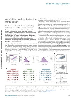
  • 1. nature neuroscience  VOLUME 20 | NUMBER 3 | MARCH 2017 389 B r i e f com mu n i cat i on s An important question in systems neuroscience is how behavioral state modulates the processing of sensory signals in large networks of neurons. To gain further insights into the impact of behavioral state on interactions between different cell types in local cortical circuitry, we employed a new approach to functional circuit mapping based on examining the covariance of correlation coefficients between responses of genetically defined neurons and changes in locomotion and pupil diameter. Responses of neurons expressing the calcium indicator GCaMP6 (ref. 1) were measured in alert, head-fixed mice in a premotor region of mouse frontal cortex referred to as the frontal association area (FrA, 2.8 mm from bregma and 0.75 mm from the midline (Supplementary Fig. 1). We chose this region because the estimation of correlation coefficients is facilitated when cells show elevated spontaneous acti­ vity, and pyramidal neurons in this region show spontaneous activity that is substantially higher than observed in primary sensory cortices. Behavior was monitored by recording pupil diameter and running speed while imaging. To measure the degree of association between behavior and neural response, we computed correlation coefficients between the activity of pyramidal cells in layer 2/3 with pupil size and running speed, which reflect the level of arousal2–5. We found that 33% of excitatory neurons imaged were significantly (P < 0.0001) negatively correlated to both pupil diameter and running, while 43% were significantly positively correlated with both measures (Fig. 1a–c). Similar response profiles were observed using GCaMP6f (Supplementary Fig. 2), and longitudinal imaging showed that neurons stably maintain the sign of their coefficients (Fig. 1d). Suppression of layer 2/3 pyramidal neurons during running has not been reported in visual cortex2,5–8. In agreement with these studies, An inhibitory pull–push circuit in frontal cortex Pablo Garcia-Junco-Clemente1,2, Taruna Ikrar3, Elaine Tring1, Xiangmin Xu3, Dario L Ringach1,4 & Joshua T Trachtenberg1 Push–pull is a canonical computation of excitatory cortical circuits. By contrast, we identify a pull–push inhibitory circuit in frontal cortex that originates in vasoactive intestinal polypeptide (VIP)-expressing interneurons. During arousal, VIP cells rapidly and directly inhibit pyramidal neurons; VIP cells also indirectly excite these pyramidal neurons via parallel disinhibition. Thus, arousal exerts a feedback pull–push influence on excitatory neurons—an inversion of the canonical push–pull of feedforward input. 1Department of Neurobiology, David Geffen School of Medicine, University of California, Los Angeles, Los Angeles, California, USA. 2Instituto de Biomedicina de Sevilla, IBiS, Hospital Universitario Virgen del Rocío/CSIC/Universidad de Sevilla and Departamento de Fisiología Médica y Biofísica, Universidad de Sevilla, and CIBERNED, Seville, Spain. 3Department of Anatomy & Neurobiology, University of California, Irvine, Irvine, California, USA. 4Department of Psychology, University of California, Los Angeles, Los Angeles, California, USA. Correspondence should be addressed to P.G.-J.-C. (pgarciajunco@gmail.com). Received 9 August 2016; accepted 16 December 2016; published online 23 January 2017; doi:10.1038/nn.4483 0 2,000 4,000 6,000 8,000 10,000 Frame number Pyr–neurons 0 2,000 4,000 6,000 8,000 10,000 Frame number Pyr+neurons a 0 2,000 4,000 6,000 8,000 10,000 Frame number Low correlation neurons 4s.d. 1 0.6 0.2 –0.2 –0.6 –1 –1 0.6–0.6 –0.2 10.2 Correlation coefficient of pupil to signals Correlationcoefficient ofballtosignals Pyr neurons Pyr– Pyr+ Low correlation 400 300 200 100 0 –0.5 0 0.5 Correlation coefficient of running to signals 1 0.8 0.6 0.4 0.2 Cumulativecorrelation 0 1 0.8 0.6 0.4 0.2 Cumulativecorrelation 0 300 200 100 0 –0.5 0 0.5 Correlation coefficient of pupil to signals Numberofexccells Numberofexccells 1 –1 0.2 0.6 –0.2 –0.6 –1 1–0.2–0.6 0.2 0.6 Correlation coefficient of pupil to signals Correlationcoefficient ofrunningtosignals Correlation coefficient of pupil to signals –0.6 –0.2 0.2 0.6 1 1 0.5 0 0 Cumulativeprobability Prefrontal cortex Visual cortex 1 –1 0.2 0.6 –0.2 –0.6 –1 1–0.2–0.6 0.2 0.6 Estimated coefficient: 0.59 Correlationcoefficient ofpupiltosignals,Day3 Correlation coefficient of pupil to signals, day 1 b c d e f ©2017NatureAmerica,Inc.,partofSpringerNature.Allrightsreserved.
  • 2. 390 VOLUME 20 | NUMBER 3 | MARCH 2017  nature neuroscience b r i e f com mu n i cat i on s we found that only ~1% of excitatory neurons in V1 were negatively correlated to both locomotion and pupil diameter (Fig. 1e), suggest- ing either that the suppression of pyramidal cells we see in frontal cortex arises from a unique impact of inhibition in this region or that suppression is not easily detected in V1 because spontaneous rates are low—that is, a difference in operating setpoint in V1 versus frontal cortex (Fig. 1f). VIP cells are thought to regulate cortical excitability during arousal and locomotion9 by inhibiting somatostatin-expressing (SOM) interneurons10–12; we too found evidence for this interaction in FrA (Supplementary Fig. 3). While this disinhibition may account for the increased excitatory responsiveness of some pyramidal cells dur- ing periods of heightened arousal, it cannot drive the suppression of almost 40% of the excitatory network. To investigate functional interactions between VIP cells, SOM cells and pyramidal neurons in the same network, we expressed a flexed GCaMP6s in VIP and SOM cells and GCaMP6f in all neurons and again imaged their activities in head-fixed, freely moving mice (Fig. 2a and Online Methods). VIP and SOM cells also expressed the red fluorescent protein tdTomato. We calculated the mean response for each of the four groups of neu- rons identified in these studies: VIP cells, SOM cells, and pyramidal neurons whose activity increased (Pyr+) or decreased (Pyr−) during locomotion (Fig. 2b). We then calculated the correlation coefficient between these responses and pupil diameter and locomotion (Fig. 2c). These associations co-varied across mice, with the strongest association being between VIP and Pyr− neurons (Fig. 2d). This means that if in a mouse the correlation between VIP and pupil size was higher than average (across our population), then the correlation between Pyr− and pupil size was correspondingly more negative. This relationship is sug- gestive of a strong, direct connection between VIP and Pyr− neurons. We confirmed this connection using channelrhodopsin-assisted circuit mapping13 (Fig. 3a–e). We found that all pyramidal neurons received direct VIP input, but that the strength of this input varied over a wide range (by almost two orders of magnitude; Fig. 3f). This heterogeneity it is consistent with the distribution of positive and negative correlations between pyramidal cell responses and behavioral state that we see, but it may also reflect differences between brain slices that we cannot control. Figure 1  A stable subset of excitatory neurons is inhibited during arousal. (a) Ordinate: plot of the correlation coefficients between excitatory response and locomotion. Abscissa: plot of the correlation coefficients between those same neurons and pupil diameter. 3,108 neurons, 11 mice, 17 fields of view. Neurons with a P value <0.0001 for each coefficient: 1,175 negatively correlated to pupil diameter; 1,193 negatively correlated to running; 1,559 positively correlated to pupil diameter; 1,643 positively correlated to running; 1,350 positively correlated with both pupil and running; 1,020 negatively correlated with both pupil and running. Remaining cells were not significantly correlated with either measure. (b) Histograms plotting excitatory correlation coefficients to running (left) and pupil diameter (right). A Gaussian distribution was fitted to each histogram (black dashed line). Gray lines in both show the cumulative distributions. (c) Example traces from five excitatory neurons whose activities were negatively correlated (red, left), positively correlated (green, middle), or uncorrelated (blue, right) with locomotion and pupil diameter. Cells were taken from the pool of neurons shown in the colored circles in a. (d) Plot of correlation coefficients between pyramidal cell responses and pupil diameter for the same 440 pyramidal neurons imaged at two time points 3 d apart. 440 neurons, 5 mice, 5 fields of view. Error d.f. = 438; r.m.s. error = 0.198; R2 = 0.375 (where R2 is the variance); t = 16.2; F = 263 versus constant model, P = 1.17 × 10−46. Significance of the linear regression was determined using a two-sided t-test with n − 2 d.f. (e) Same plot as in a, but for pyramidal neurons imaged in primary visual cortex. 518 neurons, 5 mice, 5 fields of view. 53 negatively correlated to pupil diameter; 31 negatively correlated to running; 5 negatively correlated to both pupil diameter and running. (f) Cumulative distributions of correlation coefficients to pupil diameter for neurons in V1 and FrA. 80 60 40 20 40 20 40 20 2,000 4,000 6,000 8,000 10,000 2,000 4,000 6,000 8,000 10,000 PyrVIPSOM Frame number Cellnumber Frame number 1 0.9 0.8 0.7 0.6 –0.55 –0.45 –0.35 Pupil,VIP Pupil, Pyr– Pupil, Pyr+ Pupil, all groups 1 0.9 0.8 0.7 0.6 0.5 –0.8 –0.4 0 Slope: –0.23 Pupil,VIP Pupil, SOM 1 0.9 0.8 0.7 0.6 0.5 0.5 0.50–0.50.6 0.7 Pupil,VIP –0.8 –0.4 0 0.4 0.8 Correlationcoefficient, signalvs.pupil VIP SOMPyr+ Pyr– Pupil,VIP 1 0.8 0.6 0.4 0.2 0 Slope: 0.84 Slope: –0.89 DilatedConstricted Pupildiameter VIP SOMPyr+ Pyr– 2s.d. dc a b Figure 2  Covariation of coefficients between VIP and Pyr cells across mice. (a) Three panels showing z-scored time series responses of 95 excitatory neurons (top), 50 VIP cells (middle) and 48 SOM cells (bottom) imaged in the same mouse over 13 min. For the excitatory neurons, responses above and below the white lines are significantly positively and negatively correlated, respectively, with pupil size. Mice are triple transgenic SOM-Cre;VIP-Cre;Ai9. (b) Top: z-scored time series for the pupil diameter in the same mouse shown in a. Regions of the trace representing constricted or dilated pupil are color coded and represent regions that remained significantly above or below the mean for a duration of greater than 3 s. Bottom: mean response of the Pyr+ and Pyr− neurons, VIP and SOM cells in a. (c) Box plots of the distribution of correlation coefficients of mean responses for each group of cells to pupil. Whiskers define median, upper quartile, lower quartile, upper adjacent value, and lower adjacent value. n = 747 pyramidal neurons, 357 VIP cells, 204 SOM cells, 4 mice, 8 fields of view. (d) Covariation across animals of correlation coefficients among population activity and pupil size. VIP−Pyr+: t = 3.44; r.m.s. error = 0.05; R2 = 0.608; F = 11.9 versus constant model, P = 0.0139; 6 d.f.; VIP−Pyr−: t = −3.9; r.m.s. error = 0.045; R2 = 0.671; F = 15.3 versus constant model, P = 0.0079; 6 d.f.; VIP−SOM: t = −1.57; r.m.s. error = 0.072; R2 = 0.173; F = 2.47 versus constant model, P = 0.167; 6 d.f.. Plots are of fields of view. Significance of the linear regression was determined using a two-sided t test with n − 2 d.f. ©2017NatureAmerica,Inc.,partofSpringerNature.Allrightsreserved.
  • 3. nature neuroscience  VOLUME 20 | NUMBER 3 | MARCH 2017 391 b r i e f com mu n i cat i on s In vivo, we found a significant suppression in the aggregate response of Pyr− neurons when VIP cells were optogenetically stimulated (Fig. 3g,h). In contrast, Pyr+ cells showed a significant increase in aggregate response (Fig. 3h), which peaked approximately 10 frames (645 ms) after the dip seen in the Pyr− neurons. This delayed response may be due to the multisynaptic, disinhibitory relay from VIP to SOM to Pyr11,14,15. Light-activated responses were not observed in the absence of ChR2 (Fig. 3i). These data identify a pull–push inhibitory circuit that is active during arousal. Pyramidal neurons likely receive both direct VIP cell-mediated inhibition and indirect VIP → SOM cell-mediated disinhibition. This inhibitory circuitry is an inversion of canonical excitatory push–pull circuitry16,17. An expected outcome from this circuitry is that variabi­ lity in the net balance of inhibition and disinhibition generates a hetero- geneous response of excitatory neurons, some of which are enhanced during arousal as others are suppressed. The mean balance between the strengths of direct inhibition and disinhibition appears to vary across cortical areas: in our measurements, the strength of direct VIP inhibition of pyramidal neurons was most pronounced in frontal cortex and weak in occipital cortex. The net effect of these two pathways on individual cells is expected to shift their operating point and, therefore, modulate the gain of pyramidal neurons during arousal8. Methods Methods, including statements of data availability and any associated accession codes and references, are available in the online version of the paper. Note: Any Supplementary Information and Source Data files are available in the online version of the paper. VIP-Cre;Ai32 ChR2+ VIP neuron Pyr + Pyr– Pyramidal neuron Blue LED ON Blue LED OFF RunningStop Blue LED L2 L5/6 VIP-Cre;Ai32 L3 ChR2 photoactivation via blue laserIPSC recording a c d b e f ChR2+ VIP cell 500 µm 200 ms 50 pA 200 pA 200 ms 30 24 18 12 6 0 pA 200 µm WeakVIPinput StrongVIPinput Averagepeakamplitude(pA) 0 2 4 6 8 10 12 14 16 18 20 100 80 60 40 20 0 Cell ID (sorted) ** * Time (s) 0 1 2 3 5 1.00 1.02 0.98 0.96 1.04 1.06 ∆F/F∆F/F Pyr+ Pyr– Pyr+ Pyr– 64 ChR2 in vitro ChR2 in vivo 1.00 1.02 0.98 0.96 1.04 1.06 Time (s) 0 1 2 3 5 64–1–2 200 µm g h i Figure 3  VIP cells act as a pull–push circuit in vivo. (a) Schematic of the approach used to map VIP cell–derived inhibitory postsynaptic currents (IPSCs) in pyramidal neurons (dark blue) recorded by patch clamp in layer 2/3 of FrA. (b) Brightfield image of a brain slice. The patch pipette can be seen entering from the left. The position of the recorded cell is indicated by the black dot. The blue dots show the positions of the photostimulation sites. (c) Inhibitory postsynaptic currents evoked from each photostimulation site. Responses evoked from stimulation at the two sites outlined by the blue squares are shown to the right. (d) Fluorescence image of section showing expression of YFP-tagged ChR2 in VIP interneurons. Scale bar, 200 µm. (e) Examples of a weak and strong heat map of VIP cell–derived inhibitory currents to two different pyramidal neurons. (f) Amplitude of the mean inhibitory current to each of 20 pyramidal neurons from all local VIP cells in the slice. (g) Schematic of experimental setup. Top: VIP cells are represented as yellow-green circles, pyramidal cells as blue triangles. The green and red triangles represent pyramidal cells that are excited or inhibited during running, respectively. Bottom: photostimulation through the objective lens. Stimulation was applied when mice were quiescent. (h) Mean z-score of Pyr− responses to ChR2-VIP cell stimulation. Shaded areas define ±1 s.e.m. of the responses. Vertical blue lines delineate the time when the LED was turned on and the photomultiplier tube was gated off. A significant suppression of GCaMP6s response in the Pyr− cells occurs in ~575 ms. Wilcoxon rank-sum test; 345 cells, 4 fields of view, 2 mice; Pyr+ P = 6.14 × 10⁻¹⁴; Pyr− P = 0.024. (i) Same measure as in h, but in a mouse lacking ChR2 in VIP cells. 168 cells, 2 fields of view, 1 mouse. ©2017NatureAmerica,Inc.,partofSpringerNature.Allrightsreserved.
  • 4. 392 VOLUME 20 | NUMBER 3 | MARCH 2017  nature neuroscience b r i e f com mu n i cat i on s Acknowledgments This work was funded by R01 EY023871 (JTT), R01 EY018322 (DLR) and R01 EB022915 (DLR). PGJC was supported by postdoctoral Fellowship EX2009-0750 from the Spanish Ministry of Education, Culture and Sport and by postdoctoral contract from Junta de Andalucía (P12-CTS-2232). AUTHOR CONTRIBUTIONS P.G.-J.-C. collected and analyzed the data in Figures 1 and 2 and Supplementary Figures 1–3. D.L.R. analyzed the covariance data in Figure 2. E.T., D.L.R. and J.T.T. contributed to the in vivo photostimulation experiments in Figure 3. T.I. and X.X. contributed to the slice ChR2 mapping data in Figure 3. COMPETING FINANCIAL INTERESTS The authors declare no competing financial interests. Reprints and permissions information is available online at http://www.nature.com/ reprints/index.html. 1. Chen, T.W. et al. Nature 499, 295–300 (2013). 2. McGinley, M.J., David, S.V. & McCormick, D.A. Neuron 87, 179–192 (2015). 3. Ebitz , R.B. & Platt, M.L. Neuron 85, 628–640 (2015). 4. Zekveld, A.A., Heslenfeld, D.J., Johnsrude, I.S., Versfeld, N.J. & Kramer, S.E. Neuroimage 101, 76–86 (2014). 5. Vinck, M., Batista-Brito, R., Knoblich, U. & Cardin, J.A. Neuron 86, 740–754 (2015). 6. Niell, C.M. & Stryker, M.P. Neuron 65, 472–479 (2010). 7. Polack, P.O., Friedman, J. & Golshani, P. Nat. Neurosci. 16, 1331–1339 (2013). 8. Mineault, P.J., Tring, E., Trachtenberg, J.T. & Ringach, D.L. J. Neurosci. 36, 6382–6392 (2016). 9. Letzkus, J.J., Wolff, S.B. & Lüthi, A. Neuron 88, 264–276 (2015). 10. Pfeffer, C.K., Xue, M., He, M., Huang, Z.J. & Scanziani, M. Nat. Neurosci. 16, 1068–1076 (2013). 11. Lee, S., Kruglikov, I., Huang, Z.J., Fishell, G. & Rudy, B. Nat. Neurosci. 16, 1662–1670 (2013). 12. Pi, H.J. et al. Nature 503, 521–524 (2013). 13. Petreanu, L., Huber, D., Sobczyk, A. & Svoboda, K. Nat. Neurosci. 10, 663–668 (2007). 14. Karnani, M.M. et al. J. Neurosci. 36, 3471–3480 (2016). 15. Fu, Y. et al. Cell 156, 1139–1152 (2014). 16. Anderson, J.S., Carandini, M. & Ferster, D. J. Neurophysiol. 84, 909–926 (2000). 17. Douglas, R.J. & Martin, K.A. J. Physiol. (Lond.) 440, 735–769 (1991). ©2017NatureAmerica,Inc.,partofSpringerNature.Allrightsreserved.
  • 5. nature neurosciencedoi:10.1038/nn.4483 ONLINE METHODS Animals. All procedures were approved by UCLA’s Office of Animal Research Oversight (the Institutional Animal Care and Use Committee, IACUC) and UCI’s IACUC, and were in accord with guidelines set by the US National Institutes of Health. A total of 50 mice from Jackson Lab, both male (23) and female (27), aged P45–56, were used for in vivo imaging in this study as follow: 7 Viptm1(cre)Zjh/J (stock number 010908), 7 B6N.Cg-Ssttm2.1(cre)Zjh/J (stock number 018973), 27 Gad2tm2(cre)Zjh/J (stock number 010802), 4 Viptm1(cre)Zjh/J/Ssttm2.1(cre)Zjh/J and 2 (in vivo) + 4 (in vitro) Viptm1(cre)Zjh/J/ Ai32 (stock number 012569). All Cre lines were previously crossed with B6;129S6-Gt(ROSA)26Sortm9(CAG-tdTomato)Hze/J (stock number 007905) to express td-Tomato in a Cre-dependent manner (except for the Vip-ChR2 mice). All lines were crossed into a C57Bl6 background for seven generations or longer. Mice were housed in groups of two or three per cage in a normal 12/12 light dark cycle. Animals were naive subjects with no prior history of participation in research studies. Numbers of mice used, numbers of fields of view, and numbers of cells imaged are all provided in the appropriate figure legends. Surgery. Carprofen (an anti-inflammatory) was administered preoperatively. Mice were then anesthetized with isoflurane (4–5% induction, 1.5–2% surgery). Core body temperature was maintained at 37.5 °C using a feedback heating sys- tem. Eyes were coated with a thin layer of ophthalmic ointment to prevent desic- cation. Anesthetized mice were mounted in a stereotaxic apparatus. Blunt ear bars were placed in the external auditory meatus to immobilize the head. A portion of the scalp overlying the two hemispheres of the cortex (approximately 8 mm × 6 mm) was then removed to expose the underlying skull. After the skull was exposed it was dried and covered by a thin layer of Vetbond. After the Vetbond dries (approximately 15 min) it provides a stable and solid surface to affix the aluminum bracket with dental acrylic. The bracket was then affixed to the skull and the margins sealed with Vetbond and dental acrylic to prevent infections. Virus injection. A 2.5-mm-diameter region of skull overlying the FrA cor- tex (right hemisphere) was removed. Care was taken to leave the dura intact. Depending on the experiment, GCaMP6-slow, GCaMP6-fast or GCaMP6s- Floxed (UPenn Vector Core: AAV1.Syn.GCaMP6f.WPRE.SV40; stock number AV-1-PV2822; AAV1.Syn.GCaMP6s.WPRE.SV40, number AV-1-PV2824; AAV1.Syn.Flex.GCaMP6s.WPRE.SV40,numberAV-1-PV2821)wasexpressedin cortical neurons using adeno-associated virus (AAV-GCaMP6 titer: ~13 genomes ml−1). The virus was loaded into a glass micropipette and slowly inserted into the FrA using a micromanipulator. One injection was made 2.8 mm from the bregma and 0.75 mm from the midline. AAV-GCaMP6 was pressure injected using a PicoSpritzer III (Parker, Hollis, NH) (4 puffs at 15–20 p.s.i. with a duration of 10 ms, each puff separated by 4 s) starting at a depth of 350 µm below the pial sur- face and making injections every 10 µm moving up, with the last injection made at 100 µm below the pial surface. The total volume injected across all depths was approximately 1 µl. In some cases, GCaMP6s-Flex and GCaMPf were injected at the same time (0.5 µl each). The injections were made by a computer program in control of the micromanipulator and the PicoSpritzer. A square coverslip 1 mm on a side, previously cut with a diamond tip, was glued to a 2.5-mm-diameter cover glass using a UV-curing optical adhesive18. UV light was used for gluing and sterilizing both coverslips, and then they were placed directly on the dura and sealed around the edges with VetBond. When dry, the edges of the cover glass were further sealed with dental acrylic. At the end of the surgery, all exposed skull and wound margins were sealed with VetBond and dental acrylic. Mice were then removed from the stereotaxic apparatus, given a subcutaneous bolus of warm sterile saline, and allowed to recover on the heating pad. When fully alert they were placed back in their home cage. Imaging. Once expression of GCaMP6 was observed in FrA, typically between 12 and 15 days after the injection, imaging sessions took place. Imaging was per- formed using a resonant scanning, two-photon microscope (Neurolabware, Los Angeles, CA) controlled by Scanbox acquisition software (Scanbox, Los Angeles, CA). The light source was a Coherent Chameleon Ultra II laser (Coherent Inc, Santa Clara, CA) running at 920 nm. The objective was a 16× water immersion lens (Nikon, 0.8 NA, 3 mm working distance). The microscope frame rate was 15.6 Hz (512 lines with a resonant mirror at 8 kHz). Eye movements and pupil size were recorded via a Dalsa Genie M1280 camera (Teledyne Dalsa, Ontario, Canada) fitted with a 740-nm long-pass filter. Images were captured at an average depth of 210 µm (90% of imaging fields within the range 125–300 µm). During imaging a substantial amount of light exits the brain through the pupil. Thus, no additional illumination was required to image the pupil. Mice were head-fixed on a floating, spherical treadmill on which they had been trained to balance and run. All mice were trained on the spherical treadmill for 3 d, 20 min per day before experiments were started. Movement of the spherical treadmill was recorded via a Dalsa Genie M640 camera. Both locomotion and eye movement data were synchronized to the microscope frames. We first summed images over a 30-s period to obtain a high signal-to-noise image of Cre-dependent tdTomato expression, using 1,000-nm excitation, to identify specific populations of inhibitory cells, depending on the experiment. We then changed the wavelength to 920 nm and imaged GCaMP6(f/s) dynam- ics during 10–15 min per field. Mice were head fixed but able to run during the entire imaging session. Data processing. Motion stabilization. Calcium images were aligned to cor- rect for motion artifacts in a two-step process. First, we aligned images rigidly in a recursive fashion to correct for slow drifts in the imaging plane. Pairs of neighboring images in time were aligned by finding the peak of their cross- correlation; then pairs of averages of such pairs were aligned; and so on. In the second step, we aligned images nonrigidly to a reference mean image to correct for fast in-plane movements, which are frequently observed during grooming. We iteratively applied the Lucas-Kanade algorithm19 to nonrigidly match a refer- ence mean image, refining the estimate of this reference mean image after each alignment iteration. Segmentation. Following motion stabilization, we used a Matlab graphical user interface tool developed in our laboratory to manually define regions of interest corresponding to putative cell bodies. We used correlation and kurtosis images to identify cell candidates20. The correlation image, corresponding to the average correlation of a pixel and its eight neighbors across time, highlights regions of space that co-vary in time. The kurtosis image highlights regions in space with signals composed of large, infrequent deviations—putative spikes. These images were computed after subtracting a rank-2 factor from the calcium movies, corresponding to mean and linear temporal trends. We used these images to visually identify approximately circular regions of space of an appropriate radius with high correlation and high kurtosis. Clicking a seed pixel at the center of such a candidate patch allowed the definition of a region of interest by flood-filling an image corresponding to the correlation of the highlighted pixel and every other pixel in the image field21. The interface then allowed the user to dynamically grow or shrink the region of interest to a desired size. Red and green channels were segmented separately and the result- ant masks were matched, enabling us to assign GCaMP signals to inhibitory or excitatory neurons appropriately. Analysis. For data in Figures 1 and 2 and Supplementary Figures 1 and 2, we determined the correlation coefficient between each cell’s activity and both locomotion and pupil diameter. For data in Figure 3, we first calculated the correlation coefficient for each excitatory neuron’s activity and pupil diameter. Temporal responses of all cells that had significantly positive correlations coef- ficients were averaged together (Pyr+), and temporal responses of cells with sig- nificantly negative correlations were averaged (Pyr−). Mean time series were also derived for the VIP and SOM cells. These mean time series were then correlated with pupil diameter and their coefficients plotted in Figure 3c. In Figure 3d, for each mouse we plotted the coefficients for each cell type relative to the coeffi- cients obtained for VIP cells. The slope, r-value, and P-value of linear regression lines of covariance were measured. Analysis of VIP and SOM cells expressing GCaMP in the same mouse. In this experiment, both SOM-expressing and VIP-expressing cells also express tdTo- mato and GCaMP6s. Because we could not distinguish SOM cells from VIP cells on the basis of waveform, we assigned all tdTomato-expressing cells that had a positive correlation coefficient to pupil diameter as VIP cells and all those with negative coefficients as SOM cells. From our data in Supplementary Figure 3, we know that of the 875 VIP cells that were imaged, 105 were negatively correlated with pupil diameter, and of the 417 SOM cells imaged, 70 were positively corre- lated with pupil diameter. That is, of the 1,292 cells in the VIP and SOM pool, 175 (13.5%) would be incorrectly assigned using the strategy we employed here. ©2017NatureAmerica,Inc.,partofSpringerNature.Allrightsreserved.
  • 6. nature neuroscience doi:10.1038/nn.4483 ChR2-assisted circuit mapping. Briefly, whole cell recordings were obtained from pyramidal neurons of layer 3 in FrA of VIP-Cre+/−;Ai32+/− mice at an age of ~7 weeks. Spatial maps of VIP connectivity strength to each patched pyramidal neuron were derived by systematically stimulating ChR2-expressing VIP cells at a mapping grid of 256 different sites arranged in a 16 × 16 matrix spanning all cortical layers (Fig. 3). Spatially restricted optogenetic activation of ChR2-expressing VIP cells was accomplished using a 473-nm blue laser (30 mW, 2.5 ms, laser spot diameter ~50 µm). Voltage and current clamp con- trols were performed to normalize light stimulation protocols between slices and across animals. During optogenetic stimulation experiments, the ionotropic glutamate receptor antagonists CNQX (10 µM) and CCP (5 µM) were added to the bath solution to block excitatory synaptic input (GABAergic transmission is unaffected) and avoid any potential disinhibition effects. Because interneurons can also be connected by electrical synapses, we blocked gap junctions using 100 µM carbenoxolone. Whole-cell voltage-clamp recordings were made from the recorded pyramidal neurons to measure photoactivation-evoked inhibitory postsynaptic currents at the empirically determined holding potential of +5 mV in voltage clamp mode with cesium-containing internal solution. Post hoc GFP staining (Abcam ab6556; 1:500 dilution) of the recorded slices verified ChR2-YFP expression in VIP neurons. In vivo ChR2-VIP photostimulation with pyramidal cell imaging. VIP-Cre mice were crossed with the Ai32 line of mice housing a flexed ChR2 driven by a CAG promoter targeted to the Rosa26 locus (JAX Stock number: 012569). Offspring of these crosses expressed ChR2 in VIP interneurons; they were verified by genotyping. AAV-GCaMP6f was injected into frontal cortex of these mice as described above. Craniotomies were made as described above. Following strong GCaMP6f expression, mice were head fixed under the resonant scanning micro- scope and were imaged, head fixed and free to run, to identify pyramidal neurons whose activities increased or decreased with running. A blue light-emitting diode (ThorLabs M470L3) focused onto the objective back aperture was used to excite ChR2 in the imaging field during periods of behavioral quiescence (not running). The LED was controlled using custom Matlab software to generate 5-V TTL sig- nals. Each light pulse was 40 ms in duration. During the blue light flash, the PMTs were rapidly gated. For this experiment, we changed the PMT on our custom microscope to a Hamamatsu H11706-40, which has built-in circuitry to rapidly gate the gain of the PMT within 400 ns (rise and fall). These PMTs have a maxi- mum shutter width of 10 ms; therefore, we triggered the PMT with four successive TTLs to obtain an effective open time of 40 ms. Using this approach, we avoided light contamination from the LED in our GCaMP6 signals. GCaMP6f responses were recorded simultaneously. Movies of cell responses were motion corrected as described, cells were identified, and signals extracted. Because pyramidal cell firing is sparse, only a small fraction of the pyramidal neurons imaged were fir- ing within the 40-ms light flash. Therefore, we repeated the light flash 21 times at random intervals while imaging continuously. Cell responses were averaged across the 21 trials and across cells to produce the trace shown in Figure 3. Statistics. Data collection and analysis was not blinded. No randomization was used to assign subject to the experimental groups. Experiments required mice of known genotype, making randomization impossible. No statistical methods were used to predetermine sample sizes, but our sample sizes are like those reported in previous publications18–20. Statistical tests are indicated in the figure legends with accompanying P values. Numbers of cells, numbers of fields of view, and numbers of mice are given in the appropriate figure legends. A Supplementary Methods Checklist is available. Data and code availability. The data that support the findings of this study are available from the corresponding author upon reasonable request. Matlab scripts can be requested from the corresponding author. 18. Huber, D. et al. Nature 484, 473–478 (2012). 19. Greenberg, D.S. & Kerr, J.N. J. Neurosci. Methods 176, 1–15 (2009). 20. Smith, S.L. & Häusser, M. Nat. Neurosci. 13, 1144–1149 (2010). 21. Ozden, I., Lee, H.M., Sullivan, M.R. & Wang, S.S. J. Neurophysiol. 100, 495–503 (2008). ©2017NatureAmerica,Inc.,partofSpringerNature.Allrightsreserved.