mapping functional brain activation using iodoantipyrine in male serotonin tr...
Ronan Talty - RISE 2015 Poster
1. Undergraduate
Category: Physical and Life Sciences
Degree Level: B.S. Behavioral Neuroscience
Abstract ID# 748
Estrogen-Dopamine Interactions in Female Rats During Extinction Learning
Ronan Talty, Mollee Farrell, Katelyn Flick, Sam Cohen, Rebecca Shansky
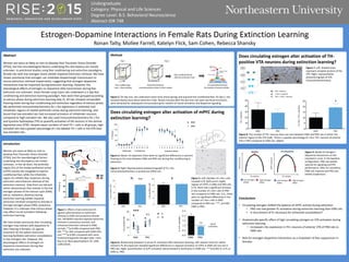
PL IL
Figure 7. Left: Shaded areas
represent sampled sections of the
VTA. Right: representative
photomicrograph of VTA
immunohistochemistry.
Figure 8. The number of TH+ neurons does not vary between EMD and PRO rats in either the
anterior region of the VTA (Left). There is a greater percentage of c-fos+ TH+ neurons of anterior
VTA in PRO compared to EMD rats. (Right).
Does circulating estrogen alter activation of TH-
positive VTA neurons during extinction learning?
Conclusions
• Circulating estrogen shifted the balance of mPFC activity during extinction
• PRO rats had greater PL activation during extinction learning than EMD rats
• Is recruitment of PL necessary for enhanced consolidation?
• Anatomically specific effect of high circulating estrogen on VTA activation during
extinction learning
• Increased c-fos expression in TH+ neurons of anterior VTA of PRO rats vs
EMD rats
• Role for estrogen-dopamine interactions as a modulator of fear suppression in
females
Figure 9. Model of estrogen–
dopamine interactions on the
inverted-U curve. In the baseline
configuration, PRO rats exhibit
optimal D1 signaling and PFC
performance. After D1 simulation,
EMD rats improve and PRO rats
exhibit impairment.
D1 activity
mPFCfunction
Low estrogen
(EMD)
High estrogen
(PRO)
D1 Stimulation
mPFCfunction
Low estrogen
(EMD)
High estrogen
(PRO)
Baseline
D1 activity
Abstract
Women are twice as likely as men to develop Post Traumatic Stress Disorder
(PTSD), but the neurobiological factors underlying this discrepancy are mostly
unknown. In preclinical studies using fear conditioning and extinction paradigms,
female rats with low estrogen levels exhibit impaired extinction retrieval. We have
shown previously that estrogen can modulate dopaminergic transmission to
rescue extinction retrieval impairments, suggesting that estrogen-dopamine
interactions may be important during extinction learning. However, the
physiological effects of estrogen on dopamine (DA) transmission during fear
extinction are unknown. Intact female Long-Evans rats underwent a 2-day fear
conditioning and extinction learning paradigm. Rats were then grouped according
to estrous phase during extinction learning (day 2). All rats showed comparable
freezing levels during fear conditioning and extinction regardless of estrous phase.
We performed immunohistochemistry for c-fos expression in prelimbic and
infralimbic regions of medial prefrontal cortex during extinction learning, and
found that low-estradiol rats had increased activation of infralimbic neurons
compared to high-estradiol rats. We also used immunohistochemistry for c-fos
and tyrosine hydroxylase (TH) to quantify activation of DA neurons in the ventral
tegmental area (VTA). Despite equal numbers of total TH + cells in all groups, high-
estradiol rats had a greater percentage of c-fos labeled TH + cells in the VTA than
low-estradiol rats.
Introduction
Women are twice as likely as men to
develop Post Traumatic Stress Disorder
(PTSD), but the neurobiological factors
underlying this discrepancy are mostly
unknown. In the rat brain, the prelimbic
region (PL) of the medial prefrontal cortex
(mPFC) excites the amygdala to express
conditioned fear, while the infralimbic
region (IL) inhibits fear responses during
extinction and enhances retrieval of the
extinction memory. Data from our lab and
others demonstrate that animals in the low
estrogen phases of the estrous cycle (EMD;
estrus, metestrus, diestrus) during
extinction learning exhibit impaired
extinction retrieval compared to animals in
the high estrogen phase (PRO; proestrus).
However, it is unknown how estrous phase
may affect neural activation following
extinction learning.
We have shown previously that circulating
estrogen may interact with dopamine to
alter freezing in females. D1 agonist
treatment 30 min before extinction
learning facilitates extinction consolidation
in low-estrogen rats. However, the
physiological effects of estrogen on
dopamine transmission during fear
extinction are unknown.
Figure 1. Effects of pre-extinction D1
agonist administration on extinction
retrieval in EMD and proestrous female
rats. SKF38393 induced impaired extinction
retrieval in proestrous animals, and
enhanced extinction retrieval in EMD
animals. **p=0.006 compared with PRO-
SKF, ***p=.002 compared with EMD-VEH,
and ****p<0.001 compared with same
treatment/opposite estrogen state. From
Rey et al, Neuropsychopharm 39, 1282-
1289 (2014).
Figure 2. On day one, rats underwent seven tone-shock parings and acquired the conditioned fear. On day 2, rats
received twenty tone-alone extinction trials. Ninety minutes after the last tone rats were sacrificed and brains
were extracted for subsequent immunostaining for markers of neural activation and dopamine signaling.
Methods
Does circulating estrogen alter activation of mPFC during
extinction learning?
Figure 3. Above: As expected, there were no significant differences in percent
freezing to the tone between EMD and PRO rats during fear conditioning or
extinction.
Figure 4. Right: Representative photomicrograph of PL c-fos
immunohistochemistry in proestrous (PRO) rats.
Figure 5. Left: Number of c-fos+ cells
counted in PL (left) and IL (right)
regions of mPFC in EMD and PRO rats.
In PL, there was a significant increase
in the number of c-fos+ cells in PRO
rats compared to EMD rats. In IL, there
were no significant differences in the
number of c-fos+ cells in EMD
compared to PRO rats. ***, p<0.001
EMD vs PRO.
Figure 6. Relationship between IL versus PL activation after extinction learning. Left: paired t-tests for within-
animal IL-PL discrepancies revealed significant differences in regional activation of mPFC in EMD rats but not in
PRO rats. Right: quantification of IL/PL activation demonstrated IL dominance in EMD rats. ***p<0.001 IL vs PL or
EMD vs. PRO.
PL IL