This document provides protocols for immunohistochemistry techniques. It describes the principle of using labeled antibodies to identify specific tissue antigens through antigen-antibody reactions. It outlines the direct and indirect detection methods and key steps including fixation, tissue processing, paraffin embedding, sectioning, antigen retrieval, staining, and mounting. Procedures include deparaffinization, hydration, primary and secondary antibody incubation, DAB substrate reaction, counterstaining, and dehydration prior to slide mounting. The goal is to preserve tissue components and cellular morphology for identification of specific tissues.
Real Sex Provide In Goa ✂️ Call Girl (9316020077) Call Girl In Goa
Alipathan123456 (1)
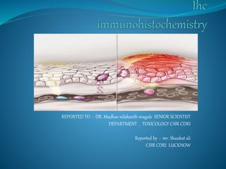
1. REPORTED TO : DR. Madhav nilakanth mugale SENIOR SCIENTIST
DEPARTMENT _ TOXICOLOGY CSIR CDRI
Reported by : mr. Shaukat ali
CSIR CDRI LUCKNOW
2. INTRODUCTION
Immunohistochemistry
(IHC) Combines histological, immunological
and biochemical techniques for the identification of specific
tissue component by means of a specific antigen/antibody
reaction tagged with a visible label.
Principle
The principle of immunohistochemistry is the localization of
antigen in tissue section by the use labeled antibodies as
specific antigen. Antigen antibody interaction that are
visualized by a marker such as fluorophore and
chromophore.
3. Antigen detection method
Direct method
In this, enzyme linked or fluorescent tagged antibody
directly bind with antigen which is present in tissue.
4.
5. Indirect method
In this, enzyme linked or fluoroscent tagged
antibody are indirectly bind with antigen.
In this method, enzyme linked antibody
Which is called as secondary antibody, it bind
with primary antibody , which is directly bind
to antigen.
9. Fixation
The broad objective of tissue fixation is to preserve cells
and tissue components in a “life-like state,for example
cell morphology.
Goals of Fixation
Prevent autolysis by rapidly terminating enzymatic and
metabolic activity.
Prevent bacterial decomposition .
The purpose of formalin fixation is to produce chemical
cross-linking of proteins within the tissue.
10. Tissue Processing
Tissue processing consists of three steps :-
Dehydration – making Hydrophobic condition using
alcohol and acetone
Clearing-using xylene to remove dehydrating agent
Infiltration- using paraffin wax to remove xylene
11. Tissues processed into paraffin will have wax in the
cassettes; in order to create smooth wax blocks, the wax
first needs to be melted away placing the entire cassette in
58°C paraffin bath for 15 minutes. Turn the heat block on
to melt the paraffin one hour before adding the tissue
cassettes.
Open cassette to view tissue sample and choose a mold
that best corresponds to the size of the tissue. A margin of
at least 2 mm of paraffin surrounding all sides of the tissue
gives best cutting support. Discard cassette lid.
Put small amount of molten paraffin in mold, dispensing
from paraffin reservoir.
Using warm forceps, transfer tissue into mold, placing cut
side down, as it was placed in the cassette.
Transfer mold to cold plate, and gently press tissue
flat. Paraffin will solidify in a thin layer which holds the
tissue in position.
12. When the tissue is in the desired orientation add the
labeled tissue cassette on top of the mold as a
backing. Press firmly.
Hot paraffin is added to the mold from the paraffin
dispenser. Be sure there is enough paraffin to cover the face
of the plastic cassette.
If necessary, fill cassette with paraffin while cooling,
keeping the mold full until solid.
Paraffin should solidify in 30 minutes. When the wax is
completely cooled and hardened (30 minutes) the paraffin
block can be easily popped out of the mold; the wax blocks
should not stick. If the wax cracks or the tissues are not
aligned well, simply melt them again and start over.
The tissue and paraffin attached to the cassette has formed
a block, which is ready for sectioning.
13. Sectioning tissues
Tissues are sectioned using a microtome. Turn on the water
bath and check that the temp is 35-37ºC. Use fresh
deionized water. Blocks to be sectioned are placed face
down on an ice block or heat sink for 10 minutes. Place a
fresh blade on the microtome; blades may be used to
section up to 10 blocks, but replace if sectioning becomes
problematic. Insert the block into the microtome chuck so
the wax block faces the blade and is aligned in the vertical
plane.
Set the dial to cutting smoothly, set to 3 - 5 µM
sections . .
14. The blade should angled at 5º. Face the block by
cutting it down to the desired tissue plane and
discard the paraffin ribbon. If the block is
ribboning well then cut another four sections and
pick them up with forceps or a fine paint brush
and float them on the surface of the 37ºC water
bath. Float the sections onto the surface of clean
glass slides. If the block is not ribboning well then
place it back on the ice block to cool off firm up
the wax. If the specimens fragment when placed
on the water bath then it may be too hot.
15.
Place the slides with paraffin sections on 65°C hot
plate for 20 minutes (so the wax just starts to melt)
to bond the tissue to the glass. Slides can be stored
overnight at room temperature.
Deparaffinization or Hydration process
1 Slide dipped in xylene I for about 10 min.
2 Slide dipped in xylene II for 10 min.
3 Slide dipped in 100% alcohol for 5min.
4 90% alcohol for 5min.
5 70 % alcohol for 5min.
6 50% alcohol for 5min.
7 Wash slide with running tap water for 5-10min.
16. 8 Antigen retrieval (boiling in sodium citrate at 90 ֯c
for 15 -20 min.pH-6.0)(70-80degree maintain
temp.)
9 Cool the slides at room temp. for 20 min.
10 Rinse with water 2 time for 5 min.
11 Endogenous peroxidase quenching for 10min.
With (PBS+H2O2 30% (4:1))
12 Wash with PBS *2times for 5min.
13 Permiabilization with 0.3% Triton x100 in PBS for
10min.
17. Blocking with 5%BSA in 0.3% PBST(T-20) for 2
hours.
1. Incubated with primary antibody 4 degree
(overnight) (dilution -1:200 in PBST(0.5%BSA).
2. Washing with PBST *3 times for 5min.
3. Incubated with HRP labelled secondary
antibody (dilution 1:300 in 0.5%BSA PBST) for
2hr.
4. Washing with PBS *3times for 5min.
5. Incubated with DAB substrate liquid for 1.5min.
18. 1. Wash with water *3 times.
2. Counter staining with hematoxylin (30 sec-
2min.)
Rinse section with water *2times.
1. 50% alcohol (3min.)
2. 70%alcohol(3min.)
3. 90%alcohol(3min)
Dehydration process
4. 100%alcohol (3min.)
5. Xylene *2 times (10min.)
6. Mounting slides with DPX.