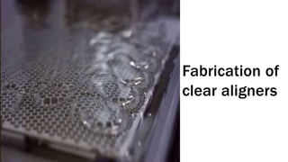
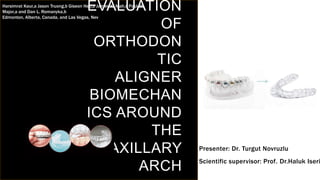
1. The study evaluated the forces and moments exerted by clear plastic aligners on individual teeth around the maxillary arch.
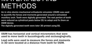
2. An electro-mechanical orthodontic simulator was used to measure the forces and moments generated when the central incisor, canine, and second premolar were each moved lingually by 0.2mm.
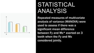
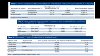
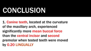
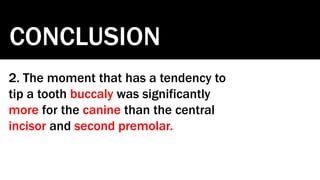
3. The results found that the canine experienced significantly more buccal force and moments tending to tip it buccally compared to the central incisor and second premolar. Clinically relevant reactionary forces were also exerted on adjacent teeth.
![SAMPLE FOOTER TEXT
INTRODUCT
ION
Clear thermoplastic appliances can exert forces upon teeth and
result in various movements of teeth.
Aim of this study is to evaluate the forces and moments
imposed from aligners.
Assessing the effect of thermoplastic material choice, thickness
and amount of activation on single tooth models is important.
This exact study mainly focuses on the forces and moment
systems imposed on central incisor, canine and second premolar
teeth around the arch using a clinically representative glycol
modified polyethylene terephthalate thermoplastic aligner
material
]
20XX 2](https://image.slidesharecdn.com/evaluationoforthodonticalignerbiomechanicsaroundthemaxillary-220313070559/85/Aligner-Biomechanics-2-320.jpg)