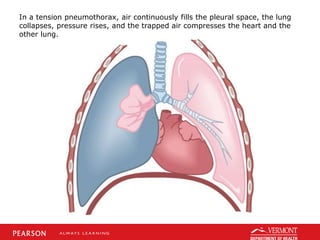
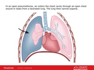
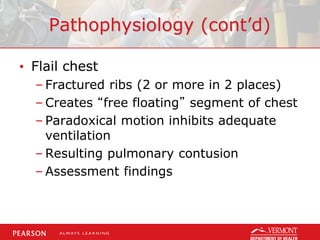
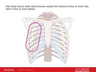
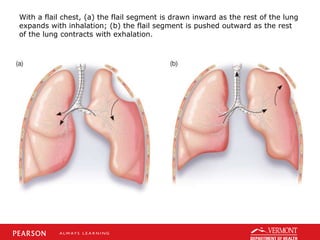
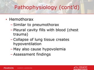
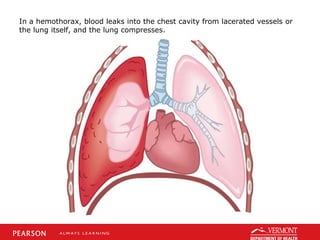
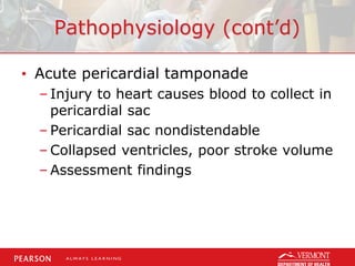
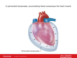
This document discusses chest trauma, including epidemiology, pathophysiology, assessment findings, differential diagnosis, and emergency medical care. It provides case studies to illustrate key points. Regarding pathophysiology, it describes how conditions like tension pneumothorax, open pneumothorax, flail chest, and hemothorax can occur from chest trauma and affect ventilation and circulation. Emergency medical care focuses on maintaining the airway, breathing, and circulation while providing full immobilization and rapid transport. Case studies demonstrate assessing and managing a potential chest trauma patient.
2. Objectives
• Review annual injury and death rates
for chest trauma victims.
• Understand pathophysiologic changes
that occur with chest trauma.
• Discuss common signs and symptoms.
• Identify current treatment modalities
for the patient with chest trauma.
3. Introduction
• Chest injuries can be obvious and
dramatic, or small and easy to miss.
• Likewise, they may be critical injuries
threatening life, or minor injuries of
relative unimportance.
• To identify the difference, the Advanced
EMT must understand the physiology of
the chest wall and its response to
trauma.
4. Epidemiology
• 20% to 25% of trauma deaths each
year are due to thoracic trauma.
• The most common mechanism is MVC.
• Immediate deaths are due to
myocardial or aorta rupture.
• Early deaths are due to tension and
open pneumothorax, tamponade, flail
segments, and hemothorax.
5. Pathophysiology
• Chest trauma distorts the normal
thoracic anatomy.
• Distortion injures body system and
causes a change in physiology.
• V/Q ratio disturbances, hypoxemia,
hypercapnea ensue.
• Ultimately, cellular death occurs.
6. Pathophysiology (cont’d)
• Tension pneumothorax
– Disruption of visceral pleura
– Accumulation of intrathoracic air
– Collapse of lung tissue
– Shifting of mediastinum
– Changes in hemodynamics
– Assessment
Early findings
Late findings
7. In a tension pneumothorax, air continuously fills the pleural space, the lung
collapses, pressure rises, and the trapped air compresses the heart and the
other lung.
8. Pathophysiology (cont’d)
• Open pneumothorax
– Disruption of parietal pleura from hole
in chest
– Accumulation of intrathoracic air
– Collapse of lung tissue
– Injury may turn into tension
pneumothorax
– Assessment findings
9. In an open pneumothorax, air enters the chest cavity through an open chest
wound or leaks from a lacerated lung. The lung then cannot expand.
10. Pathophysiology (cont’d)
• Flail chest
– Fractured ribs (2 or more in 2 places)
– Creates “free floating” segment of chest
– Paradoxical motion inhibits adequate
ventilation
– Resulting pulmonary contusion
– Assessment findings
11. Flail chest occurs when blunt trauma causes the fracture of two or more ribs,
each in two or more places.
12. With a flail chest, (a) the flail segment is drawn inward as the rest of the lung
expands with inhalation; (b) the flail segment is pushed outward as the rest
of the lung contracts with exhalation.
13. Pathophysiology (cont’d)
• Hemothorax
– Similar to pneumothorax
– Pleural cavity fills with blood (chest
trauma)
– Collapse of lung tissue creates
hypoventilation
– May also cause hypovolemia
– Assessment findings
14. In a hemothorax, blood leaks into the chest cavity from lacerated vessels or
the lung itself, and the lung compresses.
19. Emergency Medical Care
• Spinal immobilization considerations
• Assess and maintain the airway.
• Determine breathing adequacy.
– High-flow via NRB with adequate
breathing.
– High-flow via PPV @ 10-12/min if
inadequate.
– Occlude any punctures to chest wall.
20. Emergency Medical Care (cont’d)
• Assess circulatory components
– Check pulse, skin characteristics
– Control major bleeds
• Provide full immobilization
• Initiate safe and expeditious transport
21. Emergency Medical Care (cont’d)
• Do not delay transport to start an IV
line.
• Use a large-bore catheter (14 or 16
gauge).
• Run the fluids to maintain a systolic
blood pressure of 80 to 90 mmHg or
until radial pulses are regained.
22. Emergency Medical Care (cont’d)
• Once this is achieved, reduce the fluid
infusion and titrate to maintain the
systolic blood pressure at 80 to 90
mmHg or to maintain radial pulses.
23. Case Study
• Your EMS unit is summoned for a
patient who was injured while hunting.
Upon your arrival, you find a male
patient holding his hand over his right
thorax. Some blood is seeping past his
fingers, and the breathing looks
labored. Friends report he was
accidently shot with an arrow.
24. Case Study (cont’d)
• Scene Size-Up
– Standard precautions taken.
– Scene is safe, no sign of struggle.
– Young male, 18 years old.
– Patient found sitting along edge of road.
– No patient entry nor egress problems.
– No additional resources needed
presently.
25. Case Study (cont’d)
• Primary Assessment Findings
– Patient responsive.
– Airway open and maintained by self.
– Breathing is rapid, patient is dyspneic.
– Carotid and radial pulses present but
radial gets weaker with inhalation.
– Peripheral skin cool, pale, sweaty.
– No other major bleeds or concerns.
26. Case Study (cont’d)
• Is this patient a high or low priority?
Why?
• What interventions should be provided
at this time?
27. Case Study (cont’d)
• What are your differentials thus far that
the patient could be suffering from?
• Do you think that this patient will have
a problem with the ventilation or
perfusion side of the V/Q ratio?
28. Case Study (cont’d)
• Medical History
– Patient denies any
• Medications
– Patient denies any
• Allergies
– Patient denies any
29. Case Study (cont’d)
• Pertinent Secondary Assessment
Findings
– Pupils dilated but reactive, membranes
pale.
– Airway patent, breathing tachypneic.
– Peripheral perfusion diminishing.
– Absent breath sounds to right thorax.
– Patient's mental status still continuing
to deteriorate.
31. Case Study (cont’d)
• What would be key clinical indications
the patient is deteriorating despite
treatment?
• What advantage does “burping” the
occlusive dressing have?
• Why would PPV possibly be detrimental
to the patient?
32. Case Study (cont’d)
• Care provided:
– Patient immobilized.
– High-flow oxygen via NRB mask, switched
to PPV due to failing ventilations.
– Occlusive dressing to chest injury.
– Rapid transport to hospital initiated.
– Minimize Scene Time
– Established intravenous access (en-route)
to ED
33. Summary
• Chest wall injuries can result in significant
disturbances to the V/Q ratio.
• Although the injury typically can't be
“fixed” in the prehospital setting, the
patient can have supportive treatment
provided that will support lost function.
• With any severe trauma patient, minimize
scene time and perform interventions en-
route to ED.
Editor's Notes
Discuss the objectives.
Discuss that most every major body system is represented in the thorax.
Injuries to this location can cause mild or significant injury, including death.
Relate epidemiology to the frequency with which the Advanced EMT will have patients with chest trauma.
With chest trauma, the change in physiology is due to a structural change in the thoracic cavity.
Discuss how these changes ultimately produce changes in cellular integrity.
Discuss the etiology, pathophysiology, and basic assessment findings for a tension pneumothorax.
Discuss the etiology, pathophysiology, and basic assessment findings for an open pneumothorax.
Discuss the etiology, pathophysiology, and basic assessment findings for a flail chest.
Discuss the etiology, pathophysiology, and basic assessment findings for a hemothorax.
Discuss the etiology, pathophysiology, and basic assessment findings for an acute pericardial tamponade.
With chest trauma, there is not a specific sign or symptom that identifies one clinical syndrome from another, rather, it is a collection of symptoms coupled with the mechanism of injury that will identify the underlying pathology.
Review and discuss the slide. Stress the need to ensure life threats are properly managed.
The most immediately life-threatening thoracic injuries that require rapid recognition and intervention and expeditious transport are:
Tension pneumothorax
Open pneumothorax
Flail chest
Massive hemothorax
Acute pericardial tamponade
Some protocols require that the systolic blood pressure be maintained at 70 mmHg to reduce the incidence of hemodilution and increased bleeding associated with aggressive fluid administration in the trauma patient with uncontrolled bleeding. Follow your local protocol.
Discuss the case study.
This is a potentially unstable patient for two reasons.
First, the soft-tissue injury the patient is applying pressure to may be deep enough to pierce into the pleural cavity of the thorax. Or, it may be a significant bleed, or it may be neither and it's just a soft-tissue wound.
Treatment at this time would be high-flow oxygen, and the application of an occlusive dressing over the chest wall injury and then reapplication of direct pressure to help minimize the bleeding.
At this time, the differentials include:
An open pneumothorax
Major bleed
Tension pneumothorax
Flailed chest wall
If the underlying injury is an open pneumothorax, then there is a disturbance on the
ventilation side of the equation.
If the patient has a hemothorax, the patient will have perfusion disturbances as well.
Discuss the case progression.
Changes to the mental status, drop in pulse oximeter, declining breath sounds, increased agitation, worsening lung compliance.
Burping the dressing during exhalation would allow the expulsion of any accumulating air in the chest, which should allow the subsequent breaths to start inflating the lung.
In a patient with a penetrating chest wall injury, the provision of PPV could allow air to escape the lung tissue (if the visceral pleura is also damaged), and accumulate in the pleural cavity. This then will collapse the lung and interfere with normal ventilation.