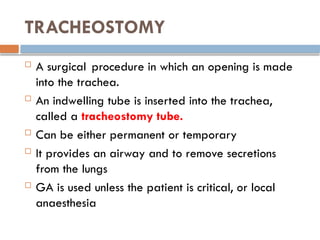
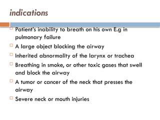
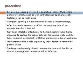
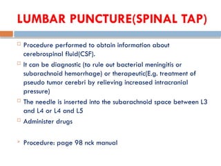
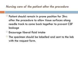
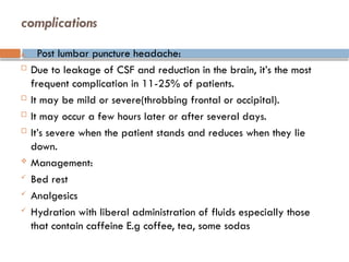
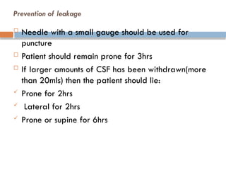
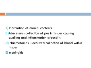
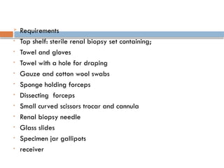
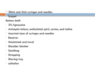
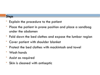
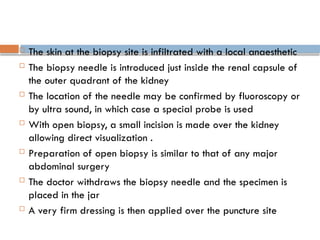
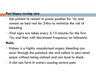
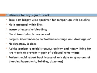
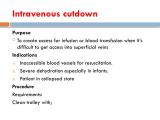
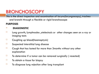
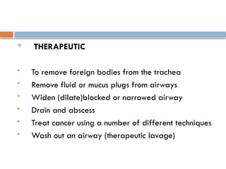
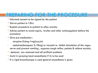
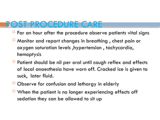
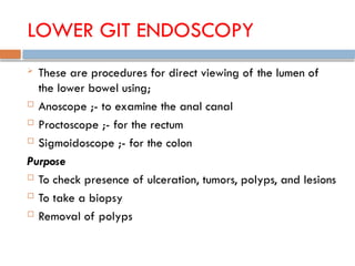
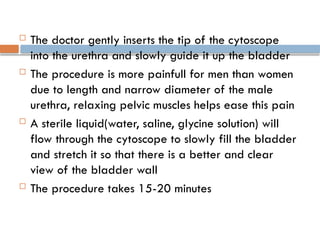
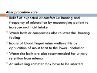
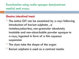
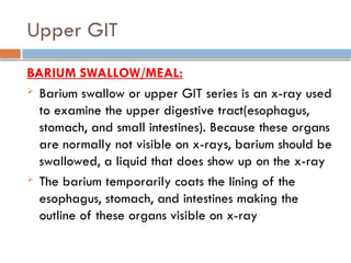
ADVANCED NURSING
PROCEDURES
BY ABDIRAHMAN IBRAHIM KADIYE
COURSE OUTLINE
 Aseptic technique:
 Tracheostomy
 Abdominal parecentesis
 Lumbar puncture
 Bone marrow puncture
 Liver biopsy
 Renal biopsy
 Venous cutdown
 Incision and drainage
 Endoscopy
 bronchoscopy
 Upper GIT: oesophagoscopy
gastroscopy
 Lower GIT: anoscopy
Proctoscopy
Colonoscopy/sigmoidoscopy
 cytoscopy
Examination with radio opaque dyes and x-rays
 Barrium swallow/barrium meal
 Barium enema
 Cholecystogram/ cholangiogram
 Angiography/ venogram
 Electroencephalogram (EEG)
 Myelogram
 Others
 electrocardiography
Specific objectives
By the end of the course the student will be able to:
 Describe the various aseptic nursing procedures
 Describe the nursing management before, during
and after the procedure
 Demonstrate ideal nursing skills involved in the care
of the patient during and after the procedures
TRACHEOSTOMY
 A surgical procedure in which an opening is made
into the trachea.
 An indwelling tube is inserted into the trachea,
called a tracheostomy tube.
 Can be either permanent or temporary
 It provides an airway and to remove secretions
from the lungs
 GA is used unless the patient is critical, or local
anaesthesia
indications
 Patient’s inability to breath on his own E.g in
pulmonary failure
 A large object blocking the airway
 Inherited abnormality of the larynx or trachea
 Breathing in smoke, or other toxic gases that swell
and block the airway
 A tumor or cancer of the neck that presses the
airway
 Severe neck or mouth injuries
procedure
 Surgical procedure performed in operating room or I.C.U. where
patient’s ventilation can be well controlled and optimal asceptic
technique can be maintained
 A surgical opening is made between 2nd
and 3rd
tracheal rings,
 After trachea is exposed, a cuffed tracheostomy tube of an
appropriate size is inserted
 Cuff is an inflatable attachment to the tracheostomy tube that is
designed to occlude the space between the trachea walls and the
tube, to permit mechanical ventilation and minimize risk of aspiration.
 Tracheostomy tube is held in place by tapes fastened around the
patient’s neck
 Sterile gauze is usually placed between the tube and the skin to
absorb drainage and reduce the risk of infection.
Nursing care
 Patient requires continous monitoring and assessment
 Opening(stoma) must be kept patent by proper sunctioning of
secretions
 After vital signs stabilize, put patient in semi-fowlers position to
facilitate ventilation, promote drainage, minimize edema, and
prevent strain on the suture lines.
 Analgesia and sedative should be administered with caution due to
the risk of suppressing the cough reflex
 Alleviate patients apprehension and provide effective means of
communication. Place a paper and pen or functional call light at all
times near the patient’s bed to ensure communication
 The nurse can also advise patient to occlude stoma when they want
to speak
Changing the dressing
 Requirements:
 Sterile gloves and clean gloves
 Hydrogen peroxide
 Normal saline solution/ sterile water
 Cotton-tipped applicators
 Twill tape(and type of tube prescribe if it’s to be changed)
Procedure
 Wash hands.
 Explain procedure to the patient.
 Put on clean gloves, remove and discard soiled dressings.
 Prepare sterile supplies: H2O2, normal saline solution, or sterile water, cotton tipped
applicators, dressing and tape.
 Don sterile gloves.
 Cleanse the wound and plate of the tracheostomy tube with sterile cotton tipped
applicators moistened with H2O2.
 Rinse with sterile saline solution
 Soak inner cannula in peroxide or sterile saline per manufacturers
instructions, rinse with saline solution and ensure all dried secretions
have been removed
 Dry and re-insert inner cannula or replace with new disposable
inner cannula.
 Place clean twill tape in position to secure the tracheostomy tube
by inserting one end of the tape through the other side opening of
the outer cannula.
 Take the tape around the back of patient’s neck and thread it
through the opposite opening of the outer cannula. Bring both ends
around so that they meet on one side of the neck. Tighten the tape
until only 2 fingers can be comfortably inserted under it. Secure
with a knot.
 Remove soiled twill tape after a new tape is in place.
 NOTE:
 Some long term tracheostomies with healed stomas may not require a
dressing, other tracheostomies do. In such cases, use of sterile tracheostomy
dressing, fitting it securely under the twill tapes and flange of tracheostomy
tube so that the incision is covered
Tracheostomy sunctioning
 It’s done to maintain a clear airway since patient’s own cough mechanism is
deficient
Indications
 Accumulation of secretions in the lungs
 Increased production of secretions
 Mechanically ventilated patients
 Procedure and requirements pg 190-191 green NCK manual
Humidifying and filtering the air
 Soak a thin piece of gauze in sterile normal saline
and place it across the opening
 Tape the gauze in position
 Change the gauze regularly as it will quickly dry out
Diet
 Give the patient fluid or soft diet initially until he/she
is accustomized to the tube, then they can have normal
diet.
Removal of tracheostomy sutures
 They are removed on the 7th
day post operatively
Removal of tracheostomy tube
 Before removal, the patient is weaned from the tube
by;
a) The tube is covered with a dressing for increased
periods of time to monitor how the patient breaths
and tolerates
b) Corking(use of corks) with wide holes and reducing
to ones with smaller holes instead of application of
dressings.then the tube is eventually removed, the
hole closes over and a dressing is applied.
 Observe patient carefully for signs of respiratory
distress.
On discharge

If permanent, teach patient how to remove the tracheostomy tube, how to clean it and how to
replace it.

When outside, advise patient to wear loose covering E.g. scarf for protection over the stoma.
Complications of tracheostomy

May occur early or late in tracheostomy tube management. They include:

Bleeding

Pneumothorax

Air embolism

Recurrent laryngeal nerve damage

Posterior tracheal wall penetration

Long term:

Airway obstruction from obstructions by secretions

Protrusion of the cuff over the opening of the tube

Infection

Dysphagia

Tracheal dilatation, ischaemia, and necrosis

Tracheoesophageal fistula
ABDOMINAL PARACENTESIS/ ABDOMINAL TAPPING
 This is the puncture of the abdomen and the withdrawal of fluid that has
collected in the peritoneal cavity.
 The fluid build up is called ascites
 It relieves abdominal pressure and obtains specimen for laboratory analysis.
Indications
i. To find the cause of ascites
ii. To diagnose infection in peritoneal fluid
iii. To check for certain types of cancer E.g liver cancer
iv. To remove large amounts of fluid that causes pain or difficulty in breathing
or that affects kidney or bowel(intestine) functioning.
 Procedure: page 4 NCK procedure manual
preparation
 Signed consent form
 Prepare patient by providing the necessary
information and instructions by offering re-assurance.
 Instruct the patient to void.
 Gather appropriate sterile equipment and collection
of receptacles.
 Place the patient in upright position on the edge of
the bed or in a chair with feet supported on a stool.
 If the patient is confined in bed use fowlers position.
 Place sphygmomanometer cuff around patients arm
Nursing action post procedure
 Check rate of drainage and adjust to the required rate of flow
 Return patient to bed or to a comfortable sitting position
 Measure, describe and record the fluid collected
 Label samples of fluid and send them to lab
 Monitor vital signs every 25 minutes for 1hr, every 30 minutes for 2hrs, every hour for
2hrs and then every 4hrs
NOTE:
 BP and pulse may change as fluid shift occurs after removal of fluid.
 An elevated temperature is a sign of infection
 Assess for hypovolemia, electrolyte shifts, changes in mental status and encephalopathy
which may occur with removal of fluid and fluid shifts and should be reported.
 Check puncture site for leakage or bleeding , leakage of fluid may occur because of
changes in abdominal pressure and may contribute to further loss of fluid if undetected
 Leakage suggest a possible site of infection and bleeding may occur in patients with
altered clotting secondary to liver disease.
Provide patient teaching regarding:
 need to monitor for bleeding or excessive drainage
from puncture site.
 Importance of avoiding heavy lifting or straining.
 Need to change position slowly
 Frequency of monitoring for fever
LUMBAR PUNCTURE(SPINAL TAP)
 Procedure performed to obtain information about
cerebrospinal fluid(CSF).
 It can be diagnostic (to rule out bacterial meningitis or
subarachnoid hemorrhage) or therapeutic(E.g. treatment of
pseudo tumor cerebri by relieving increased intracranial
pressure)
 The needle is inserted into the subarachnoid space between L3
and L4 or L4 and L5
 Administer drugs
 Procedure: page 98 nck manual
Nursing care of the patient after the procedure
 Patient should remain in prone position for 3hrs
after the procedure to allow tissue surfaces along
needle track to come back together to prevent CSF
leakage
 Encourage liberal fluid intake
 The specimen should be labelled and sent to the lab
with the request form.
complications
A. Post lumbar puncture headache:
 Due to leakage of CSF and reduction in the brain, it’s the most
frequent complication in 11-25% of patients.
 It may be mild or severe(throbbing frontal or occipital).
 It may occur a few hours later or after several days.
 It’s severe when the patient stands and reduces when they lie
down.
 Management:
 Bed rest
 Analgesics
 Hydration with liberal administration of fluids especially those
that contain caffeine E.g coffee, tea, some sodas
Prevention of leakage
 Needle with a small gauge should be used for
puncture
 Patient should remain prone for 3hrs
 If larger amounts of CSF has been withdrawn(more
than 20mls) then the patient should lie:
 Prone for 2hrs
 Lateral for 2hrs
 Prone or supine for 6hrs
B) Herniation of cranial contents
C)Abscesses : collection of pus in tissues causing
swelling and inflammation around it.
D) Haematomas : localized collection of blood within
tissues
E) meningitis
Bone marrow puncture(aspiration)
 Bone marrow is the soft tissue inside bones that helps form
blood cells, it’s found in the hollow part of most bones.
 Bone marrow aspiration is the removal of a small amount
of this tissue in liquid form for examination.
Sites
 Pelvic bone
 Breast bone i.e. over the sternum
Procedure: NCK manual pg 197-198
Indications
Patients in which the following conditions are
suspected;
• Leukaemia
• Anaemia
• Thrombocytopenia
• Hodgkin lymphoma
• Multiple myeloma and non-hodgkin lymphoma
After care:
• Confine the patient in bed for an hour or longer as ordered by
the doctor
• Observe the puncture site for bleeding, if present apply
pressure and bandage
• Record the procedure in the patient’s cardex
• Label the specimen and take them to lab with request form
• Advise the patient not to make the biopsy site wet by showering
until 24 hrs are over
• If the bleeding soaks through the dressing, the patient should
report to hospital
• They should avoid vigorous activity or exercises for a day or
two- this helps minimize bleeding and discomfort
Liver biopsy
 Is a procedure where small pieces of liver tissue are removed
in order to be sent to lab for examination
 It’s helpful in the diagnosis of diseases that affect the liver E.g
liver cirrhosis
Purpose
i. Diagnosis: this allows establishment of very specific
diagnosis
ii. Monitoring: monitoring effectiveness of therapy that the
patient is receiving for a liver disease. It can also provide
warning if certain therapies the patients are receiving are
damaging to the liver
Indications
 Alcoholic liver
 Elevated liver enzymes of unknown origin
 Jaundice and billiary tract obstruction
 Hemochromatosis
 Hepatomegally of undetermined cause
 Autoimmune liver disease
 Cancers of the liver and non cancerous tumor
 Chronic viral hepatitis
 Liver transplantation- to rule out rejection
Procedure
Requirements on trolley:
As for bone marrow biopsy with addition of the following
• Scapel or blade
• Specimen jar
• Menghini liver biopsy needle
Preparation
a) Ascertain the results of coagulation tests- prothrombin
time, partial thromboplastin time, and platelet count
b) Check for signed consent- confirm that informed
consent has been provided
c) Take and record patient’s vital signs immediately before biopsy
d)Explain the procedure to the patient- steps of the procedure, sensations
expected, after-effects expected, restrictions of activity and monitoring
procedures to follow
Steps
• Support the patient during the procedure
• Expose the right side of the patients upper abdomen(right hypochondriac)
• Instruct the patient to inhale and exhale deeply several times finally to
exhale and to hold breath at the end of expiration(prevents puncturing of
the diaphragm and risk of lacerating the liver is minimized)
• The physician promptly introduces the biopsy needle by way of the trans
thoracic (inter costal) or trans abdominal (sub costal) route, penetrates the
liver, aspirates and withdraws. The entire procedure is completed within 5-
10 seconds.
• Instruct the patient to resume breathing
Nursing care post procedure
1)Immediately after biopsy, assist the patient to turn onto
the right side; place a pillow under costal margin, and
caution the patient to remain in this position, recumbent
and immobile, for several hours.
• Instruct the patient to avoid coughing or straining.
• In this position, the liver capsule at the site of penetration
is compressed against the chest wall, and the escape of
blood or bile through the perforation is prevented.
2) Measure and record patients pulse, respiratory rate and
BP at 10-15 minutes intervals for the first hour , then 30
minutes for the next 2hrs, or until the patient stabilizes.
 Changes in vital signs may indicate; bleeding, severe haemorrhage
or bile peritonitis, most frequent complications of liver biopsy.
3)If the patient is discharged after the procedure, instruct the patient
to avoid heavy lifting and strenuous activity for one week.
Complication
 Fever
 Pain, swelling, redness or discharge around needle insertion site
 Chest pains
 Shortness of breathing
 Fainting or dizziness- sign of possible blood loss
 Nausea and vomiting
 Worsening abdominal pains – bleeding, leakage of bile
Renal biopsy(needle biopsy of the kidney)
 This is removal of a small piece of kidney tissue for
examination. It can be done through;
• Ultra sound guided kidney biopsy
• CT-guided kidney biopsy
• Or surgical biopsy
Indications
1) Protein in urine
2) Unexplained acute renal failure
3) Glomerulopathies
4) Transplant rejection
Contraindications
 Bleeding tendencies
 Uncontrolled hypertension
 Solitary kidney
 Morbid obesity
procedure
 Patient preparation
• Coagulation tests to identify patients at risk of post biopsy
bleeding
• Starve the patient for 6-8hrs(incase of nephrectomy)
• Establish an intravenous line
• Urine specimen is taken before biopsy for baseline data
• Sedation, if ordered is given
• Instruct the patient to hold his breath(to stop movement of
the kidney) during the insertion of biopsy needle. If sedated
place in prone with a sandbag under the abdomen
 Requirements
• Top shelf: sterile renal biopsy set containing;
• Towel and gloves
• Towel with a hole for draping
• Gauze and cotton wool swabs
• Sponge holding forceps
• Dissecting forceps
• Small curved scissors trocar and cannula
• Renal biopsy needle
• Glass slides
• Specimen jar gallipots
• receiver
 20mls and 5mls syringes and needles
 Scapel
Bottom shelf:
• 2% lignocaine
• Antiseptic lotions; methylated spirit, savlon, and iodine
• Assorted sizes of syringes and needles
• Receiver
• Mackintosh and towel
• Shoulder blanket
• Sandbag
• Strapping
• Shaving tray
• collodion
Steps
 Explain the procedure to the patient
 Place the patient in prone position and place a sandbag
under the abdomen
 Fold down the bed clothes and expose the lumbar region
 Cover patient with shoulder blanket
 Protect the bed clothes with mackintosh and towel
 Wash hands
 Assist as required
 Skin is cleaned with antiseptic
 The skin at the biopsy site is infiltrated with a local anaesthetic
 The biopsy needle is introduced just inside the renal capsule of
the outer quadrant of the kidney
 The location of the needle may be confirmed by fluoroscopy or
by ultra sound, in which case a special probe is used
 With open biopsy, a small incision is made over the kidney
allowing direct visualization .
 Preparation of open biopsy is similar to that of any major
abdominal surgery
 The doctor withdraws the biopsy needle and the specimen is
placed in the jar
 A very firm dressing is then applied over the puncture site
Post biopsy nursing care
 Ask patient to remain in prone position for 1hr and
remain on bed rest for 24hrs to minimize the risk of
bleeding
 Vital signs are taken every 5-15 minutes for the first
1hr, and then with decreased frequency as indicated.
Note;
 Kidney is a highly vascularised organ, bleeding can
occur through the puncture site and collect in peri-renal
space without being noticed and can lead to shock.
 A clot can form in ureters causing severe pain
 Observe for any signs of shock
 Take post biopsy urine specimen for comparison with baseline
 Hb is assessed within 8hrs
 Incase of excessive bleeding;
• Blood transfusion is commenced
• Surgical intervention to control haemorrhage and drainage or’
• Nephrectomy is done
 Advise patient to avoid strenuous activity and heavy lifting for
two weeks to prevent trigger of delayed hemorrhage
 Patient should report back incase of any signs or symptoms of
bleeding(haematuria, fainting, dizzyness)
Intravenous cutdown
Purpose
 To create access for infusion or blood transfusion when it’s
difficult to get access into superficial veins
Indications
1) Inaccessible blood vessels for resuscitation.
2) Severe dehydration especially in infants.
3) Patient in collapsed state
Procedure
Requirements:
Clean trolley with;
 Sterile pack with;
 Bowl with cotton wool
 Gauze swabs
 Hand towel
 ‘O’ towel
 Scapel handle
 2 mosquito forceps
 2 blunt hooks
 1 toothed dissecting forceps
 Gloves
 Bottom shelve;
 Strapping
 bandage
 Splint
 scissors
 Antiseptic lotion
 Small mackintosh and draw sheet
 Pair of sterile and non sterile gloves
 Local anaesthetics
 Blood giving set/ infusion set
 In addition have;
 Sterile syringes and needles
 Scapel blades
 Sutures and needles
 IV cannulas(appropriate sizes)
 Dripstand, and fluid charts
Steps:
 Wheel the equipment to the bedside
 Explain the procedure to the patient
 Assist doctor or clinician as appropriate
 Support client during procedure(positioning and reassuring)
 Regulate fluids accordingly
 After the procedure clear equipment and dispose waste according to
the institutions policy of waste management
Post procedure
 Immobilize the limb by splinting and advise the patient to immobilize
it as well
 Evaluate patient’s condition and tolerance to procedure
 Document the procedure
Incision and drainage
 This is a minor surgical procedure for treating abscesses and boils
 They can form in any loose tissue or subcutaneous tissue
 They release pus or pressure built up under the skin from abscesses, boils
or infected paranasal sinus
Incision
 This is the surgical cutting of soft tissues E.g skin or muscle with a scapel
and blade
 It should be large enough to allow free drainage
 The skin has to be cleaned first with antiseptic before an incision is made
 A drainage tube is stitched to the skin after incision and and instructions
given when to shorten or remove
 Skin around the wound is dressed with antiseptic
Drainage
 This is removal of fluid that has accumulated abnormally
from a cavity in the body E.g. serous fluid from a swollen
joint, pus from an internal abscess.
A drain
 This is a device which is rubber, tube or wick used to draw
fluid or pus from internal cavity to the surface
 It can also be inserted during an operation to ensure any
fluid formed passes to the surface preventing accumulation
and infection or pressure in the operation site
Indications
 Soft tissue abscesses
Contraindications
 Infections without abscess formation
 Premature incision before localization of pus will not be curative
and bacteremia from manipulation may result
Requirements
 A standard suture tray with a scapel and packing gauzes added
 Sterile gauzes
 Antiseptic solution
 Local anaesthesia
 Syringe and needles for anaesthesia infiltration
 Blade/ scapel
 Dressing supplies
Steps
 The area around the abscess or swelling is cleaned
with antiseptic.
 The abscess is then incised so that the pus or fluid
can drain away
 A rubber tube or wick or ribbon gauge is placed in
the incision to assist in the drainage to allow healing
to take place. suturing is done ( depending). The
wound is then cleaned with antiseptic and sterile
dressing of the right size applied. If too deep and
doesn’t need a tube, its packed, then dressed.
AFTER CARE
 Advice the patient to keep the area elevated
 Instruct patient not to disturb the dressing or splint(to
immobilize arm or leg)until the first follow up visit
 Provide appropriate analgesia especially
immediately post-operatively though drainage
relieves most of the pain of an abscess
 Therapeutic antibiotics are administered
Complications
 Transient bacteremia
 Scar formation
 Recurrence of infection
 Neurovascular injury
ENDOSCOPY
 This is the use of a hollow instrument to look inside the
body cavities or organs . It uses flexible tube that has a
small (camera)on the end of it.
 The instrument is referred to as an endoscope
 An endoscope is passed thru a natural body opening or
via a small incision. For example a laparascope is
inserted thru small surgical incision in the pelvic or
abdominal area
 If an ultra-sound probe is added to endoscope E.g. GIT
endoscope this is called endoscopic ultrasound
BRONCHOSCOPY
This is the direct inspection and examination of larynx(laryngoscopy), trachea
and bronchi through a flexible or rigid bronchoscope
PURPOSES
 DIAGONISTIC
 Lung growth, lymphnodes ,atelectasis or other changes seen on x-ray or
imaging tests
 Coughing up blood(haemoptysis)
 Suspected interstitial lung disease
 Cough that has lasted for more than 3months without any other
explanation
 To determine if a tumor can be removed surgically ( resected)
 To obtain a tissue for biopsy
 To diagnose lung rejection after lung transplant
 THERAPEUTIC
 To remove foreign bodies from the trachea
 Remove fluid or mucus plugs from airways
 Widen (dilate)blocked or narrowed airway
 Drain and abscess
 Treat cancer using a number of different techniques
 Wash out an airway (therapeutic lavage)
PREPARING FOR THE PROCEDURE
 Informed consent to be signed by the patient
 Starve patient 6-12hrs
 Explain procedure to patient to allay anxiety
 Advise patient to avoid asprin, brufen and other anticoagulants before the
procedure
 Give pre-medication :
• atropine 0.6mg-1mg(iv,sc,im)
• sedative(diazepam 5-10mg) or narcotic to inhibit stimulation of the vagus
nerve and prevent vomiting , suppress cough reflex ,sedate & relieve anxiety.
 dentures are removed and all artificial prothesis
 Assist in spraying local anaesthesia if its to be used
 If a rigid bronchoscope is used general anaesthesia is given
POST PROCEDURE CARE
 For an hour after the procedure observe patients vital signs
 Monitor and report changes in breathing , chest pain or
oxygen saturation levels ,hypertension , tachycardia,
hemoptysis
 Patient should be nil per oral until cough reflex and effects
of local anaesthesia have worn off. Cracked ice is given to
suck, later fluid.
 Observe for confusion and lethargy in elderly
 When the patient is no longer experiencing effects off
sedation they can be allowed to sit up
complications
 Aspiration
 Bronchospasm
 Hypoxaemia
 Pneumothorax
 Bleeding
 Abrasion of lining of airways leading to swelling,
inflammation and infection
UPPER GIT FIBRE (ENDOSCOPY)
 Also called esophagogastroduodenoscopy. It visualizes the
upper part of the GI tract up to duodenum
 It’s a minimally invasive procedure since it doesn’t require an
incision into one of the major body cavities and doesn’t
require significant recovery after procedure (unless sedation
or anaesthesia have been used)
 It allows direct visualization of gastric mucosa thru a lighted
endoscope for suspected gastric tumors and diseases , colored
photos or motion pictures can also be taken
 Mouth guard are used during the procedure to prevent the
patient from biting the scope
OESOPHAGOSCOPY AND GASTROSCOPY
 An esophagoscopy is a procedure to view the inside
of the esophagus
 Gastroscopy is a procedure to view inside of
stomach
 Duodenoscopy is a procedure to view duodenum .
 These procedures are performed as a single
procedure and are collectively referred to as an
upper endoscopy or
oesophagogastroduodenoscopy(OGD)
Purposes
 Examine esophagus for ulcers and tumors
 Taking biopsy
 Removal of foreign bodies
 Dysphagia or odynophagia
 Persistent nausea and vomiting
 Dyspepsia
 Acute upper GIT bleeding
 Chronic anaemia/or iron deficiency anaemia after non
gastro-intestinal origin has been eliminated.
 Gastro-esophageal reflux with warning signs i.e. weight
loss, dysphagia, bleeding and anaemia
 Peptic ulcer disease
Preparation of the patient
 Starve 6-8hrs(stomach must be empty for clear view)
 Narcotic analgesia is given 30minutes before
 Patients throat maybe sprayed with local anaesthesia to
help prevent discomfort, or they gargle local anaesthetic
agent for mouth and throat or
 IV diazepam 5-10mg is given just before the procedure
 Atropine 0.6-1.0mg IM/IV is also given to reduce
secretions
 Organon 0.5-1.0mg IM is given to relax smooth muscles
Procedure
 Local anaesthesia is sprayed in mouth or throat or
sedation is done
 Endoscope is then passed smoothly and slowly in the
areas being examined
 The procedure takes 10-20minutes
 The doctor may inject moderate amount of air to
expand the stomach allowing better visualizaton
 Biopsy is taken for examination or images of the
digestive tract are taken for documentation of any
abnormality; esophageal varices can be banded,
resecting or ablating mucosal tissue
After procedure care
 Patient should not eat 3-4hrs after procedure until
gag reflex returns to prevent aspiration
 Observe for vital signs post procedure
 Observe for signs of perforation E.g. pain,
hematemesis
 Minor throat discomfort may occur after procedure
and last upto 24hrs, it can be relieved with
lozenges, cool saline gargle or analgesics
 Advise the patient to report any unusual or severe
abdominal pain or bleeding following the procedure
Complications
 Bleeding
 Infection
 Perforation
 Cardiopulmonary problems
 Adverse reactions to medications
LOWER GIT ENDOSCOPY
 These are procedures for direct viewing of the lumen of
the lower bowel using;
 Anoscope ;- to examine the anal canal
 Proctoscope ;- for the rectum
 Sigmoidoscope ;- for the colon
Purpose
 To check presence of ulceration, tumors, polyps, and lesions
 To take a biopsy
 Removal of polyps
Indications
 Gastrointestinal problems such as unexplained bleeding
 Persistent changes in bowel habits
 Anaemia
 Age 50yrs; 5yearly then 3 yearly
 To diagnose colitis or colon/rectal cancer
Preparation
 The patient should take only clear oral fluids and not to
consume any food for at least 24hrs before the exam
 Laxatives and enemas may be required before the start
of exam to clear the lower bowel
Anoscopy/proctoscopy
Procedure
 Explain the procedure to the patient
 Put the patient in knee chest position with feet
beyond the edge of the bed, knees apart to give
support, head resting on the couch, fore arms on
either side of the head and hands placed one on
top of the other above the head
 The patient is told of the progress, the possibility he
might feel like moving his bowel and thanked for
co-operation
 Suction may be done to remove secretions
 Biopsy may be taken
 Polyps may be removed and the area cauterized to
prevent bleeding
 The tissue removed is placed on moist gauze and
then in the appropriate container, labelled , and
sent to the pathology lab with the request form.
 After each use the tubes are washed thoroughly
 Disposable tubes should be disposed safely
Colonoscopy/sigmoidoscopy
 This is endoscopic examination of the large bowel and distal
part of the small bowel with an endoscope passed through the
anus.(colonoscope)
 The colonoscope has a small camera attached to a flexible
tube that can reach and examine the entire length of the
colon.
Indications
 Diagnostic:
 Colon cancer, inflammatory bowel disease, GIT haemorrhage,
ulceration, polyps, and removal of biopsy; changes in bowel
habit(unexplained).
 Prophylactic:
 Removal of polyps
 Therapeutic:
 Removal of foreign bodies
 Screening every 10yrs from 50yrs for colorectal cancer
Patient preparation
 Explain the procedure to the patient
 Starve the patient of solid foods for 3 days to empty the GIT
 Laxatives are given for two days
 On day of examination an enema is given until rectum is clear
 Narcotic analgesic may be ordered and administered
 Diazepam is given to sedate the patient
Procedure
 Sigmoidoscopy; position the patient in knee chest
 colonoscopy; the patient lies on left side with legs
drawn up
 The first step is usually a digital rectal examination
to examine the tone of the anal sphincter and to
determine if the preparation is adequate
 Patient is sedated, the endoscope is then passed
through the anus up the rectum and colon(sigmoid,
descending, transverse, and ascending colon, the
caecum) and ultimately the terminal ileum
 The bowel is ocassionally inflated with air to maximize
visibility(procedure that may give false sensation of need to
have a bowel movement)
 Biopsies are then taken for histology or examination and
diagnostic evaluation of colon is done.
Post procedure care
 Advise the patient to refrain from operating heavy
machinery until a day after the procedure
 The patient resumes other normal activities after effects of
sedation wear off E.g. eating and drinking normally
 Advise patient to report signs of, chills, fever, rectal
bleeding(more than a tablespoon) swelling or redness at IV
site or severe abdominal pain or bloating
 Mild abdominal pain and bloating is expected after
the procedure
Complications
 Perforation
 Haemorrhage
 Inflammation of the bowel
cystoscopy
 This is endoscopy of the urinary bladder via the
urethra
 A cytoscope is used
 Diagnostic cytoscopy is carried out using local
anaesthesia, GA is sometimes used for operative
cytoscopic procedures.
 To enable clear visualization, sterile irrigation
solution is instilled to distend the bladder and wash
away any clots.
Indications
 Urinary tract infections
 To assess the ureter and kidney pelves
 Haematuria
 Incontinence( loss of bladder control)
 Unusual cells found in urine sample
 To remove renal calculi(kidney stones)
 Urinary blockage E.g. prostate enlargement and
stricture
 Unusual growth, polyp, tumor or cancer and obtain
biopsy
Patient preparation
 Explain the procedure to the patient
 Patient may take 1-2 glasses of water (or as instructed) before
going for the examination
 They should not urinate for a sufficient period of time, such that
they are able to urinate prior to the procedure
Procedure
 Patient lies on their back(supine) with their knees slightly apart,
they may also need to flex their knees especially when doing
rigid cytoscopy examination.
 For flexible cytoscopy , local anaesthesia is used
 Local anaesthesia is applied directly from a tube or needleless
syringe into the urinary tract
 The doctor gently inserts the tip of the cytoscope
into the urethra and slowly guide it up the bladder
 The procedure is more painfull for men than women
due to length and narrow diameter of the male
urethra, relaxing pelvic muscles helps ease this pain
 A sterile liquid(water, saline, glycine solution) will
flow through the cytoscope to slowly fill the bladder
and stretch it so that there is a better and clear
view of the bladder wall
 The procedure takes 15-20 minutes
After procedure care
 Relief of expected discomfort i.e burning and
frequency of micturation by encouraging patient to
increase oral fluid intake
 Warm bath or compresses also relieves the burning
feeling
 Incase of blood tinged urine –relieve this by
application of moist heat to the lower abdomen
 Warm sitz bath are also recommended for urinary
retention from edema
 An indwelling catheter may have to be inserted
Examination using radio opaque dyes(contrast
media) and xrays.
Gastro intestinal tract:
 The entire GIT can be examined by x-rays following
introduction of barium sulphate , a
tasteless,odourless, non-grannular absolutely
insoluble and non-absorbable powder opaque to
x-rays, ingested in form of a thin aqueous
suspension
 The dyes take the shape of the organ
 Barium sulphate is used as a contrast media
Upper GIT
BARIUM SWALLOW/MEAL:
 Barium swallow or upper GIT series is an x-ray used
to examine the upper digestive tract(esophagus,
stomach, and small intestines). Because these organs
are normally not visible on x-rays, barium should be
swallowed, a liquid that does show up on the x-ray
 The barium temporarily coats the lining of the
esophagus, stomach, and intestines making the
outline of these organs visible on x-ray
Indications
 To check position, patency, and calibre of the
esophagus
 Presence or absence of right atrial enlargement
causing pressure on the esophagus
 Esophageal varices
 Ulcers, tumors, and abnormalities in the stomach
 Patency of the pylorus and duodenum
 To assess the rate of gastric emptying and activity
of small intestines
 Check for causes of inflammation of intestines and
swallowing problems
Patient preparation
 Starve the patient from midnight
 A laxative is given to clear the intestinal tract
 Smoking is discouraged the morning of the examination because it
stimulates gastric motility
Procedure
 The patient is given barium meal to swallow. He may also be asked to
swallow some tablets that ‘fizz’(cause air bubbles to be released in the
stomach)
 The x-ray technician asks the patient to stand or lie in different
positions over the next few minutes, to help spread around the barium
sulphate.
 Most x-ray pictures are taken when the patient lies on the back on the
table, the x-ray machine or table is moved few times so it can take
pictures of all internal structures
 The radiologist observes the passage of barium
through the esophagus and stomach and notes
abnormalities of the outline and in filling and
emptying of the stomach.
 As the barium passes through the small intestine a
further x-ray examination is made to assess the rate
of gastric emptying and the activity of the small
intestines
 Advice the patient to hold breath during x-ray
After care
 Advice the patient to drink more water than usual to
help clear out the barium and to prevent
constipation, which may be a side effect of the test
 Tell the patient that their stool may appear light in
color for a couple of days but it clears off
spontaneously.
 After the test, the patient should eat normally and
resume their normal activities
Lower GIT:
Barium enema
 This is a special x-ray of the large intestine which
includes colon and rectum.(barium enema-white
chalky material)
Indications
 Diagnose and evaluate the extent of inflammatory
bowel disease such as ulcerative colitis and crohn’s
disease
 Polyps can be diagnosed though can’t be removed
like in colonoscopy.
 Other bowel problems diagnosed are:
 Diverticulosis(small pouches formed on colon wall
that can get inflammed)
 Intussusception
 Acute appendicitis or twisted loop of bowel(volvulus)
 Intestinal obstruction
 Colon cancer
Patient preparation
 Clear fluids are given the day before operation or
keep the patient nil per oral after midnight
 A laxative is given E.g. magnesium citrate and warm
water enemas to clear the bowels
Procedure
 Explain the procedure to the patient
 The patient lies on x-ray table and preliminary x-ray is taken,
the pt is asked to lie on the side while a well lubricated enema
tube is inserted into the rectum
 As the enema enters the body, pt might have the sensation that
they have to have a bowel movement
 The barium sulfate enema is then allowed to flow into the colon
 A small balloon at the tip of the enema tube may be inflated
to the help keep the barium sulfate inside.
 The flow of the barium sulfate is monitored by radiologist on
x-ray fluoroscope screen(like TVmonitor)
 Air may be puffed into the colon to distend it and
provide better images (known as double contrast
exam)
 The patient is asked to move to different positions
and table is slightly tipped to get different views
Note
 If there is suspected bowel perforation, a water
soluble contrast is used instead of barium. This is
because contrast may leak to the peritoneal cavity
and water soluble material, compared to barium is
less obscuring at laparatomy
After care
 An evacuating enema or laxative is given to
facilitate barium removal
 Stool softeners may be given incase of constipation
Complications
 Bowel perforation(rare)
 constipation
Investigations of the gall bladder
CHOLECYSTOGRAM/CHOLECTOGRAPHY
 This is an x-ray procedure used to help evaluate gall bladder, for the
procedure, a special contrast media is given in form of tablets which
are swallowed to help visualize the gall bladder on x-ray.
 The test helps diagnose disorders of the liver and gall bladder
including tumors and gall stones
Contrast media used:
 It’s oral and it’s given according to the manufacturer’s instructions. They
include:
 Orablix
 Telepaque
 biloptin
Procedure preparation
 Contrast media is given the evening before, ensure it’s
not vomited(usually 6 tablets and shouldn’t drink anything
there after)
 An enema is given the night before to clear gas and
feaces
 A day before test give fatty meal, evening give low fat
meals
Procedure
 When contrast media tablets are given they are
absorbed from intestines into the blood stream, removed
from blood by liver, and excreted by liver into the bile.
 The contrast media together with the bile is highly
concentrated in the gall bladder, contrast media
outlines the gall stones that are radiolucent(x-ray
pass through them), and that are usually invisible on
standard x-ray
 Bile ducts themselves can not be seen on the x-ray in
oral cholecystogram(OCG) because the contrast
media is not concentrated on the ducts, therefore ,
any gallstones lodged in the ducts will go
undetected on OCG
 Depending on how well the contrast dye is
absorbed, polyps and tumors may also be visible on
Note
 Due to development of improved technology,
cholecystogram is no longer performed routinely,
ultrasound and CT scan are faster and often more
accurate in diagnosing conditions of the gall
bladder
 Gall stones are usually seen as dark spots on x-ray
films
After procedure care
 Advise patient to drink a lot of water to flush out the
dye from their system
cholangiogram
 This is a radiologic procedure that is primarily to
examine the larger bile ducts within the liver and
the bile ducts outside the liver
 The contrast media is given intravenously
 The procedure is used is to locate gall stones within
the bile ducts and identify other causes of
obstruction to the flow of bile. E.g strictures of bile
ducts and cancers that may impair normal flow of
bile
Procedure
 To do an intravenous cholangiogram, an iodine
containing dye is injected intravenously into the
blood
 The dye is removed from the blood by the liver
which excretes it into bile.
 The dye is concentrated enough just as it’s secreted
into the bile that it does not need to be further
concentrated by the gall bladder in order to outline
bile ducts and any gall stones that may form within
them.
 several x-rays (radiographs) are taken as the liver
angiography
 This is the radiographic study of the blood vessels
 An angiogram uses a radio-opaque substance or
contrast medium to make the blood vessels visible
under x-ray
 The key ingredient in most radiographic contrast
media is iodine
Purpose
 Detect narrowing (stenosis) or blockages in blood
vessels(occlusions)
 Diagnose atherosclerosis
 To reveal site of aneurysm, cerebral tumors, valvular defects
etc.
 To map renal anatomy in transplant donors
 Tumor, blood clot or arterio-venous malformations(abnormal
tangles of arteries and veins) in the brain
 After penetrating trauma E.g. stab wound an gunshots to detect
blood vessels injury.
 To check position of shunts and stents placed by physicians into
blood vessels
Precautions
 Patient’s with kidney disease or injury may suffer
further kidney damage from the contrast media for
angiography
 Patient who have blood clotting problems
 Known allergy to contrast media
 Pregnant woman to avoid procedure
arteriography
 This is a procedure used to create an image of the
inside of the arteries using radio opaque contrast
media
 It involves injection of a contrast dye into the artery
of interest. The dye shows up clearly on x-ray films
allowing the radiologist to follow the movement of
the dye through the arteries
 The path of the dye reveals occlusions blockages
narrowing of arteries and other problems such as
malformations in arteries
 The procedure starts with patient lying on the x-ray table,
with assistance of fluoroscopy, local anaesthesia is infiltrated,
a catheter is threaded into the body , usually through the
femoral artery and directed to the area of interest
 Once in place, contrast dye is injected and a series of x-rays
taken
Patient preparation
 Admit the patient a day before or done in OPD or radiology
department
 Keep nil per oral for 2 hours
 Explain the procedure to the patient and that he/she
may experience feeling of warmth when the dye is
injected. Local irritation at the site of injection may occur
 Possibility of reaction to the dye is assessed before
 The signs of reaction include:
1. Dyspnea
2. Tachycardia
3. Nausea
4. Vomiting
5. Sweating
6. Numbness of extremities
After care
 Advise the patient to take lots of fluids for the next
2hrs to aid in the excretion of the dye
 Keep the patient under observation for 24hrs
 Observe the catheter site for bleeding
 Vital signs taken frequently to note any change
incase of reaction
Venography/phlebography
 Is a procedure in which radiograph of the veins is
taken after special dye is injected into the veins.
 Venogram examines the condition of veins and the
valves.
 Venogram can show the veins in the legs, pelvis,
arms, veins leading to the heart and veins leaving
kidneys.
Sites
1. Dorsal vein of the foot into deep veins
2. Femoral vein to the pelvis
Purpose
 Check blood clots in the veins
 Assess varicose veins before surgery
 Help physician insert medical device such as stent in
a vein
 Guide in treatment of diseased veins
 Complete or partial obstruction of veins
Intravenous pyelography(ivp)/excretory
urogram/pyelography/urography
 Is a special x-ray examinaton of the kidneys,
bladder, and ureters.
 A radio opaque contrast agent(sodium diatrizoate
or meglumine diatrizoate) is administered
intravenously through the vein in the arm to show the
kidneys, ureters and bladder through x-ray imaging
as dye moves through upper and lower urinary
system
Purpose
 Providing rough estimate of renal function
 To detect problems of kidneys, ureters and bladder
especially obstruction to urine flow through the
collecting system, most common cause of kidney
stone and also detect bladder and kidney infection
 Incase of haematuria
 tumors
Procedure
 Patient preparation
 Contrast media is given to the patient through
needle or cannula into vein, the contrast media is
excreted via kidneys and it becomes visible on x-
rays almost immediately after injection.
 X-rays are taken at specific time intervals to capture
the contrast as it travels through the different parts
of the urinary system
 This gives a comprehensive view of the patient
anatomy and some information on functioning of the
renal system
Note
 The patient may feel burning or flushing sensation in
the arm as the dye is injected, they also feel metallic
taste in the mouth but these are normal and
disappear quickly
Patient preparation for IVP and infusion drip
pyelography
 Take history of any allergies to the dye, especially
iodine or shellfish or other sea food and notify the
doctor or radiologist
 Note conditions such as diabetes, multiple myeloma,
renal insufficiency or dehydration
 Age , specially the elderly
 A laxative is given the night before to eliminate faeces and
gas
 Restriction of fluids 8-10hrs to promote concentrated urine,
except for those with other noted problems E.g. DM who
may not withstand dehydration, special instructions are
given
 Explain the procedure to the patient and the sensations it
may produce i.e. feeling of warmth and flushing of the
face which is temporary
 If patient is allergic a test dose of the dye is given intra-
dermally; if no reaction occurs within 15 minutes the regular
test dose is given
NOTE
 Anaphylactic reaction(nephrotoxicity) may occur, though rare,
even when the skin test is negative hence monitor the patient
for reaction and urinary output
 Emergency drugs and emergency must always be ready E.g.
adrenaline, corticosteroids, vasopressors, O2, tracheostomy set,
airway and sunction
Post procedure care
 Advise the patient to take lots of fluids to clear the dye as it
may cause headache, nausea, or vomiting
 Anti-emetics incase of vomiting
 Analgesics for the headache
 Vital signs monitoring
Infusion drip pyelography
 Is IV infusion of a large volume of dilute contrast agent to
opacify the renal parencyma and fill the urinary tract, when
prolonged opacification of the drainage is required, so that
tomograms(body section radiography) can be made at
specified intervals after the start of the infusion to show the
filled and distended connection system
 The IV infusion is allowed to drip rapidly(unrestricted) through
an 18 gauge needle. It takes 6-10mins, in older patients and
those with cardiac disease slower infusion is prudent
 It shows delays in excretion in cases of ureteral obstruction or
renal failure.
Retrograde pyelograghy
 Is a radiologic technique of examining strictures of the collecting
system of the kidney
 A radio opaque contrast medium is injected through a urinary
catheter into the ureter and the calyces of the pelvis of the
kidney using cytoscope
Indications
 Incases where IVP provides inadequate visualization
 Before extracorporeal shock-wave lithotripsy(to break kidney
stones)
 Identification of filling defects E.g. kidney stones and tumors
 During replacement of ureteral stents
Contraindication
 Presence of infected urine
 Pregnancy
 Allergy to contrast media
Complications
 Infection
 Perforation of ureter
 haematuria
Myelogram/myelography
 This is a radiologic examination that uses contrast
medium to detect pathology of the spinal cord,
including location of spinal cord injury, cysts and
tumors
 The procedure involves injection of radio opaque
dye into cervical or lumbar spine followed by
several x-ray projection
 A myelogram may help find the cause of pain not
found by an MRI or CT scan
Indications
 The cause of leg or arm numbness, weakness or pain
 Narrowing of the spinal canal(spinal stenosis)
 Tumor or infection causing problems with the spinal
cord or nerve roots
 A spinal disc that has ruptured (herniated disc)
 Inflammation of the membrane covering the brain
and spinal cord
 Problems with blood vessels to the spine
Pre procedure care
 Explain the procedure to the patient
 Obtain a signed consent
 Starve the patient before the procedure for the prescribed
time
 Remove dentures and metal ornaments or objects
Procedure
 Patient lies prone on the x-ray table with lower extremities
tightly secured with straps to the table
 Skin area is infiltrated with local anaesthesia, dye is then
injected into the spinal sac, then the table is slowly rotated
into circular motion
 First down at the head end for 4-6 minutes then
rotated up at the head end for the same duration
 Several more minutes the process is completed. This
movement ensures the contrast has sufficiently
worked it’s way through the spinal cord, followed by
x-rays, CT or MRI scans
 If the dye introduced in spinal tap was oil based,
the physician conducting the procedure will remove
it after the procedure. If water based dye is used
it’s not removed as it is eventually absorbed by the
body
Post procedure care
 Patient should be in recumbent position for 24hrs
with head raised
 Observe for neurologic signs especially headache
as a result of CSF leakage
 Encourage the patient to increase fluid intake to
enhance dye excretion
 Instruct the patient to avoid strenuous activities and
heavy lifting at least a day after test
Electro-encephalogram(EEG)
 Is recording of electrical activity along the scalp
 The brain’s spontaneous electrical activity is
recorded over a short period of time, 20-40
minutes from multiple electrodes placed on the
scalp
 The electrodes are connected to wires then a
machine which records electrical impulses, the results
are printed out or displayed on a computer monitor
 Different patterns of electrical impulses can denote
various form of neurological problems E.g. epilepsy
Indications
 Evaluation of brain disorders
 Determine brain death
 To determine whether to wean off anti-epileptic
medications
NOTE
 most EEG show the type and location of the activity
in the brain during a seizure thus evaluates people
with problems associated with brain function E.g.
confusion, coma, tumors, long term difficulties with
thinking or memory, or weakness of specific parts of
the body like in coma
 Used to prove if some one on life support machine
has no chance of recovery
Patient preparation
 Head is shaved or patient avoids hair styling
products on the day of exam
 Tranquilizers and stimulants are with held for 24-48
hours
 Drinks that are stimulants are also withheld E.g.
coffee, tea, cola etc.
 Explain the procedure to the patient i.e. takes 45-
60 minutes, doesn’t cause electric shock, it’s a test
not a form of treatment
Procedure
 Patient lies on the table, about 16-20 electrodes are attached
to the scalp with a conductive gel or paste
 Each electrode is attached to an individual wire
 Electrode location and names are specified by the
international 10-20 system
 The patient is asked to relax, lies with eyes open, then later
with eyes closed
 The patient may be asked to breath deeply and rapidly or
stare at a flashing light, these activities produces changes in
the brain wave patterns.
 If being evaluated for sleep disorder, EEG may be performed
continouosly through out the night when the patent is sleeping.
 The brain’s electrical activity is recorded continously
through out the exam on special EEG paper.
 EEG results are interpreted by a neurologist
After procedure care
 Instruct the patient when to resume medications
especially anti-seizure medications that had been
with held
 Patient doesn’t require recovery time but instruct
them to report to hospital if;
I. Number of seizures increase
II. An altered mental status
III. Having new loss of function
Others: ELECTROCARDIOGRAPHY(ECG)
 Is a test that records the electrical activity of the heart, it’s used
to detect and locate the source of heart problems
 It shows the heart rate, rhythm and records the strength and
timing of the electrical signals as they pass through each part of
the heart
Purpose
 Measure the rate and regularity of the heart beats
 Size and position of the heart chambers
 Presence of any damage to the heart
 Effects of drugs or devices used to regulate the heart E.g
pacemaker
Indications
 Symptoms of myocardial infarction
 Symptoms of pulmonary embolism
 Cardiac murmurs
 Syncope or collapse
 Seizures
 Perceived cardiac dysrhythmias
 Assessing patients with systemic diseases
 Monitoring during anaesthesia and critically ill
patients
Patient preparation
 Explain the procedure to the patient
 Patient should avoid exercising or taking cold water immediately
before ECG as this may give false results
 Shaving hair where electrode will attach
Procedure
 The technician attaches 12 soft patches called electrodes to the skin
on the chest arms and legs
 The patient lies still on the table in supine position while electrodes
detect the electrical signals of the heart
 A machine then records these signals on graph paper or displays
them on the monitor
 Entire test takes about 10 minutes , after the test the electrodes are
removed from the skin and discarded
END

ADVANCED NURSING PROCEDURES.pptxmmnbbbnn

  • 1.
  • 2.
    COURSE OUTLINE  Aseptictechnique:  Tracheostomy  Abdominal parecentesis  Lumbar puncture  Bone marrow puncture  Liver biopsy  Renal biopsy  Venous cutdown  Incision and drainage
  • 3.
     Endoscopy  bronchoscopy Upper GIT: oesophagoscopy gastroscopy  Lower GIT: anoscopy Proctoscopy Colonoscopy/sigmoidoscopy  cytoscopy
  • 4.
    Examination with radioopaque dyes and x-rays  Barrium swallow/barrium meal  Barium enema  Cholecystogram/ cholangiogram  Angiography/ venogram  Electroencephalogram (EEG)  Myelogram  Others  electrocardiography
  • 5.
    Specific objectives By theend of the course the student will be able to:  Describe the various aseptic nursing procedures  Describe the nursing management before, during and after the procedure  Demonstrate ideal nursing skills involved in the care of the patient during and after the procedures
  • 6.
    TRACHEOSTOMY  A surgicalprocedure in which an opening is made into the trachea.  An indwelling tube is inserted into the trachea, called a tracheostomy tube.  Can be either permanent or temporary  It provides an airway and to remove secretions from the lungs  GA is used unless the patient is critical, or local anaesthesia
  • 7.
    indications  Patient’s inabilityto breath on his own E.g in pulmonary failure  A large object blocking the airway  Inherited abnormality of the larynx or trachea  Breathing in smoke, or other toxic gases that swell and block the airway  A tumor or cancer of the neck that presses the airway  Severe neck or mouth injuries
  • 8.
    procedure  Surgical procedureperformed in operating room or I.C.U. where patient’s ventilation can be well controlled and optimal asceptic technique can be maintained  A surgical opening is made between 2nd and 3rd tracheal rings,  After trachea is exposed, a cuffed tracheostomy tube of an appropriate size is inserted  Cuff is an inflatable attachment to the tracheostomy tube that is designed to occlude the space between the trachea walls and the tube, to permit mechanical ventilation and minimize risk of aspiration.  Tracheostomy tube is held in place by tapes fastened around the patient’s neck  Sterile gauze is usually placed between the tube and the skin to absorb drainage and reduce the risk of infection.
  • 9.
    Nursing care  Patientrequires continous monitoring and assessment  Opening(stoma) must be kept patent by proper sunctioning of secretions  After vital signs stabilize, put patient in semi-fowlers position to facilitate ventilation, promote drainage, minimize edema, and prevent strain on the suture lines.  Analgesia and sedative should be administered with caution due to the risk of suppressing the cough reflex  Alleviate patients apprehension and provide effective means of communication. Place a paper and pen or functional call light at all times near the patient’s bed to ensure communication  The nurse can also advise patient to occlude stoma when they want to speak
  • 10.
    Changing the dressing Requirements:  Sterile gloves and clean gloves  Hydrogen peroxide  Normal saline solution/ sterile water  Cotton-tipped applicators  Twill tape(and type of tube prescribe if it’s to be changed) Procedure  Wash hands.  Explain procedure to the patient.  Put on clean gloves, remove and discard soiled dressings.  Prepare sterile supplies: H2O2, normal saline solution, or sterile water, cotton tipped applicators, dressing and tape.  Don sterile gloves.  Cleanse the wound and plate of the tracheostomy tube with sterile cotton tipped applicators moistened with H2O2.  Rinse with sterile saline solution
  • 11.
     Soak innercannula in peroxide or sterile saline per manufacturers instructions, rinse with saline solution and ensure all dried secretions have been removed  Dry and re-insert inner cannula or replace with new disposable inner cannula.  Place clean twill tape in position to secure the tracheostomy tube by inserting one end of the tape through the other side opening of the outer cannula.  Take the tape around the back of patient’s neck and thread it through the opposite opening of the outer cannula. Bring both ends around so that they meet on one side of the neck. Tighten the tape until only 2 fingers can be comfortably inserted under it. Secure with a knot.  Remove soiled twill tape after a new tape is in place.
  • 12.
     NOTE:  Somelong term tracheostomies with healed stomas may not require a dressing, other tracheostomies do. In such cases, use of sterile tracheostomy dressing, fitting it securely under the twill tapes and flange of tracheostomy tube so that the incision is covered Tracheostomy sunctioning  It’s done to maintain a clear airway since patient’s own cough mechanism is deficient Indications  Accumulation of secretions in the lungs  Increased production of secretions  Mechanically ventilated patients  Procedure and requirements pg 190-191 green NCK manual
  • 13.
    Humidifying and filteringthe air  Soak a thin piece of gauze in sterile normal saline and place it across the opening  Tape the gauze in position  Change the gauze regularly as it will quickly dry out Diet  Give the patient fluid or soft diet initially until he/she is accustomized to the tube, then they can have normal diet. Removal of tracheostomy sutures  They are removed on the 7th day post operatively
  • 14.
    Removal of tracheostomytube  Before removal, the patient is weaned from the tube by; a) The tube is covered with a dressing for increased periods of time to monitor how the patient breaths and tolerates b) Corking(use of corks) with wide holes and reducing to ones with smaller holes instead of application of dressings.then the tube is eventually removed, the hole closes over and a dressing is applied.  Observe patient carefully for signs of respiratory distress.
  • 15.
    On discharge  If permanent,teach patient how to remove the tracheostomy tube, how to clean it and how to replace it.  When outside, advise patient to wear loose covering E.g. scarf for protection over the stoma. Complications of tracheostomy  May occur early or late in tracheostomy tube management. They include:  Bleeding  Pneumothorax  Air embolism  Recurrent laryngeal nerve damage  Posterior tracheal wall penetration  Long term:  Airway obstruction from obstructions by secretions  Protrusion of the cuff over the opening of the tube  Infection  Dysphagia  Tracheal dilatation, ischaemia, and necrosis  Tracheoesophageal fistula
  • 16.
    ABDOMINAL PARACENTESIS/ ABDOMINALTAPPING  This is the puncture of the abdomen and the withdrawal of fluid that has collected in the peritoneal cavity.  The fluid build up is called ascites  It relieves abdominal pressure and obtains specimen for laboratory analysis. Indications i. To find the cause of ascites ii. To diagnose infection in peritoneal fluid iii. To check for certain types of cancer E.g liver cancer iv. To remove large amounts of fluid that causes pain or difficulty in breathing or that affects kidney or bowel(intestine) functioning.  Procedure: page 4 NCK procedure manual
  • 17.
    preparation  Signed consentform  Prepare patient by providing the necessary information and instructions by offering re-assurance.  Instruct the patient to void.  Gather appropriate sterile equipment and collection of receptacles.  Place the patient in upright position on the edge of the bed or in a chair with feet supported on a stool.  If the patient is confined in bed use fowlers position.  Place sphygmomanometer cuff around patients arm
  • 18.
    Nursing action postprocedure  Check rate of drainage and adjust to the required rate of flow  Return patient to bed or to a comfortable sitting position  Measure, describe and record the fluid collected  Label samples of fluid and send them to lab  Monitor vital signs every 25 minutes for 1hr, every 30 minutes for 2hrs, every hour for 2hrs and then every 4hrs NOTE:  BP and pulse may change as fluid shift occurs after removal of fluid.  An elevated temperature is a sign of infection  Assess for hypovolemia, electrolyte shifts, changes in mental status and encephalopathy which may occur with removal of fluid and fluid shifts and should be reported.  Check puncture site for leakage or bleeding , leakage of fluid may occur because of changes in abdominal pressure and may contribute to further loss of fluid if undetected  Leakage suggest a possible site of infection and bleeding may occur in patients with altered clotting secondary to liver disease.
  • 19.
    Provide patient teachingregarding:  need to monitor for bleeding or excessive drainage from puncture site.  Importance of avoiding heavy lifting or straining.  Need to change position slowly  Frequency of monitoring for fever
  • 20.
    LUMBAR PUNCTURE(SPINAL TAP) Procedure performed to obtain information about cerebrospinal fluid(CSF).  It can be diagnostic (to rule out bacterial meningitis or subarachnoid hemorrhage) or therapeutic(E.g. treatment of pseudo tumor cerebri by relieving increased intracranial pressure)  The needle is inserted into the subarachnoid space between L3 and L4 or L4 and L5  Administer drugs  Procedure: page 98 nck manual
  • 21.
    Nursing care ofthe patient after the procedure  Patient should remain in prone position for 3hrs after the procedure to allow tissue surfaces along needle track to come back together to prevent CSF leakage  Encourage liberal fluid intake  The specimen should be labelled and sent to the lab with the request form.
  • 22.
    complications A. Post lumbarpuncture headache:  Due to leakage of CSF and reduction in the brain, it’s the most frequent complication in 11-25% of patients.  It may be mild or severe(throbbing frontal or occipital).  It may occur a few hours later or after several days.  It’s severe when the patient stands and reduces when they lie down.  Management:  Bed rest  Analgesics  Hydration with liberal administration of fluids especially those that contain caffeine E.g coffee, tea, some sodas
  • 23.
    Prevention of leakage Needle with a small gauge should be used for puncture  Patient should remain prone for 3hrs  If larger amounts of CSF has been withdrawn(more than 20mls) then the patient should lie:  Prone for 2hrs  Lateral for 2hrs  Prone or supine for 6hrs
  • 24.
    B) Herniation ofcranial contents C)Abscesses : collection of pus in tissues causing swelling and inflammation around it. D) Haematomas : localized collection of blood within tissues E) meningitis
  • 25.
    Bone marrow puncture(aspiration) Bone marrow is the soft tissue inside bones that helps form blood cells, it’s found in the hollow part of most bones.  Bone marrow aspiration is the removal of a small amount of this tissue in liquid form for examination. Sites  Pelvic bone  Breast bone i.e. over the sternum Procedure: NCK manual pg 197-198
  • 26.
    Indications Patients in whichthe following conditions are suspected; • Leukaemia • Anaemia • Thrombocytopenia • Hodgkin lymphoma • Multiple myeloma and non-hodgkin lymphoma
  • 27.
    After care: • Confinethe patient in bed for an hour or longer as ordered by the doctor • Observe the puncture site for bleeding, if present apply pressure and bandage • Record the procedure in the patient’s cardex • Label the specimen and take them to lab with request form • Advise the patient not to make the biopsy site wet by showering until 24 hrs are over • If the bleeding soaks through the dressing, the patient should report to hospital • They should avoid vigorous activity or exercises for a day or two- this helps minimize bleeding and discomfort
  • 28.
    Liver biopsy  Isa procedure where small pieces of liver tissue are removed in order to be sent to lab for examination  It’s helpful in the diagnosis of diseases that affect the liver E.g liver cirrhosis Purpose i. Diagnosis: this allows establishment of very specific diagnosis ii. Monitoring: monitoring effectiveness of therapy that the patient is receiving for a liver disease. It can also provide warning if certain therapies the patients are receiving are damaging to the liver
  • 29.
    Indications  Alcoholic liver Elevated liver enzymes of unknown origin  Jaundice and billiary tract obstruction  Hemochromatosis  Hepatomegally of undetermined cause  Autoimmune liver disease  Cancers of the liver and non cancerous tumor  Chronic viral hepatitis  Liver transplantation- to rule out rejection
  • 30.
    Procedure Requirements on trolley: Asfor bone marrow biopsy with addition of the following • Scapel or blade • Specimen jar • Menghini liver biopsy needle Preparation a) Ascertain the results of coagulation tests- prothrombin time, partial thromboplastin time, and platelet count b) Check for signed consent- confirm that informed consent has been provided
  • 31.
    c) Take andrecord patient’s vital signs immediately before biopsy d)Explain the procedure to the patient- steps of the procedure, sensations expected, after-effects expected, restrictions of activity and monitoring procedures to follow Steps • Support the patient during the procedure • Expose the right side of the patients upper abdomen(right hypochondriac) • Instruct the patient to inhale and exhale deeply several times finally to exhale and to hold breath at the end of expiration(prevents puncturing of the diaphragm and risk of lacerating the liver is minimized) • The physician promptly introduces the biopsy needle by way of the trans thoracic (inter costal) or trans abdominal (sub costal) route, penetrates the liver, aspirates and withdraws. The entire procedure is completed within 5- 10 seconds. • Instruct the patient to resume breathing
  • 32.
    Nursing care postprocedure 1)Immediately after biopsy, assist the patient to turn onto the right side; place a pillow under costal margin, and caution the patient to remain in this position, recumbent and immobile, for several hours. • Instruct the patient to avoid coughing or straining. • In this position, the liver capsule at the site of penetration is compressed against the chest wall, and the escape of blood or bile through the perforation is prevented. 2) Measure and record patients pulse, respiratory rate and BP at 10-15 minutes intervals for the first hour , then 30 minutes for the next 2hrs, or until the patient stabilizes.
  • 33.
     Changes invital signs may indicate; bleeding, severe haemorrhage or bile peritonitis, most frequent complications of liver biopsy. 3)If the patient is discharged after the procedure, instruct the patient to avoid heavy lifting and strenuous activity for one week. Complication  Fever  Pain, swelling, redness or discharge around needle insertion site  Chest pains  Shortness of breathing  Fainting or dizziness- sign of possible blood loss  Nausea and vomiting  Worsening abdominal pains – bleeding, leakage of bile
  • 34.
    Renal biopsy(needle biopsyof the kidney)  This is removal of a small piece of kidney tissue for examination. It can be done through; • Ultra sound guided kidney biopsy • CT-guided kidney biopsy • Or surgical biopsy Indications 1) Protein in urine 2) Unexplained acute renal failure 3) Glomerulopathies 4) Transplant rejection
  • 35.
    Contraindications  Bleeding tendencies Uncontrolled hypertension  Solitary kidney  Morbid obesity
  • 36.
    procedure  Patient preparation •Coagulation tests to identify patients at risk of post biopsy bleeding • Starve the patient for 6-8hrs(incase of nephrectomy) • Establish an intravenous line • Urine specimen is taken before biopsy for baseline data • Sedation, if ordered is given • Instruct the patient to hold his breath(to stop movement of the kidney) during the insertion of biopsy needle. If sedated place in prone with a sandbag under the abdomen
  • 37.
     Requirements • Topshelf: sterile renal biopsy set containing; • Towel and gloves • Towel with a hole for draping • Gauze and cotton wool swabs • Sponge holding forceps • Dissecting forceps • Small curved scissors trocar and cannula • Renal biopsy needle • Glass slides • Specimen jar gallipots • receiver
  • 38.
     20mls and5mls syringes and needles  Scapel Bottom shelf: • 2% lignocaine • Antiseptic lotions; methylated spirit, savlon, and iodine • Assorted sizes of syringes and needles • Receiver • Mackintosh and towel • Shoulder blanket • Sandbag • Strapping • Shaving tray • collodion
  • 39.
    Steps  Explain theprocedure to the patient  Place the patient in prone position and place a sandbag under the abdomen  Fold down the bed clothes and expose the lumbar region  Cover patient with shoulder blanket  Protect the bed clothes with mackintosh and towel  Wash hands  Assist as required  Skin is cleaned with antiseptic
  • 40.
     The skinat the biopsy site is infiltrated with a local anaesthetic  The biopsy needle is introduced just inside the renal capsule of the outer quadrant of the kidney  The location of the needle may be confirmed by fluoroscopy or by ultra sound, in which case a special probe is used  With open biopsy, a small incision is made over the kidney allowing direct visualization .  Preparation of open biopsy is similar to that of any major abdominal surgery  The doctor withdraws the biopsy needle and the specimen is placed in the jar  A very firm dressing is then applied over the puncture site
  • 41.
    Post biopsy nursingcare  Ask patient to remain in prone position for 1hr and remain on bed rest for 24hrs to minimize the risk of bleeding  Vital signs are taken every 5-15 minutes for the first 1hr, and then with decreased frequency as indicated. Note;  Kidney is a highly vascularised organ, bleeding can occur through the puncture site and collect in peri-renal space without being noticed and can lead to shock.  A clot can form in ureters causing severe pain
  • 42.
     Observe forany signs of shock  Take post biopsy urine specimen for comparison with baseline  Hb is assessed within 8hrs  Incase of excessive bleeding; • Blood transfusion is commenced • Surgical intervention to control haemorrhage and drainage or’ • Nephrectomy is done  Advise patient to avoid strenuous activity and heavy lifting for two weeks to prevent trigger of delayed hemorrhage  Patient should report back incase of any signs or symptoms of bleeding(haematuria, fainting, dizzyness)
  • 43.
    Intravenous cutdown Purpose  Tocreate access for infusion or blood transfusion when it’s difficult to get access into superficial veins Indications 1) Inaccessible blood vessels for resuscitation. 2) Severe dehydration especially in infants. 3) Patient in collapsed state Procedure Requirements: Clean trolley with;
  • 44.
     Sterile packwith;  Bowl with cotton wool  Gauze swabs  Hand towel  ‘O’ towel  Scapel handle  2 mosquito forceps  2 blunt hooks  1 toothed dissecting forceps  Gloves  Bottom shelve;  Strapping  bandage
  • 45.
     Splint  scissors Antiseptic lotion  Small mackintosh and draw sheet  Pair of sterile and non sterile gloves  Local anaesthetics  Blood giving set/ infusion set  In addition have;  Sterile syringes and needles  Scapel blades  Sutures and needles  IV cannulas(appropriate sizes)  Dripstand, and fluid charts
  • 46.
    Steps:  Wheel theequipment to the bedside  Explain the procedure to the patient  Assist doctor or clinician as appropriate  Support client during procedure(positioning and reassuring)  Regulate fluids accordingly  After the procedure clear equipment and dispose waste according to the institutions policy of waste management Post procedure  Immobilize the limb by splinting and advise the patient to immobilize it as well  Evaluate patient’s condition and tolerance to procedure  Document the procedure
  • 47.
    Incision and drainage This is a minor surgical procedure for treating abscesses and boils  They can form in any loose tissue or subcutaneous tissue  They release pus or pressure built up under the skin from abscesses, boils or infected paranasal sinus Incision  This is the surgical cutting of soft tissues E.g skin or muscle with a scapel and blade  It should be large enough to allow free drainage  The skin has to be cleaned first with antiseptic before an incision is made  A drainage tube is stitched to the skin after incision and and instructions given when to shorten or remove  Skin around the wound is dressed with antiseptic
  • 48.
    Drainage  This isremoval of fluid that has accumulated abnormally from a cavity in the body E.g. serous fluid from a swollen joint, pus from an internal abscess. A drain  This is a device which is rubber, tube or wick used to draw fluid or pus from internal cavity to the surface  It can also be inserted during an operation to ensure any fluid formed passes to the surface preventing accumulation and infection or pressure in the operation site Indications  Soft tissue abscesses
  • 49.
    Contraindications  Infections withoutabscess formation  Premature incision before localization of pus will not be curative and bacteremia from manipulation may result Requirements  A standard suture tray with a scapel and packing gauzes added  Sterile gauzes  Antiseptic solution  Local anaesthesia  Syringe and needles for anaesthesia infiltration  Blade/ scapel  Dressing supplies
  • 50.
    Steps  The areaaround the abscess or swelling is cleaned with antiseptic.  The abscess is then incised so that the pus or fluid can drain away  A rubber tube or wick or ribbon gauge is placed in the incision to assist in the drainage to allow healing to take place. suturing is done ( depending). The wound is then cleaned with antiseptic and sterile dressing of the right size applied. If too deep and doesn’t need a tube, its packed, then dressed.
  • 51.
    AFTER CARE  Advicethe patient to keep the area elevated  Instruct patient not to disturb the dressing or splint(to immobilize arm or leg)until the first follow up visit  Provide appropriate analgesia especially immediately post-operatively though drainage relieves most of the pain of an abscess  Therapeutic antibiotics are administered
  • 52.
    Complications  Transient bacteremia Scar formation  Recurrence of infection  Neurovascular injury
  • 53.
    ENDOSCOPY  This isthe use of a hollow instrument to look inside the body cavities or organs . It uses flexible tube that has a small (camera)on the end of it.  The instrument is referred to as an endoscope  An endoscope is passed thru a natural body opening or via a small incision. For example a laparascope is inserted thru small surgical incision in the pelvic or abdominal area  If an ultra-sound probe is added to endoscope E.g. GIT endoscope this is called endoscopic ultrasound
  • 54.
    BRONCHOSCOPY This is thedirect inspection and examination of larynx(laryngoscopy), trachea and bronchi through a flexible or rigid bronchoscope PURPOSES  DIAGONISTIC  Lung growth, lymphnodes ,atelectasis or other changes seen on x-ray or imaging tests  Coughing up blood(haemoptysis)  Suspected interstitial lung disease  Cough that has lasted for more than 3months without any other explanation  To determine if a tumor can be removed surgically ( resected)  To obtain a tissue for biopsy  To diagnose lung rejection after lung transplant
  • 55.
     THERAPEUTIC  Toremove foreign bodies from the trachea  Remove fluid or mucus plugs from airways  Widen (dilate)blocked or narrowed airway  Drain and abscess  Treat cancer using a number of different techniques  Wash out an airway (therapeutic lavage)
  • 56.
    PREPARING FOR THEPROCEDURE  Informed consent to be signed by the patient  Starve patient 6-12hrs  Explain procedure to patient to allay anxiety  Advise patient to avoid asprin, brufen and other anticoagulants before the procedure  Give pre-medication : • atropine 0.6mg-1mg(iv,sc,im) • sedative(diazepam 5-10mg) or narcotic to inhibit stimulation of the vagus nerve and prevent vomiting , suppress cough reflex ,sedate & relieve anxiety.  dentures are removed and all artificial prothesis  Assist in spraying local anaesthesia if its to be used  If a rigid bronchoscope is used general anaesthesia is given
  • 57.
    POST PROCEDURE CARE For an hour after the procedure observe patients vital signs  Monitor and report changes in breathing , chest pain or oxygen saturation levels ,hypertension , tachycardia, hemoptysis  Patient should be nil per oral until cough reflex and effects of local anaesthesia have worn off. Cracked ice is given to suck, later fluid.  Observe for confusion and lethargy in elderly  When the patient is no longer experiencing effects off sedation they can be allowed to sit up
  • 58.
    complications  Aspiration  Bronchospasm Hypoxaemia  Pneumothorax  Bleeding  Abrasion of lining of airways leading to swelling, inflammation and infection
  • 59.
    UPPER GIT FIBRE(ENDOSCOPY)  Also called esophagogastroduodenoscopy. It visualizes the upper part of the GI tract up to duodenum  It’s a minimally invasive procedure since it doesn’t require an incision into one of the major body cavities and doesn’t require significant recovery after procedure (unless sedation or anaesthesia have been used)  It allows direct visualization of gastric mucosa thru a lighted endoscope for suspected gastric tumors and diseases , colored photos or motion pictures can also be taken  Mouth guard are used during the procedure to prevent the patient from biting the scope
  • 60.
    OESOPHAGOSCOPY AND GASTROSCOPY An esophagoscopy is a procedure to view the inside of the esophagus  Gastroscopy is a procedure to view inside of stomach  Duodenoscopy is a procedure to view duodenum .  These procedures are performed as a single procedure and are collectively referred to as an upper endoscopy or oesophagogastroduodenoscopy(OGD)
  • 61.
    Purposes  Examine esophagusfor ulcers and tumors  Taking biopsy  Removal of foreign bodies  Dysphagia or odynophagia  Persistent nausea and vomiting  Dyspepsia  Acute upper GIT bleeding  Chronic anaemia/or iron deficiency anaemia after non gastro-intestinal origin has been eliminated.  Gastro-esophageal reflux with warning signs i.e. weight loss, dysphagia, bleeding and anaemia
  • 62.
     Peptic ulcerdisease Preparation of the patient  Starve 6-8hrs(stomach must be empty for clear view)  Narcotic analgesia is given 30minutes before  Patients throat maybe sprayed with local anaesthesia to help prevent discomfort, or they gargle local anaesthetic agent for mouth and throat or  IV diazepam 5-10mg is given just before the procedure  Atropine 0.6-1.0mg IM/IV is also given to reduce secretions  Organon 0.5-1.0mg IM is given to relax smooth muscles
  • 63.
    Procedure  Local anaesthesiais sprayed in mouth or throat or sedation is done  Endoscope is then passed smoothly and slowly in the areas being examined  The procedure takes 10-20minutes  The doctor may inject moderate amount of air to expand the stomach allowing better visualizaton  Biopsy is taken for examination or images of the digestive tract are taken for documentation of any abnormality; esophageal varices can be banded, resecting or ablating mucosal tissue
  • 64.
    After procedure care Patient should not eat 3-4hrs after procedure until gag reflex returns to prevent aspiration  Observe for vital signs post procedure  Observe for signs of perforation E.g. pain, hematemesis  Minor throat discomfort may occur after procedure and last upto 24hrs, it can be relieved with lozenges, cool saline gargle or analgesics  Advise the patient to report any unusual or severe abdominal pain or bleeding following the procedure
  • 65.
    Complications  Bleeding  Infection Perforation  Cardiopulmonary problems  Adverse reactions to medications
  • 66.
    LOWER GIT ENDOSCOPY These are procedures for direct viewing of the lumen of the lower bowel using;  Anoscope ;- to examine the anal canal  Proctoscope ;- for the rectum  Sigmoidoscope ;- for the colon Purpose  To check presence of ulceration, tumors, polyps, and lesions  To take a biopsy  Removal of polyps
  • 67.
    Indications  Gastrointestinal problemssuch as unexplained bleeding  Persistent changes in bowel habits  Anaemia  Age 50yrs; 5yearly then 3 yearly  To diagnose colitis or colon/rectal cancer Preparation  The patient should take only clear oral fluids and not to consume any food for at least 24hrs before the exam  Laxatives and enemas may be required before the start of exam to clear the lower bowel
  • 68.
    Anoscopy/proctoscopy Procedure  Explain theprocedure to the patient  Put the patient in knee chest position with feet beyond the edge of the bed, knees apart to give support, head resting on the couch, fore arms on either side of the head and hands placed one on top of the other above the head  The patient is told of the progress, the possibility he might feel like moving his bowel and thanked for co-operation
  • 69.
     Suction maybe done to remove secretions  Biopsy may be taken  Polyps may be removed and the area cauterized to prevent bleeding  The tissue removed is placed on moist gauze and then in the appropriate container, labelled , and sent to the pathology lab with the request form.  After each use the tubes are washed thoroughly  Disposable tubes should be disposed safely
  • 70.
    Colonoscopy/sigmoidoscopy  This isendoscopic examination of the large bowel and distal part of the small bowel with an endoscope passed through the anus.(colonoscope)  The colonoscope has a small camera attached to a flexible tube that can reach and examine the entire length of the colon. Indications  Diagnostic:  Colon cancer, inflammatory bowel disease, GIT haemorrhage, ulceration, polyps, and removal of biopsy; changes in bowel habit(unexplained).
  • 71.
     Prophylactic:  Removalof polyps  Therapeutic:  Removal of foreign bodies  Screening every 10yrs from 50yrs for colorectal cancer Patient preparation  Explain the procedure to the patient  Starve the patient of solid foods for 3 days to empty the GIT  Laxatives are given for two days  On day of examination an enema is given until rectum is clear  Narcotic analgesic may be ordered and administered  Diazepam is given to sedate the patient
  • 72.
    Procedure  Sigmoidoscopy; positionthe patient in knee chest  colonoscopy; the patient lies on left side with legs drawn up  The first step is usually a digital rectal examination to examine the tone of the anal sphincter and to determine if the preparation is adequate  Patient is sedated, the endoscope is then passed through the anus up the rectum and colon(sigmoid, descending, transverse, and ascending colon, the caecum) and ultimately the terminal ileum
  • 73.
     The bowelis ocassionally inflated with air to maximize visibility(procedure that may give false sensation of need to have a bowel movement)  Biopsies are then taken for histology or examination and diagnostic evaluation of colon is done. Post procedure care  Advise the patient to refrain from operating heavy machinery until a day after the procedure  The patient resumes other normal activities after effects of sedation wear off E.g. eating and drinking normally  Advise patient to report signs of, chills, fever, rectal bleeding(more than a tablespoon) swelling or redness at IV site or severe abdominal pain or bloating
  • 74.
     Mild abdominalpain and bloating is expected after the procedure Complications  Perforation  Haemorrhage  Inflammation of the bowel
  • 75.
    cystoscopy  This isendoscopy of the urinary bladder via the urethra  A cytoscope is used  Diagnostic cytoscopy is carried out using local anaesthesia, GA is sometimes used for operative cytoscopic procedures.  To enable clear visualization, sterile irrigation solution is instilled to distend the bladder and wash away any clots.
  • 76.
    Indications  Urinary tractinfections  To assess the ureter and kidney pelves  Haematuria  Incontinence( loss of bladder control)  Unusual cells found in urine sample  To remove renal calculi(kidney stones)  Urinary blockage E.g. prostate enlargement and stricture  Unusual growth, polyp, tumor or cancer and obtain biopsy
  • 77.
    Patient preparation  Explainthe procedure to the patient  Patient may take 1-2 glasses of water (or as instructed) before going for the examination  They should not urinate for a sufficient period of time, such that they are able to urinate prior to the procedure Procedure  Patient lies on their back(supine) with their knees slightly apart, they may also need to flex their knees especially when doing rigid cytoscopy examination.  For flexible cytoscopy , local anaesthesia is used  Local anaesthesia is applied directly from a tube or needleless syringe into the urinary tract
  • 78.
     The doctorgently inserts the tip of the cytoscope into the urethra and slowly guide it up the bladder  The procedure is more painfull for men than women due to length and narrow diameter of the male urethra, relaxing pelvic muscles helps ease this pain  A sterile liquid(water, saline, glycine solution) will flow through the cytoscope to slowly fill the bladder and stretch it so that there is a better and clear view of the bladder wall  The procedure takes 15-20 minutes
  • 79.
    After procedure care Relief of expected discomfort i.e burning and frequency of micturation by encouraging patient to increase oral fluid intake  Warm bath or compresses also relieves the burning feeling  Incase of blood tinged urine –relieve this by application of moist heat to the lower abdomen  Warm sitz bath are also recommended for urinary retention from edema  An indwelling catheter may have to be inserted
  • 80.
    Examination using radioopaque dyes(contrast media) and xrays. Gastro intestinal tract:  The entire GIT can be examined by x-rays following introduction of barium sulphate , a tasteless,odourless, non-grannular absolutely insoluble and non-absorbable powder opaque to x-rays, ingested in form of a thin aqueous suspension  The dyes take the shape of the organ  Barium sulphate is used as a contrast media
  • 81.
    Upper GIT BARIUM SWALLOW/MEAL: Barium swallow or upper GIT series is an x-ray used to examine the upper digestive tract(esophagus, stomach, and small intestines). Because these organs are normally not visible on x-rays, barium should be swallowed, a liquid that does show up on the x-ray  The barium temporarily coats the lining of the esophagus, stomach, and intestines making the outline of these organs visible on x-ray
  • 82.
    Indications  To checkposition, patency, and calibre of the esophagus  Presence or absence of right atrial enlargement causing pressure on the esophagus  Esophageal varices  Ulcers, tumors, and abnormalities in the stomach  Patency of the pylorus and duodenum  To assess the rate of gastric emptying and activity of small intestines  Check for causes of inflammation of intestines and swallowing problems
  • 83.
    Patient preparation  Starvethe patient from midnight  A laxative is given to clear the intestinal tract  Smoking is discouraged the morning of the examination because it stimulates gastric motility Procedure  The patient is given barium meal to swallow. He may also be asked to swallow some tablets that ‘fizz’(cause air bubbles to be released in the stomach)  The x-ray technician asks the patient to stand or lie in different positions over the next few minutes, to help spread around the barium sulphate.  Most x-ray pictures are taken when the patient lies on the back on the table, the x-ray machine or table is moved few times so it can take pictures of all internal structures
  • 84.
     The radiologistobserves the passage of barium through the esophagus and stomach and notes abnormalities of the outline and in filling and emptying of the stomach.  As the barium passes through the small intestine a further x-ray examination is made to assess the rate of gastric emptying and the activity of the small intestines  Advice the patient to hold breath during x-ray
  • 85.
    After care  Advicethe patient to drink more water than usual to help clear out the barium and to prevent constipation, which may be a side effect of the test  Tell the patient that their stool may appear light in color for a couple of days but it clears off spontaneously.  After the test, the patient should eat normally and resume their normal activities
  • 86.
    Lower GIT: Barium enema This is a special x-ray of the large intestine which includes colon and rectum.(barium enema-white chalky material) Indications  Diagnose and evaluate the extent of inflammatory bowel disease such as ulcerative colitis and crohn’s disease  Polyps can be diagnosed though can’t be removed like in colonoscopy.  Other bowel problems diagnosed are:
  • 87.
     Diverticulosis(small pouchesformed on colon wall that can get inflammed)  Intussusception  Acute appendicitis or twisted loop of bowel(volvulus)  Intestinal obstruction  Colon cancer Patient preparation  Clear fluids are given the day before operation or keep the patient nil per oral after midnight  A laxative is given E.g. magnesium citrate and warm water enemas to clear the bowels
  • 88.
    Procedure  Explain theprocedure to the patient  The patient lies on x-ray table and preliminary x-ray is taken, the pt is asked to lie on the side while a well lubricated enema tube is inserted into the rectum  As the enema enters the body, pt might have the sensation that they have to have a bowel movement  The barium sulfate enema is then allowed to flow into the colon  A small balloon at the tip of the enema tube may be inflated to the help keep the barium sulfate inside.  The flow of the barium sulfate is monitored by radiologist on x-ray fluoroscope screen(like TVmonitor)
  • 89.
     Air maybe puffed into the colon to distend it and provide better images (known as double contrast exam)  The patient is asked to move to different positions and table is slightly tipped to get different views Note  If there is suspected bowel perforation, a water soluble contrast is used instead of barium. This is because contrast may leak to the peritoneal cavity and water soluble material, compared to barium is less obscuring at laparatomy
  • 90.
    After care  Anevacuating enema or laxative is given to facilitate barium removal  Stool softeners may be given incase of constipation Complications  Bowel perforation(rare)  constipation
  • 91.
    Investigations of thegall bladder CHOLECYSTOGRAM/CHOLECTOGRAPHY  This is an x-ray procedure used to help evaluate gall bladder, for the procedure, a special contrast media is given in form of tablets which are swallowed to help visualize the gall bladder on x-ray.  The test helps diagnose disorders of the liver and gall bladder including tumors and gall stones Contrast media used:  It’s oral and it’s given according to the manufacturer’s instructions. They include:  Orablix  Telepaque  biloptin
  • 92.
    Procedure preparation  Contrastmedia is given the evening before, ensure it’s not vomited(usually 6 tablets and shouldn’t drink anything there after)  An enema is given the night before to clear gas and feaces  A day before test give fatty meal, evening give low fat meals Procedure  When contrast media tablets are given they are absorbed from intestines into the blood stream, removed from blood by liver, and excreted by liver into the bile.
  • 93.
     The contrastmedia together with the bile is highly concentrated in the gall bladder, contrast media outlines the gall stones that are radiolucent(x-ray pass through them), and that are usually invisible on standard x-ray  Bile ducts themselves can not be seen on the x-ray in oral cholecystogram(OCG) because the contrast media is not concentrated on the ducts, therefore , any gallstones lodged in the ducts will go undetected on OCG  Depending on how well the contrast dye is absorbed, polyps and tumors may also be visible on
  • 94.
    Note  Due todevelopment of improved technology, cholecystogram is no longer performed routinely, ultrasound and CT scan are faster and often more accurate in diagnosing conditions of the gall bladder  Gall stones are usually seen as dark spots on x-ray films After procedure care  Advise patient to drink a lot of water to flush out the dye from their system
  • 95.
    cholangiogram  This isa radiologic procedure that is primarily to examine the larger bile ducts within the liver and the bile ducts outside the liver  The contrast media is given intravenously  The procedure is used is to locate gall stones within the bile ducts and identify other causes of obstruction to the flow of bile. E.g strictures of bile ducts and cancers that may impair normal flow of bile
  • 96.
    Procedure  To doan intravenous cholangiogram, an iodine containing dye is injected intravenously into the blood  The dye is removed from the blood by the liver which excretes it into bile.  The dye is concentrated enough just as it’s secreted into the bile that it does not need to be further concentrated by the gall bladder in order to outline bile ducts and any gall stones that may form within them.  several x-rays (radiographs) are taken as the liver
  • 97.
    angiography  This isthe radiographic study of the blood vessels  An angiogram uses a radio-opaque substance or contrast medium to make the blood vessels visible under x-ray  The key ingredient in most radiographic contrast media is iodine
  • 98.
    Purpose  Detect narrowing(stenosis) or blockages in blood vessels(occlusions)  Diagnose atherosclerosis  To reveal site of aneurysm, cerebral tumors, valvular defects etc.  To map renal anatomy in transplant donors  Tumor, blood clot or arterio-venous malformations(abnormal tangles of arteries and veins) in the brain  After penetrating trauma E.g. stab wound an gunshots to detect blood vessels injury.  To check position of shunts and stents placed by physicians into blood vessels
  • 99.
    Precautions  Patient’s withkidney disease or injury may suffer further kidney damage from the contrast media for angiography  Patient who have blood clotting problems  Known allergy to contrast media  Pregnant woman to avoid procedure
  • 100.
    arteriography  This isa procedure used to create an image of the inside of the arteries using radio opaque contrast media  It involves injection of a contrast dye into the artery of interest. The dye shows up clearly on x-ray films allowing the radiologist to follow the movement of the dye through the arteries  The path of the dye reveals occlusions blockages narrowing of arteries and other problems such as malformations in arteries
  • 101.
     The procedurestarts with patient lying on the x-ray table, with assistance of fluoroscopy, local anaesthesia is infiltrated, a catheter is threaded into the body , usually through the femoral artery and directed to the area of interest  Once in place, contrast dye is injected and a series of x-rays taken Patient preparation  Admit the patient a day before or done in OPD or radiology department  Keep nil per oral for 2 hours
  • 102.
     Explain theprocedure to the patient and that he/she may experience feeling of warmth when the dye is injected. Local irritation at the site of injection may occur  Possibility of reaction to the dye is assessed before  The signs of reaction include: 1. Dyspnea 2. Tachycardia 3. Nausea 4. Vomiting 5. Sweating 6. Numbness of extremities
  • 103.
    After care  Advisethe patient to take lots of fluids for the next 2hrs to aid in the excretion of the dye  Keep the patient under observation for 24hrs  Observe the catheter site for bleeding  Vital signs taken frequently to note any change incase of reaction
  • 104.
    Venography/phlebography  Is aprocedure in which radiograph of the veins is taken after special dye is injected into the veins.  Venogram examines the condition of veins and the valves.  Venogram can show the veins in the legs, pelvis, arms, veins leading to the heart and veins leaving kidneys. Sites 1. Dorsal vein of the foot into deep veins 2. Femoral vein to the pelvis
  • 105.
    Purpose  Check bloodclots in the veins  Assess varicose veins before surgery  Help physician insert medical device such as stent in a vein  Guide in treatment of diseased veins  Complete or partial obstruction of veins
  • 106.
    Intravenous pyelography(ivp)/excretory urogram/pyelography/urography  Isa special x-ray examinaton of the kidneys, bladder, and ureters.  A radio opaque contrast agent(sodium diatrizoate or meglumine diatrizoate) is administered intravenously through the vein in the arm to show the kidneys, ureters and bladder through x-ray imaging as dye moves through upper and lower urinary system
  • 107.
    Purpose  Providing roughestimate of renal function  To detect problems of kidneys, ureters and bladder especially obstruction to urine flow through the collecting system, most common cause of kidney stone and also detect bladder and kidney infection  Incase of haematuria  tumors
  • 108.
    Procedure  Patient preparation Contrast media is given to the patient through needle or cannula into vein, the contrast media is excreted via kidneys and it becomes visible on x- rays almost immediately after injection.  X-rays are taken at specific time intervals to capture the contrast as it travels through the different parts of the urinary system  This gives a comprehensive view of the patient anatomy and some information on functioning of the renal system
  • 109.
    Note  The patientmay feel burning or flushing sensation in the arm as the dye is injected, they also feel metallic taste in the mouth but these are normal and disappear quickly Patient preparation for IVP and infusion drip pyelography  Take history of any allergies to the dye, especially iodine or shellfish or other sea food and notify the doctor or radiologist  Note conditions such as diabetes, multiple myeloma, renal insufficiency or dehydration
  • 110.
     Age ,specially the elderly  A laxative is given the night before to eliminate faeces and gas  Restriction of fluids 8-10hrs to promote concentrated urine, except for those with other noted problems E.g. DM who may not withstand dehydration, special instructions are given  Explain the procedure to the patient and the sensations it may produce i.e. feeling of warmth and flushing of the face which is temporary  If patient is allergic a test dose of the dye is given intra- dermally; if no reaction occurs within 15 minutes the regular test dose is given
  • 111.
    NOTE  Anaphylactic reaction(nephrotoxicity)may occur, though rare, even when the skin test is negative hence monitor the patient for reaction and urinary output  Emergency drugs and emergency must always be ready E.g. adrenaline, corticosteroids, vasopressors, O2, tracheostomy set, airway and sunction Post procedure care  Advise the patient to take lots of fluids to clear the dye as it may cause headache, nausea, or vomiting  Anti-emetics incase of vomiting  Analgesics for the headache  Vital signs monitoring
  • 112.
    Infusion drip pyelography Is IV infusion of a large volume of dilute contrast agent to opacify the renal parencyma and fill the urinary tract, when prolonged opacification of the drainage is required, so that tomograms(body section radiography) can be made at specified intervals after the start of the infusion to show the filled and distended connection system  The IV infusion is allowed to drip rapidly(unrestricted) through an 18 gauge needle. It takes 6-10mins, in older patients and those with cardiac disease slower infusion is prudent  It shows delays in excretion in cases of ureteral obstruction or renal failure.
  • 113.
    Retrograde pyelograghy  Isa radiologic technique of examining strictures of the collecting system of the kidney  A radio opaque contrast medium is injected through a urinary catheter into the ureter and the calyces of the pelvis of the kidney using cytoscope Indications  Incases where IVP provides inadequate visualization  Before extracorporeal shock-wave lithotripsy(to break kidney stones)  Identification of filling defects E.g. kidney stones and tumors  During replacement of ureteral stents
  • 114.
    Contraindication  Presence ofinfected urine  Pregnancy  Allergy to contrast media Complications  Infection  Perforation of ureter  haematuria
  • 115.
    Myelogram/myelography  This isa radiologic examination that uses contrast medium to detect pathology of the spinal cord, including location of spinal cord injury, cysts and tumors  The procedure involves injection of radio opaque dye into cervical or lumbar spine followed by several x-ray projection  A myelogram may help find the cause of pain not found by an MRI or CT scan
  • 116.
    Indications  The causeof leg or arm numbness, weakness or pain  Narrowing of the spinal canal(spinal stenosis)  Tumor or infection causing problems with the spinal cord or nerve roots  A spinal disc that has ruptured (herniated disc)  Inflammation of the membrane covering the brain and spinal cord  Problems with blood vessels to the spine
  • 117.
    Pre procedure care Explain the procedure to the patient  Obtain a signed consent  Starve the patient before the procedure for the prescribed time  Remove dentures and metal ornaments or objects Procedure  Patient lies prone on the x-ray table with lower extremities tightly secured with straps to the table  Skin area is infiltrated with local anaesthesia, dye is then injected into the spinal sac, then the table is slowly rotated into circular motion
  • 118.
     First downat the head end for 4-6 minutes then rotated up at the head end for the same duration  Several more minutes the process is completed. This movement ensures the contrast has sufficiently worked it’s way through the spinal cord, followed by x-rays, CT or MRI scans  If the dye introduced in spinal tap was oil based, the physician conducting the procedure will remove it after the procedure. If water based dye is used it’s not removed as it is eventually absorbed by the body
  • 119.
    Post procedure care Patient should be in recumbent position for 24hrs with head raised  Observe for neurologic signs especially headache as a result of CSF leakage  Encourage the patient to increase fluid intake to enhance dye excretion  Instruct the patient to avoid strenuous activities and heavy lifting at least a day after test
  • 120.
    Electro-encephalogram(EEG)  Is recordingof electrical activity along the scalp  The brain’s spontaneous electrical activity is recorded over a short period of time, 20-40 minutes from multiple electrodes placed on the scalp  The electrodes are connected to wires then a machine which records electrical impulses, the results are printed out or displayed on a computer monitor  Different patterns of electrical impulses can denote various form of neurological problems E.g. epilepsy
  • 121.
    Indications  Evaluation ofbrain disorders  Determine brain death  To determine whether to wean off anti-epileptic medications NOTE  most EEG show the type and location of the activity in the brain during a seizure thus evaluates people with problems associated with brain function E.g. confusion, coma, tumors, long term difficulties with thinking or memory, or weakness of specific parts of the body like in coma
  • 122.
     Used toprove if some one on life support machine has no chance of recovery Patient preparation  Head is shaved or patient avoids hair styling products on the day of exam  Tranquilizers and stimulants are with held for 24-48 hours  Drinks that are stimulants are also withheld E.g. coffee, tea, cola etc.  Explain the procedure to the patient i.e. takes 45- 60 minutes, doesn’t cause electric shock, it’s a test not a form of treatment
  • 123.
    Procedure  Patient lieson the table, about 16-20 electrodes are attached to the scalp with a conductive gel or paste  Each electrode is attached to an individual wire  Electrode location and names are specified by the international 10-20 system  The patient is asked to relax, lies with eyes open, then later with eyes closed  The patient may be asked to breath deeply and rapidly or stare at a flashing light, these activities produces changes in the brain wave patterns.  If being evaluated for sleep disorder, EEG may be performed continouosly through out the night when the patent is sleeping.
  • 124.
     The brain’selectrical activity is recorded continously through out the exam on special EEG paper.  EEG results are interpreted by a neurologist After procedure care  Instruct the patient when to resume medications especially anti-seizure medications that had been with held  Patient doesn’t require recovery time but instruct them to report to hospital if; I. Number of seizures increase II. An altered mental status III. Having new loss of function
  • 125.
    Others: ELECTROCARDIOGRAPHY(ECG)  Isa test that records the electrical activity of the heart, it’s used to detect and locate the source of heart problems  It shows the heart rate, rhythm and records the strength and timing of the electrical signals as they pass through each part of the heart Purpose  Measure the rate and regularity of the heart beats  Size and position of the heart chambers  Presence of any damage to the heart  Effects of drugs or devices used to regulate the heart E.g pacemaker
  • 126.
    Indications  Symptoms ofmyocardial infarction  Symptoms of pulmonary embolism  Cardiac murmurs  Syncope or collapse  Seizures  Perceived cardiac dysrhythmias  Assessing patients with systemic diseases  Monitoring during anaesthesia and critically ill patients
  • 127.
    Patient preparation  Explainthe procedure to the patient  Patient should avoid exercising or taking cold water immediately before ECG as this may give false results  Shaving hair where electrode will attach Procedure  The technician attaches 12 soft patches called electrodes to the skin on the chest arms and legs  The patient lies still on the table in supine position while electrodes detect the electrical signals of the heart  A machine then records these signals on graph paper or displays them on the monitor  Entire test takes about 10 minutes , after the test the electrodes are removed from the skin and discarded
  • 128.