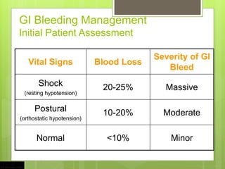
3. Core Principles in
GI Bleeding Management
Assessment and stabilization of hemodynamic status
Determine the source of bleeding
Stop active bleeding
Treatment of underlying abnormality
Prevent recurrent bleeding
4. GI Bleeding Management
Definitions
Hematemesis: bloody vomitus (bright red or
coffee-grounds)
Melena: black, tarry, foul-smelling stool
Hematochezia: bright red or maroon blood
per rectum
Occult: positive stool occult test
Symptoms of anemia: angina, dyspnea, or
lightheadedness
5. GI Bleeding Management
Patient Assessment
Hemodynamic status
Localization of bleeding source
CBC, PT, and T & C
Risk factors
Prior h/o PUD or bleeding
Cirrhosis
Coagulopathy
ASA or NSAID’s
6. GI Bleeding Management
Initial Patient Assessment
Vital Signs Blood Loss
Severity of GI
Bleed
Shock
(resting hypotension)
20-25% Massive
Postural
(orthostatic hypotension)
10-20% Moderate
Normal <10% Minor
8. GI Bleeding Management
Location of Bleeding
Upper
Proximal to Ligament of Treitz
Melena (100-200 cc of blood)
Azotemia
Nasogastric aspirate
Lower
Distal to Ligament of Treitz
Hematochezia
9. Acute UGIB
Demographics
Over 400,000 admissions annually
80% self-limited
Mortality 10-14%
Continued or recurrent bleeding - mortality 30-
40%
Nonvariceal UGIB w/o complication*
Mean LOS 2.7 days, $3402 (2008 $)
Nonvariceal UGIB with complication*
Mean LOS 4.4 days, $5632 (2008 $)
Adam V, Barkun A. Value Health. 2008;11:1-3.
21. Effect of Proton-Pump Inhibition on Peptic Ulcer
Bleeding
Gralnek et al. New Eng J Med 2008;359:928-37.
22. Management of PUD
after EGD in High Risk Pts
Proton-pump inhibitor 80 mg IV bolus dose plus continuous infusion for
72 hrs
Admit to monitored bed or ICU setting
Initiate oral intake of clear liquid diet 6 hrs after EGD in pts with
hemodynamic stability
Transition to oral PPI after completing IV course
Perform testing for H. pylori infection
For selected patients, discuss need for NSAIDs and antiplatelet therapy
Gralnek et al. New Eng J Med 2008;359:928-37.
23. Management of PUD
after EGD in Low Risk Pts
Oral proton-pump inhibitor
Initiate oral intake with a regular diet 6 hrs after EGD in pts with hemodynamic
stability
Perform testing for H. pylori infection
For selected patients, discuss need for NSAIDs and antiplatelet therapy
Consider early discharge in selected pts
Gralnek et al. New Eng J Med 2008;359:928-37.
26. Management of
Acute Variceal Bleeding
Suspected Variceal Bleeding
Endoscopy
Band ligation or sclerotherapy
Continue Octreotide for 5 days
Early rebleeding
Failure to control TIPS or surgery
Octreotide 50 ug bolus, 50 ug/hr
Conservative blood volume resuscitation
Antibiotics
27. Antibiotic Prophylaxis in GI
Bleeding in Cirrhotic Patients
Fluoroquinolones or amoxicillin + clavulinic acid
Meta-analysis 1
Decrease rates of infection
SBP, bacteremia
Increased short-term survival
RCT 2
Reduction in early rebleeding
1.Bernard et al.Hepatology. 29(6):1655-61.1999.
2.Hou et al. Hepatology. 39(3):746-53.2004.
32. Management of Ulcer Bleeding: ACG Guidelines
Initial Assessment and Risk Stratification
Hemodynamic status should be assessed immediately upon presentation
and resuscitative measures begun as needed (Strong recommendation).
Blood transfusions should target Hgb ≥ 7 g / dl, with higher Hgbs targeted in
patients with clinical evidence of intravascular volume depletion or
comorbidities, such as coronary artery disease (Conditional
recommendation).
Risk assessment should be performed to stratify patients into higher and
lower risk categories and may assist in initial decisions such as timing of
endoscopy, time of discharge, and level of care (Conditional
recommendation).
Discharge from the ED without inpatient endoscopy may be considered in
patients with urea nitrogen < 18.2 mg / dl; Hgb ≥ 13.0 g / dl for men (12.0 g /
dl for women), systolic blood pressure ≥ 110 mm Hg; pulse < 100 beats /
min; and absence of melena, syncope, cardiac failure, and liver disease, as
they have < 1 % chance of requiring intervention (Conditional
recommendation).
Laine & Jensen Am J Gastroenterol 2012; 107:345–360
33. Management of Ulcer Bleeding: ACG Guidelines
Pre-endoscopic interventions
Intravenous infusion of erythromycin (250 mg ~ 30 min before
endoscopy) should be considered to improve diagnostic yield and
decrease the need for repeat endoscopy. However, erythromycin
has not consistently been shown to improve clinical outcomes
(Conditional recommendation).
Pre-endoscopic intravenous PPI (e.g., 80 mg bolus followed by 8 mg
/ h infusion) may be considered to decrease the proportion of
patients who have higher risk stigmata of hemorrhage at endoscopy
and who receive endoscopic therapy. However, PPIs do not improve
clinical outcomes such as further bleeding, surgery, or death
(Conditional recommendation).
If endoscopy will be delayed or cannot be performed, intravenous
PPI is recommended to reduce further bleeding (Conditional
recommendation).
Nasogastric or orogastric lavage is not required in patients with
UGIB for diagnosis, prognosis, visualization, or therapeutic effect
(Conditional recommendation).
Laine & Jensen Am J Gastroenterol 2012; 107:345–360
34. Management of Ulcer Bleeding: ACG Guidelines
Timing of endoscopy
Patients with UGIB should generally undergo endoscopy
within 24 h of admission, following resuscitative efforts to
optimize hemodynamic parameters and other medical
problems (Conditional recommendation).
In patients who are hemodynamically stable and without
serious comorbidities endoscopy should be performed as
soon as possible in a non-emergent setting to identify the
substantial proportion of patients with low-risk endoscopic
findings who can be safely discharged (Conditional
recommendation).
In patients with higher risk clinical features (e.g., tachycardia,
hypotension, bloody emesis or nasogastric aspirate in
hospital) endoscopy within 12 h may be considered to
potentially improve clinical outcomes (Conditional
recommendation).
Laine & Jensen Am J Gastroenterol 2012; 107:345–360
35. Management of Ulcer Bleeding: ACG Guidelines -
Endoscopy
Stigmata of recent hemorrhage should be recorded as they predict risk of
further bleeding and guide management decisions. The stigmata, in
descending risk of further bleeding, are active spurting, non-bleeding visible
vessel, active oozing, adherent clot, fl at pigmented spot, and clean base
(Strong recommendation).
Endoscopic therapy should be provided to patients with active spurting or
oozing bleeding or a non-bleeding visible vessel (Strong recommendation).
Endoscopic therapy may be considered for patients with an adherent clot
resistant to vigorous irrigation. Benefi t may be greater in patients with
clinical features potentially associated with a higher risk of rebleeding (e.g.,
older age, concurrent illness, inpatient at time bleeding began) (Conditional
recommendation).
Endoscopic therapy should not be provided to patients who have an ulcer
with a clean base or a fl at pigmented spot (Strong recommendation).
Laine & Jensen Am J Gastroenterol 2012; 107:345–360
36. Management of Ulcer Bleeding: ACG Guidelines - Endoscopy
Epinephrine therapy should not be used alone. If used, it should be
combined with a second modality (Strong recommendation).
Thermal therapy with bipolar electrocoagulation or heater probe and
injection of sclerosant (e.g., absolute alcohol) are recommended
because they reduce further bleeding, need for surgery, and mortality
(Strong recommendation).
Clips are recommended because they appear to decrease further
bleeding and need for surgery. However, comparisons of clips vs. other
therapies yield variable results and currently used clips have not been
well studied (Conditional recommendation).
For the subset of patients with actively bleeding ulcers, thermal therapy
or epinephrine plus a second modality may be preferred over clips or
sclerosant alone to achieve initial hemostasis (Conditional
recommendation).
Laine & Jensen Am J Gastroenterol 2012; 107:345–360
37. Management of Ulcer Bleeding: ACG Guidelines - Therapy
after initial endoscopy
After successful endoscopic hemostasis, intravenous PPI therapy with 80 mg
bolus followed by 8 mg/h continuous infusion for 72 h should be given to
patients who have an ulcer with active bleeding, a non-bleeding visible vessel,
or an adherent clot (Strong recommendation).
Patients with ulcers that have flat pigmented spots or clean bases can receive
standard PPI therapy (e.g., oral PPI once daily) (Strong recommendation).
Routine second-look endoscopy, in which repeat endoscopy is performed 24 h
after initial endoscopic hemostatic therapy, is not recommended (Conditional
recommendation).
Repeat endoscopy should be performed in patients with clinical evidence of
recurrent bleeding and hemostatic therapy should be applied in those with
higher risk stigmata of hemorrhage (Strong recommendation).
If further bleeding occurs after a second endoscopic therapeutic session,
surgery or interventional radiology with transcathether arterial embolization is
generally employed (Conditional recommendation).
Laine & Jensen Am J Gastroenterol 2012; 107:345–360
38. International Consensus on Nonvariceal Upper
Gastrointestinal Bleeding:
Postdischarge ASA and NSAIDs
In patients with previous ulcer bleeding who require
an NSAID, it should be recognized that treatment
with a traditional NSAID plus PPI or a COX-2 inhibitor
alone is still associated with a clinically important risk
for recurrent ulcer bleeding.
In patients with previous ulcer bleeding who require
an NSAID, the combination of a PPI and a COX-2
inhibitor is recommended to reduce the risk for
recurrent bleeding from that of COX-2 inhibitors
alone.
Barkun AN, et al. Ann Intern Med. 2010;152:101-113.
39. International Consensus on Nonvariceal Upper
Gastrointestinal Bleeding:
Postdischarge ASA and NSAIDs
In patients who receive low-dose ASA and develop
acute ulcer bleeding, ASA therapy should be
restarted as soon as the risk for cardiovascular
complication is thought to outweigh the risk for
bleeding.
In patients with previous ulcer bleeding who require
cardiovascular prophylaxis, it should be recognized
that clopidogrel alone has a higher risk for rebleeding
than ASA combined with a PPI.
Barkun AN, et al. Ann Intern Med. 2010;152:101-113.
47. Acute LGIB
Key Points
Resuscitation
UGI source
Most bleeding ceases
Colonoscopy
No role for barium studies
48. SUMMARY
GI Bleeding Management
Assessment and stabilization of hemodynamic status
Determine the source of bleeding
Stop active bleeding
Treatment of underlying abnormality
Prevent recurrent bleeding