Activation of rat alveolar macrophage derived latent transforming growth factor b(german) 1 by plasmin requires interaction with thrombospondin-1 and its cell surface receptor, cd36 (1999)
L-arginine modulates T cell metabolism and enhances survival and anti-tumor A...Gul Muneer
L-arginine modulates T cell metabolism and enhances survival and anti-tumor Activity
How metabolic reprogramming of T cells impact T cells fate and function? Indeed, these cells enhance their metabolic throughput upon antigenic stimulation to ensure the production of sufficient biomass and energy for their proliferation, differentiation and effector functions. The naïve T cells, which utilize oxidative phosphorylation for energy supply but upon activation, these cells switch their metabolic pathways toward glycolysis. An important question arises here that whether differential metabolic reprogramming (i.e., use of oxidative phosphorylation versus glycolytic activity) directs the fate and function of a given cell? And whether this process shapes the immune response and individual cell survival? The researchers from Institute for Research in Biomedicine took advantage of high-resolution Mass Spectrometry to investigate dynamics of proteome and metabolome following activation of naïve T cells.
If the many beneficial effects of the chemokines can be preserved, such efforts hold great promise for uncovering new therapies for inflammatory and immunologic disease
If the many beneficial effects of the chemokines can be preserved, such efforts hold great promise for uncovering new therapies for inflammatory and immunologic disease
Similar to Activation of rat alveolar macrophage derived latent transforming growth factor b(german) 1 by plasmin requires interaction with thrombospondin-1 and its cell surface receptor, cd36 (1999)
Investigating the function of a novel protein from Anoectochilus formosanus w...Cây thuốc Việt
Anoectochilus formosanus is a therapeutic orchid appreciated as a traditional Chinese medicine in Asia. The extracts of A. formosanus have been reported to possess hepatoprotective, anti-inflammatory, and anti-tumor activates. A novel protein was isolated from A. formosanus, and its immunomodulatory effect on murine peritoneal macrophage was investigated. Macrophages obtained from ascites of thioglycollate-induced BALB/c were co-cultured with IPAF (0–20μg/ml) for 24h and then harvested for flow cytometry analysis. The cytokine/chemokine production was measured by real time PCR and ELISA. The interaction between IPA and toll like receptors (TLRs) was investigated by TLR gene knockout (KO) mice and fluorescence labeled IPAF. The activation of NF-κB was assessed by EMSA. IPAF stimulated the TNF-α and IL-1β production, upregulated the CD86 and MHC II expression, and enhanced the phagocytic activity of macrophages. IPAF induced gene expression of IL-12 and Th1-assosiated cytokines/chemokines. The stimulating effect of IPAF was
impaired, and the IPAF–macrophage interaction was reduced in TLR4−/− C57BL/10ScNJ mice. In addition, IPAF stimulated expressions of TLR signal-related genes and the activation of NF-κB. IPAF could induce classical activated macrophage differentiation via TLR4-dependent NF-κB activation and had potential of IPAF to modulate the Th1 response. These findings provided valuable information regarding the immune modulatory mechanism of A. formosanus, and indicated the possibility of IPAF as a potential peptide drug
Inhibition of glutathione by buthionine sulfoximine enhanced the anti-cancer ...Ashujit
Multiple myeloma (MM) is an incurable blood cancer. Melphalan is an alkylating agent given prior to stem cell transplantation to MM patients. Increased glutathione confers resistance to melphalan. This study investigate the effect of inhibition of glutathione by BSO in preclinical models of MM. Pretreatment with BSO enhanced the anti-cancer effect of melphalan in cell lines and animal models. BSO and melphalan combination was well tolerated by animals and enhanced the survival as compared to controls, BSO and melphalan alone. BSO enhanced depth and duration of responses induced by melphalan. In the combination group, majority of treated animals achieved complete response (CR) and more than 20% had maintained CR. Also, the survival of animals was doubled after combination treatment as compared to BSO or melphalan alone. Mechanistic investigation demonstrated that BSO enhanced melphalan induced DNA damage, caspase cleavage and apoptosis. The combination also achieved multi-logs of cells kills in nine human multiple myeloma cell lines and primary MM cells isolated from blood and bone marrows. Interestingly, the effect of BSO and melphalan combination was abolished when cells were treated with N-acetyl cysteine and sodium thiosulfate but not with vitamin C and vitamin E. This observation suggests that effect of BSO is primarily driven by its ability to deplete glutathione and therefore preventing melphalan detoxification. Together, this study provides framework for testing the combination in a Phase I trial.
Investigation of Intracellular Signal Transduction in Multiple Endocrine Neop...KayvonneFerguson
During the Fall 2022 semester, I took the course Biotechnology Writing and Communication to fulfill the intensive writing credit for my major. During this course, we had writing assignments that addressed the learning goals of the Biotechnology major such as basic science understanding and application, critical thinking, and communication.
Our final writing assignment was a literature review of a chosen topic that covered laboratory-based biology. In this paper, we had to utilize at least 10 primary articles with 1-2 articles within the last year as well as a 10-page minimum. In my literature review, I covered the disorder Multiple Endocrine Neoplasia Type 1 involving tumors of the pituitary gland. In this review, I cover the mechanisms of tumor progression through the intracellular signal transduction pathway involving G-protein coupled receptors and the secondary messenger cAMP. I cover genes and proteins in this pathway that have an association with cell proliferation and tumor progression.
Similar to Activation of rat alveolar macrophage derived latent transforming growth factor b(german) 1 by plasmin requires interaction with thrombospondin-1 and its cell surface receptor, cd36 (1999) (20)
Slide 1: Title Slide
Extrachromosomal Inheritance
Slide 2: Introduction to Extrachromosomal Inheritance
Definition: Extrachromosomal inheritance refers to the transmission of genetic material that is not found within the nucleus.
Key Components: Involves genes located in mitochondria, chloroplasts, and plasmids.
Slide 3: Mitochondrial Inheritance
Mitochondria: Organelles responsible for energy production.
Mitochondrial DNA (mtDNA): Circular DNA molecule found in mitochondria.
Inheritance Pattern: Maternally inherited, meaning it is passed from mothers to all their offspring.
Diseases: Examples include Leber’s hereditary optic neuropathy (LHON) and mitochondrial myopathy.
Slide 4: Chloroplast Inheritance
Chloroplasts: Organelles responsible for photosynthesis in plants.
Chloroplast DNA (cpDNA): Circular DNA molecule found in chloroplasts.
Inheritance Pattern: Often maternally inherited in most plants, but can vary in some species.
Examples: Variegation in plants, where leaf color patterns are determined by chloroplast DNA.
Slide 5: Plasmid Inheritance
Plasmids: Small, circular DNA molecules found in bacteria and some eukaryotes.
Features: Can carry antibiotic resistance genes and can be transferred between cells through processes like conjugation.
Significance: Important in biotechnology for gene cloning and genetic engineering.
Slide 6: Mechanisms of Extrachromosomal Inheritance
Non-Mendelian Patterns: Do not follow Mendel’s laws of inheritance.
Cytoplasmic Segregation: During cell division, organelles like mitochondria and chloroplasts are randomly distributed to daughter cells.
Heteroplasmy: Presence of more than one type of organellar genome within a cell, leading to variation in expression.
Slide 7: Examples of Extrachromosomal Inheritance
Four O’clock Plant (Mirabilis jalapa): Shows variegated leaves due to different cpDNA in leaf cells.
Petite Mutants in Yeast: Result from mutations in mitochondrial DNA affecting respiration.
Slide 8: Importance of Extrachromosomal Inheritance
Evolution: Provides insight into the evolution of eukaryotic cells.
Medicine: Understanding mitochondrial inheritance helps in diagnosing and treating mitochondrial diseases.
Agriculture: Chloroplast inheritance can be used in plant breeding and genetic modification.
Slide 9: Recent Research and Advances
Gene Editing: Techniques like CRISPR-Cas9 are being used to edit mitochondrial and chloroplast DNA.
Therapies: Development of mitochondrial replacement therapy (MRT) for preventing mitochondrial diseases.
Slide 10: Conclusion
Summary: Extrachromosomal inheritance involves the transmission of genetic material outside the nucleus and plays a crucial role in genetics, medicine, and biotechnology.
Future Directions: Continued research and technological advancements hold promise for new treatments and applications.
Slide 11: Questions and Discussion
Invite Audience: Open the floor for any questions or further discussion on the topic.
Cancer cell metabolism: special Reference to Lactate PathwayAADYARAJPANDEY1
Normal Cell Metabolism:
Cellular respiration describes the series of steps that cells use to break down sugar and other chemicals to get the energy we need to function.
Energy is stored in the bonds of glucose and when glucose is broken down, much of that energy is released.
Cell utilize energy in the form of ATP.
The first step of respiration is called glycolysis. In a series of steps, glycolysis breaks glucose into two smaller molecules - a chemical called pyruvate. A small amount of ATP is formed during this process.
Most healthy cells continue the breakdown in a second process, called the Kreb's cycle. The Kreb's cycle allows cells to “burn” the pyruvates made in glycolysis to get more ATP.
The last step in the breakdown of glucose is called oxidative phosphorylation (Ox-Phos).
It takes place in specialized cell structures called mitochondria. This process produces a large amount of ATP. Importantly, cells need oxygen to complete oxidative phosphorylation.
If a cell completes only glycolysis, only 2 molecules of ATP are made per glucose. However, if the cell completes the entire respiration process (glycolysis - Kreb's - oxidative phosphorylation), about 36 molecules of ATP are created, giving it much more energy to use.
IN CANCER CELL:
Unlike healthy cells that "burn" the entire molecule of sugar to capture a large amount of energy as ATP, cancer cells are wasteful.
Cancer cells only partially break down sugar molecules. They overuse the first step of respiration, glycolysis. They frequently do not complete the second step, oxidative phosphorylation.
This results in only 2 molecules of ATP per each glucose molecule instead of the 36 or so ATPs healthy cells gain. As a result, cancer cells need to use a lot more sugar molecules to get enough energy to survive.
introduction to WARBERG PHENOMENA:
WARBURG EFFECT Usually, cancer cells are highly glycolytic (glucose addiction) and take up more glucose than do normal cells from outside.
Otto Heinrich Warburg (; 8 October 1883 – 1 August 1970) In 1931 was awarded the Nobel Prize in Physiology for his "discovery of the nature and mode of action of the respiratory enzyme.
WARNBURG EFFECT : cancer cells under aerobic (well-oxygenated) conditions to metabolize glucose to lactate (aerobic glycolysis) is known as the Warburg effect. Warburg made the observation that tumor slices consume glucose and secrete lactate at a higher rate than normal tissues.
This presentation explores a brief idea about the structural and functional attributes of nucleotides, the structure and function of genetic materials along with the impact of UV rays and pH upon them.
Professional air quality monitoring systems provide immediate, on-site data for analysis, compliance, and decision-making.
Monitor common gases, weather parameters, particulates.
Seminar of U.V. Spectroscopy by SAMIR PANDASAMIR PANDA
Spectroscopy is a branch of science dealing the study of interaction of electromagnetic radiation with matter.
Ultraviolet-visible spectroscopy refers to absorption spectroscopy or reflect spectroscopy in the UV-VIS spectral region.
Ultraviolet-visible spectroscopy is an analytical method that can measure the amount of light received by the analyte.
This pdf is about the Schizophrenia.
For more details visit on YouTube; @SELF-EXPLANATORY;
https://www.youtube.com/channel/UCAiarMZDNhe1A3Rnpr_WkzA/videos
Thanks...!
Lateral Ventricles.pdf very easy good diagrams comprehensive
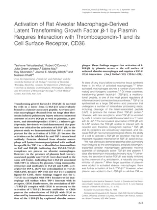
Activation of rat alveolar macrophage derived latent transforming growth factor b(german) 1 by plasmin requires interaction with thrombospondin-1 and its cell surface receptor, cd36 (1999)
2. 9further activation of L-TGF-1 occurred.5
Our findings
suggested that the generation of plasmin is important in
the posttranslational activation of alveolar macrophage-
derived L-TGF- 1 during an inflammatory pulmonary
injury response and that the activation requires the pres-
ence of intact macrophages.5
In this paper, we demonstrate that alveolar macro-
phages also secrete increased quantities of TSP-1, a
glycoprotein previously reported to activate L-TGF-1
both in the presence of cells and in cell-free solution.6
When alveolar macrophages were cultured in the pres-
ence of neutralizing antibodies to TSP-1, the posttransla-
tional activation of L-TGF-1 was abrogated. Further-
more, antibodies to the TSP-1 receptor, CD36, also
abrogated activation of alveolar macrophage-derived L-
TGF-1. Our findings support a model in which L-TGF-1
is held at the cell surface by a TSP-1/CD36 interaction
and is processed by plasmin generated by activated
alveolar macrophages.
Materials and Methods
Animals
Female Sprague-Dawley rats, which were free of respira-
tory disease and weighed between 250 and 300 g, were
obtained from the University of Manitoba vivarium. In
each experiment, all rats were matched for age and
weight.
Reagents
Bleomycin (Blenoxane) was a gift from Bristol-Myers
Squibb (Evansville, IN). Neutralizing antibody to TGF-
1–3 was obtained from Genzyme (Cambridge, MA).
Antibody to TGF-1 used for Western blot analysis was
obtained from Santa Cruz Biotechnology (Santa Cruz,
CA). The recombinant anti-human LAP antibody was ob-
tained from R&D Systems (Minneapolis, MN). TSP-1 de-
pleted of TGF- activity (sTSP-1) and monoclonal anti-
bodies to human platelet TSP-1 depleted of associated
TGF- 1 (mAb 133) used in the enzyme-linked immu-
nosorbent assay (ELISA) and TSP-1 immunoprecipitates
were either used by or obtained from Dr. Murphy-Ull-
rich.6,7
Anti-TSP-1 antibody used in experiments to neu-
tralize TSP-1 from activated alveolar macrophages was
obtained from Sigma (St. Louis, MO). The CD36 antibody,
5F1, was provided by the Fifth International Workshop on
Leukocyte Differentiation Antigens.8
Bleomycin Administration
This procedure is described in detail elsewhere.5,9,10
For
some experiments rats were sacrificed at several time
intervals after bleomycin or normal saline treatment,5,9,10
whereas for other experiments alveolar macrophages
were harvested 7 days after bleomycin administration.
The latter time point was used based on our findings that
alveolar macrophages are maximally stimulated at this
time to secrete active TGF-1.5
Macrophage Cultures
This procedure is described in detail elsewhere.5,10
The
lungs were lavaged to obtain cells for culture of alveolar
macrophages. Alveolar macrophages were maintained in
serum-free media containing Gentamicin (4 mg/100 ml;
Roussel, Montreal, PQ), Fungizone (100 l/100 ml; Gibco
BRL, Grand Island, NY) and 0.2% clotted bovine calf
plasma (BCP; National Biological Laboratory Limited, Du-
gald, MB). The macrophages were cultured in the ab-
sence or presence of a number of reagents consisting of
anti-TSP-1 antibody, 5F1 (anti-CD36 antibody), CD36
synthetic peptide (aa 93–110), or sTSP-1. In experiments
to determine the effects of exogenous sTSP-1, alveolar
macrophages were cultured with varying quantities of
sTSP-1 for 2 hours before the collection of conditioned
media (CM). In addition, CM from the same alveolar
macrophages cultured in parallel was incubated in a
cell-free solution with sTSP-1 for 2 hours. Incubation of
CM with sTSP-1 for 2 hours was chosen based on our
previous findings, that sTSP-1 can activate L-TGF-1
within 2 hours.11
In some experiments the cells were
cultured in the absence or presence of 5F1, the CD36
antibody, before the addition of sTSP-1. In experiments to
determine whether or not both plasmin and TSP-1 are
required to activate alveolar macrophage-derived L-TGF-
1, the alveolar macrophages were cultured with sTSP-1
in the absence or presence of aprotinin, an inhibitor of
plasmin activity.12
After 20 hours of incubation in 5% CO2
at 37°C, the media was collected in the presence of
protease inhibitors (leupeptin 0.5 g/ml, Amersham,
Poole, UK; aprotinin 1 g/ml, and pepstatin 1 g/ml, both
from Sigma, Oakville, ON), and frozen at Ϫ80°C until
ready for TGF- quantitation.5,10
CCL-64 Mink Lung Epithelial Growth Inhibition
Assay for TGF-
The CCL-64 growth inhibition assay to identity and quan-
titate TGF- has been described.5,9,10,13
. Briefly, to sub-
confluent cells in 0.2% BCP and resuspended in ␣-MEM,
0.2% BCP, 10 mmol/L Hepes at pH 7.4, penicillin (25
g/ml) and streptomycin (25 g/ml), and cultured as 5 ϫ
104
cells per 0.5 ml in 24-well costar dishes (Flow Labo-
ratories, Inc., Mississauga, ON) was added neutral CM or
CM that was acidified and subsequently neutralized. Af-
ter 22 hours the cells were pulsed with 0.25 Ci of 5-[125
I]
iodo 2Ј-deoxyuridine (ICN Pharmaceutical, Costa Mesa,
CA) for 2 to 3 hours at 37°C and eventually lysed with 1
ml of 1 N NaOH for 30 minutes at room temperature and
the 125
I-UdR was counted in a ␥ counter (LKB Instru-
ments, Gaithersburg, MD). A standard curve using por-
cine TGF-1 was included in each assay. For confirma-
tion of TGF- activity, neutralizing monoclonal antibody to
TGF-1–3 (Genzyme) was added before the addition of
the CM5,9,10,13
and resulted in abrogation of all TGF-
activity.
842 Yehualaeshet et al
AJP September 1999, Vol. 155, No. 3
3. Detection and Quantitation of TSP-1 by Direct
ELISA
The wells of 96-well plates (Falcon tissue culture plates)
were coated with 200 ng/well of the mAb 133 (anti-sTSP-
1). For each experiment, a standard curve containing
wells with 200 l of CM and several concentrations of
sTSP-1, ranging from 15–150 ng/well, was included. To
quantitate TSP-1 in CM, 200 l of alveolar macrophage
derived CM in carbonate buffer, pH 9.6, was incubated
overnight at 4°C. The next day, the wells were washed
three times with PBS, 0.05%, Tween Ϫ20. Nonspecific
binding sites were blocked by incubating with 250 l/well
of 1% BSA for 1 hour at 37°C. The wells were then
washed with PBS and incubated with 200 l/well of mAb
133 (7.5 g/ml) in PBS-Tween for 90 minutes at 37°C. The
wells were then washed and incubated with 30 ng/ml of
alkaline phosphatase-labeled goat anti-mouse lgG for 90
minutes at 37°C, and then assayed for color development
using the Sigma 104 AP substrate. Color development
was stopped by adding 50 l of 2N NaOH, and absor-
bency at 405 nm was read using a Bio-Tek ELISA reader.
Preparation of Synthetic CD36 Peptides
The CD36 peptide, YRVRFLAKENVTQDAEDNC(93–110)
was synthesized, based on the work of Leung et al.14
The
peptide was synthesized with an Applied Biosystems
model 431A peptide synthesizer using Fmoc (N-(9-Flu-
oreny D-methoxycarbonyl) chemistry and purified by re-
verse high pressure liquid chromatography, using a C18
column.
Localization of CD36 and TGF-1 by
Immunofluorescence
Alveolar macrophages were obtained by bronchoalveo-
lar lavage 7 days after intratracheal normal saline or
bleomycin administration, and were adjusted to 1 ϫ 106
cells/ml. For some experiments, alveolar macrophages
obtained after bleomycin administration were cultured in
the presence of anti-CD36 antibody (20 g per 106
mac-
rophages) for 30 minutes before the immunofluoresence
procedure. The immunofluorescence analysis was per-
formed as previously described.15
Briefly, cytospin
smears of 1 ϫ 105
cells suspension were fixed with 3.7%
formaldehyde for 10 minutes and washed with PBS. Non-
specific binding was blocked by 100% lamb serum
(Gibco BRL) for 5 minutes. The cells were then incubated
with anti-TGF-1 (Santa Cruz Biotechnology) and anti-
CD36 antibody, both at a concentration of 1 g/ml, for 45
minutes. After washing with PBS the cells were incubated
for 30 minutes with polyclonal anti-rabbit antibody conju-
gated to tetramethylrhodamine-isothiocyanate (TRITC) or
conjugated with fluorescein isothiocyanate (FITC) mono-
clonal anti-mouse IgM as secondary antibodies for the
detection of TGF-1 and CD36 antibodies, respectively.
Both secondary antibodies were used as 1 g/ml. The
slides were washed three times with PBS. Nuclear stain-
ing was done using 4Ј, 6Ј Diamidino-2-phenylindole
(DAPI) at a concentration of 1 g/ml for 5 minutes. The
slides were mounted in anti-bleach (12% glycerol, 4.8%
Mowiol 4–88 (Hoechst), 2.4% DABCO (1, 4 Diazabicylo
[2.2.2]-octane (Fluka) in 0.2 Mol/L Tris/HCl, pH ϭ 8.5).
Image analysis was performed using a Zeiss Axiophot
microscope, equipped with a cooled CCD camera CH
250/a (Optikon/Photometrics), driven by IPLabs Spec-
trum software version 3.1, Signal Analytics). For TRITC-
conjugated antibodies, the filter combination was BP540/
FT580/LP590, resulting in a red emission identifying the
location of TGF-1on the macrophage surface. For the
FITC-conjugated antibody, the filter combination was
450–490/FT510/515–565, resulting in a green emission
identifying the location of CD36 on the macrophage sur-
face. For DAPI, the filter combination was G365/FT395/
LP420, resulting in a blue emission. To determine
whether TGF-1 and CD36 were colocalized, the images
were obtained by a pixel overlap, which was achieved
with IPLab Spectrum/Multiprobe (V.3.1, Signal Analytics,
Fairfax, VA). In areas where the TGF-1 was localized to
the same region as CD36, the emission was yellow.
Protein Extraction and Immunoprecipitation
Alveolar macrophages obtained from rats that had been
treated with normal saline or bleomycin 7 days earlier
were lysed by incubating the cells for 20 minutes on ice
in a RIPA lysis buffer (50 mmol/L Tris-Cl, pH 7.5; 150
mmol/L NaCl, 1% Nonidet P-40, 0.1% sodium deoxy-
cholate and a cocktail of protease inhibitors; phenylmeth-
ylsulfonyl fluoride 1 mmol/L, leupeptin 1 g/ml, and apro-
tinin 0.1 g/ml, all from Sigma). In some experiments the
alveolar macrophages were cultured overnight in the ab-
sence or presence of ␣2-antiplasmin or aprotinin (Sigma),
both inhibitors of plasmin activity.12
The lysate was cen-
trifuged for 20 minutes at 12,000 ϫ g at 4°C, the super-
natant collected, and the total protein content determined
by the Bradford dye-binding assay (Bio-Rad, Missis-
sauga, ON). The total protein extract (300 g) and 10 g
of anti-sTSP-1 antibody (mAb 133) or IgG as an isotype
control for the anti-sTSP-1 antibody was incubated over-
night at 4°C. After a further incubation of 2 hours with 30
l of Protein G Plus/Protein A-Agarose (Calbiochem, San
Diego, CA) the immune complexed beads were col-
lected. The beads were washed four times with RIPA
buffer and placed in a final suspension with 25 l of
Laemmli buffer and boiled for 10 minutes. The superna-
tant containing the precipitated proteins was then used
for Western blot analysis.
Western Blot Analysis
The protein samples (25 l) were electrophoresed on
10% sodium dodecyl sulfate-polyacrylamide gel electro-
phoresis (SDS-PAGE) in a MiniPROTEAN II Electrophore-
sis Cell (Bio-Rad, Hercules, CA). Rainbow colored pro-
tein molecular weight markers (Amersham) were run
parallel to each blot as an indicator of the molecular
weight. Equality in loading of protein was evaluated using
silver staining (not shown). The separated proteins were
Activation of Macrophage-Derived L-TGF-1 by TSP-1 843
AJP September 1999, Vol. 155, No. 3
4. transferred at 50 V overnight onto nitrocellulose mem-
brane (Gibco BRL) in a Mini trans-Blot chamber with
transfer buffer (25 mmol/L Tris Cl, 192 mmol/L glycine,
and 20% methanol). The nitrocellulose membrane was
blocked for 1 hour by using 5% instant skim milk powder
in TBS. For detection of LAP a 1:500 dilution of anti-
recombinant human LAP antibody (anti-rh LAP; R&D Sys-
tems) in 1% instant skim milk powder was used. After
washing, the nitrocellulose membrane was incubated
with horseradish peroxidase linked with the secondary
antibody (Goat anti-mouse IgG; Bio-Rad) as recom-
mended by the manufacturer. Finally the washed blots
were exposed to enhanced chemiluminescence detec-
tion system (Amersham) and recorded on an autoradio-
graph (Kodak X-Omat film). Before reprobing, the nitro-
cellulose membrane was incubated at 50°C for 30
minutes with a stripping buffer (100 mmol/L 2-mercapto-
ethanol, 2% sodium dodecyl sulfate, and 62.5 mmol/L
Tris-HCl, pH 6.7). The blots were rinsed twice with TBS.
To ensure the removal of antibodies, membranes were
incubated with the enhanced chemiluminescence detec-
tion reagents and exposed to film (Kodak). No band was
detected, confirming that all antibodies were stripped off
the membrane. The same nitrocellulose membrane was
blocked using 5% instant skim milk powder in TBS. For
detection of TGF-1, 0.5 g/ml of rabbit polyclonal IgG
TGF-1 antibody (Santa Cruz Biotechnology) was used
as above.
Statistical Analysis
Statistical analysis using analysis of variance was done by
Dr. Bob Tate, Biostatistical Unit, University of Manitoba.
Results
Activation of Alveolar Macrophage-Derived L-
TGF-1 by Thrombospondin (TSP)-1
In addition to plasmin, TSP-1, a large trimeric glycopro-
tein,16
had also been demonstrated to be a physiological
substance that can activate L-TGF-1.6,17
TSP-1 was
constitutively secreted by alveolar macrophages, but af-
ter bleomycin injury, TSP-1 secretion was further in-
creased and reached maximal levels 7 days after bleo-
mycin administration (Figure 1A). The secretion of TSP-1
declined rapidly thereafter, and by 28 days after bleomy-
cin administration, the secretion returned to that of control
levels of alveolar macrophages from normal saline-
treated rats. To determine whether the presence of TSP-1
in the CM was necessary for the activation of L-TGF-1,
alveolar macrophages were cultured in the absence or
presence of anti-TSP-1 monoclonal antibody. Anti-TSP-1
antibody inhibited the activation of L-TGF- 1 but had no
effect on the secretion of the latent form of TGF-1 (Fig-
ure 1B) by the alveolar macrophages. Neutral CM of
alveolar macrophages activated by in vivo bleomycin in-
jury contained 67.6 Ϯ 7.9 pg of TGF-1 per 106
cells.
After the addition of 50 g/ml of the isotype control for
anti-TSP-1, 50.13 Ϯ 13.4 pg of TGF-1 per 106
was
present in neutral CM (P Ͻ 0.934). Because TSP-1 has
been reported to activate L-TGF-1 in solution,6
we next
determined if alveolar macrophage-derived L-TGF-1
could be directly activated in CM by sTSP-1. After 20
hours in culture, activated alveolar macrophages se-
creted large quantities of L-TGF-1.5
The addition of
sTSP-1 to this cell-free L-TGF-1 unexpectedly dimin-
Figure 1. Activation of alveolar macrophage derived L-TGF-1 by sTSP-1. A: Quantity of TSP-1 secreted by explanted alveolar macrophages obtained from rats
at varying lengths of time after bleomycin administration. Each point is the mean of samples from 3 to 6 rats. The P value is Ͻ0.0003 when the quantity of TSP-1
produced by alveolar macrophages from normal saline-treated rats is compared to TSP-1 produced by alveolar macrophages from rats treated with bleomycin 7
days earlier. B: The effects of anti-TSP-1 antibody on the activation of alveolar macrophage-derived L-TGF-1. Alveolar macrophages obtained 7 days after
bleomycin administration were cultured in the absence of anti-TSP-1 antibody or in the presence of varying concentrations. The TGF-1 present in neutral CM
(Ⅺ) represents bioactive TGF-1; that present in acidified then neutralized CM (f) represents the total TGF-1 in the same sample. Each point is the mean of
samples from 6 rats. The quantity of TGF-1 in neutral CM compared to the quantity of TGF-1 in acidified CM has a P Յ 0.001 when the anti-TSP-1 antibody
used was 50 g/ml/106
macrophages, and P Յ 0.003 when the anti-TSP-1 antibody used was 5.0 or 0.5 g/ml/106
macrophages. There was no statistical difference
in the quantity of TGF-1 in neutral and acidified CM when no anti-TSP-1 antibody was present. C: Induction of activation of L-TGF-1 by addition of sTSP-1.
Alveolar macrophages were obtained 7 days after bleomycin administration and cultured for 20 hours before the addition of sTSP-1 (0–4 g/ml) to alveolar
macrophages (f) or the CM (Ⅺ) from parallel cultures of alveolar macrophages. After a 2-hour incubation with sTSP-1, the TGF- 1 in neutral and acidified CM
(not shown) of the same sample was determined. The -fold increase in the quantity of TGF-1 present was calculated by using the quantity of active TGF-1
present in the absence of sTSP-1 as the denominator and the quantity of TGF-1 present after the addition of sTSP-1 as the numerator. Each point is the mean
of samples from 3 rats. The -fold increase in the quantity of TGF-1 in neutral CM when alveolar macrophages were treated with 0.4 or 4.0 g/ml of sTSP-1,
compared to TGF-1 in CM when no sTSP-1 was present, has P value Յ 0.05.
844 Yehualaeshet et al
AJP September 1999, Vol. 155, No. 3
5. ished the quantity of active TGF-1 that could be de-
tected by our bioassay (Figure 1C). In contrast, sTSP-1
added directly to the same CM but, in the presence of
alveolar macrophages, further activated L-TGF-1 (Fig-
ure 1C). These findings demonstrate that TSP-1 is effec-
tive in promoting the activation of alveolar macrophage-
derived L-TGF-1 in the presence of intact macrophages.
Cell Surface Association of L-TGF- 1 to CD36
Is Required for Activation of L-TGF-1
TSP-1 not only complexes with L-TGF-1,6,7,11
but it also
binds to cell surface receptors such as CD3618,19
which
are prominently expressed on macrophages.18,19
We
next determined if the CD36 binding of TSP-1 is required
for the posttranslational processing and activation of L-
TGF- 1. At the highest concentration, the presence of
antibodies specific to CD36 totally abrogated the activa-
tion of L-TGF-1 (Figure 2A). Neutral CM of alveolar
macrophages activated by in vivo bleomycin injury con-
tained 70 Ϯ 10 pg of TGF-1 per 106
cells, while after the
addition of 20 g/ml of the isotype control for anti-CD36,
55 Ϯ 1 pg of TGF-1 per 106
cells was present (P Ͻ
0.949). There was an induction of L-TGF- 1 in the pres-
ence of CD36 antibody but the mechanism of induction is
not clear. The interaction of the CD36 with its antibody
has been reported to lead to an oxidative burst in mono-
cytes20
and induction of the protein tyrosine kinases fyn,
lyn, and yes.21
We are presently determining if either of
these events results in increased production and secre-
tion of L-TGF-1 by macrophages after anti-CD36 anti-
body interaction. Alternatively it is also possible that the
L-TGF-1 is sequestered on the cell surface via the CD36
receptor. The presence of the CD36 antibody may then
displace the L-TGF-1 from the cell surface and increase
the quantity of L-TGF-1 present in the CM.
The binding of TSP-1 to the extracellular domain of
CD36 is blocked by a CD36 synthetic peptide from amino
acids 93–110 (CD3693–110
).14
When activated alveolar
macrophages were incubated with the synthetic peptide
CD3693–110
mimicking the TSP-1 binding domain, the
activation of L-TGF-1 was abrogated (Figure 2B). Al-
though the CD36 peptide 93–110 decreased activation of
L-TGF-1, unlike the CD36 antibody, the presence of the
peptide did not induce L-TGF-1 generation. The reason
for this is not clear, but it could be that the peptide does
not interact with the CD36 receptor in the same manner
as the antibody20,21
and, therefore, not cause further
activation of alveolar macrophages to release L-TGF-1.
The synthetic peptide corresponding to an area unre-
lated to CD36-TSP-1 interaction, CD36204 –288
, had no
effect on the activation of L-TGF- 1(data not shown). The
addition of sTSP-1 to alveolar macrophages increased
the activity of TGF-1 present in neutral CM (Figures 1C
and 2C). Further confirmation that the association of
TSP-1 with CD36 on alveolar macrophages was impor-
tant in activation of L-TGF- 1 was the finding that, when
the CD36 antibody was added to activated alveolar mac-
rophages in the presence of sTSP-1, there was a marked
diminution of the effects of sTSP-1 in generating mature
TGF-1 (Figure 2C).
Using indirect immunofluorescent staining, alveolar
macrophages obtained from rats previously treated with
normal saline had detectable TGF-1 (red emission; Fig-
ure 3A) and CD36 (green emission; Figure 3B) on the cell
surface. In addition, both CD36 and TGF-1 were colo-
calized (yellow emission) to a number of regions on the
same cells (Figure 3C). However, alveolar macrophages
Figure 2. Effects of anti-CD36 antibody on the activation of L-TGF-1. Alveolar macrophages obtained 7 days after bleomycin administration were cultured in the
absence or presence of different concentrations of anti-CD36 antibody or the CD36 synthetic peptide 93–110. A: The TGF-1 present in neutral CM Ⅺ and acidified
then neutralized CM f from the same sample was quantitated. Each point is the mean of samples from 4 rats. The quantity of TGF-1 present in neutral CM
compared to TGF-1 in acidified CM has a P value Յ0.0001 when the antibody was 20 g/ml/106
macrophages, and P Յ 0.01 when the antibody was 10 g/ml/106
macrophages. The other comparisons between TGF-1 in neutral and acidified CM were not significant. B: The effects of CD36 synthetic peptide aa 93–110 on
the activation of L-TGF-1. Alveolar macrophages were cultured in the absence or presence of several concentrations of the synthetic peptide of CD36 aa 93–110.
TGF-1 in neutral Ⅺ and acidified, then neutralized CM f was quantitated. All points are the mean of 4 to 6 animals. The quantity of TGF-1 in neutral CM,
compared to the TGF-1 in acidified CM of the same sample, had a P value Յ0.0006 only when CD36 peptide 93–110 was present. C: Effects of anti-CD36 antibody
on the activation of L-TGF-1 in the presence of sTSP-1. Alveolar macrophages were cultured in the absence or presence of 20 g/ml/106
macrophages of
anti-CD36 antibody, and in the absence or presence of 0.4 g/ml/106
macrophages of sTSP-1. The TGF- 1 present in neutral CM was quantitated. Each point
is the mean of samples from 4–6 rats. TGF-1 in neutral CM of alveolar macrophages from bleomycin (BLM)-treated rats compared to normal saline (N/S)-treated
rats has a P value Յ0.07. TGF-1 in neutral CM of alveolar macrophages from BLM-treated rats cultured in the absence of sTSP-1, compared to when sTSP-1 was
present, has a P value Յ0.02. TGF-1 in neutral CM of alveolar macrophages from BLM treated rats cultured with sTSP-1, compared to when sTSP-1 and anti-CD36
antibody was also present, has a P value Յ0.006.
Activation of Macrophage-Derived L-TGF-1 by TSP-1 845
AJP September 1999, Vol. 155, No. 3
6. Figure 3. Localization of TGF-1 and CD36 on the alveolar macrophage using immunofluoresence. Alveolar macrophages obtained 7 days after normal saline
administration treated with (A) polyclonal anti-rabbit lgG antibody conjugated with TRITC reacts with cell surface-associated anti-TGF-1 primary antibody, and
the location is seen as a red coloration (arrow). B: Monoclonal anti-mouse lgM antibody conjugated with FITC reacts with cell surface-associated anti-CD36 primary
antibody; the location is seen as a green coloration (arrow). C: Immunofluorescent images obtained from the above observations were used to generate a pixel
overlay of the images resulting in a yellow coloration (arrow) and demonstrating the regions of colocalization of TGF-1 and CD36. Alveolar macrophages
obtained 7 after bleomycin administration and treated as described above identify (D) TGF-1 seen as a red coloration (arrow); (E) CD36 seen as a green
coloration (arrow). F: Immunofluorescent images obtained from the above observations were used to generate a pixel overlay of images resulting in a yellow
coloration (arrow). Alveolar macrophages obtained 7 days after bleomycin administration treated in the same manner as described above but incubated with
anti-CD36 antibody before cell surface localization of (G) TGF-1 and (H) CD36. A pixel overlay of the images demonstrates either no areas of colocalization of
TGF-1 and CD36 or markedly decreased areas of colocalization (arrows) on a few cells (I).
846 Yehualaeshet et al
AJP September 1999, Vol. 155, No. 3
7. obtained 7 days after in vivo bleomycin injury had a
marked increase in the number of cells with cell surface
localization of TGF-1 (Figure 3D) and CD36 (Figure 3E),
and the colocalization of TGF-1 and CD36 was also
increased (Figure 3F). When alveolar macrophages ob-
tained 7 days after bleomycin injury were cultured in the
presence of anti-CD36 antibody before the immunofluo-
rescent staining the presence of TGF-1 (Figure 3G) and
CD36 (Figure 3H) was markedly reduced and was not
detected consistently on the same cells (Figure 3, G and
H) as observed in Figure 3, A-F. Furthermore, the colo-
calization of TGF-1 and CD36 was either not present or
markedly decreased (Figure 3I). Although alveolar mac-
rophages from normal saline-treated rats expressed little
TGF-1 and CD36 there was almost complete colocal-
ization with CD36, whereas after anti-CD36 pretreatment
of alveolar macrophages there was markedly less colo-
calization of CD36 and TGF-1. These findings suggest
that the association of TGF-1 with CD36 occurs in a
specific manner rather than randomly.
Proteins obtained from the immunoprecipitates using
alveolar macrophage cell lysates and anti-sTSP-1 anti-
bodies were identified by Western blot analysis using
anti-LAP-1 and revealed bands at 68 and 34 kd (Figure
4). The location of bands using anti-LAP-1 antibody at 68
kd has previously been confirmed to correspond to the
dimer of LAP-1, whereas the 34-kd band on the same blot
has been confirmed to correspond to the monomer of
LAP-1.22
When the same nitrocellulose membrane was
immunoblotted with anti-TGF-1 antibodies a band was
detected at 25 kd, corresponding to the dimer of TGF-1,
and another band detected at 13 kd, corresponding to
the monomer of TGF- 1. Differences of the LAP-1 and
TGF-1 band intensity between alveolar macrophages
obtained after normal saline or bleomycin treatment
could be ascertained in the bands corresponding to the
monomer and dimer of TGF-1, which were decreased in
samples from bleomycin-treated rats. The decrease was
most apparent for the band corresponding to the mono-
mer of TGF-1. Alveolar macrophages obtained after
bleomycin injury but not normal saline administration re-
lease active TGF-1 by the action of plasmin.5
In the
absence of plasmin inhibitors before protein extraction,
less TGF-1 is likely to be associated with the macro-
phages. Thus, proteins obtained from alveolar macro-
phages after immunoprecipitation with TSP-1 contain less
TGF-1 than that obtained from alveolar macrophages
after normal saline administration. The effects of plasmin
on cell-associated TGF-1 was further confirmed in the
findings reported in Figure 5A. The results in Figure 4
indicate that resting macrophages as well as those ob-
tained after bleomycin injury response have TSP-1/L-
TGF-1 complexes. Because the anti-sTSP-1 antibody
(mAb 133) used in our work only recognizes TSP-1,6,7
it
follows that the TGF-1 and LAP-1 obtained from immu-
noprecipitates using the anti-sTSP-1 antibody (mAb 133)
must have been complexed with TSP-1, and the TSP-1/
L-TGF-1 complex was present on alveolar macro-
phages.
It is of note that the immunofluorescent localization of
TGF-1 on alveolar macrophages obtained after bleomy-
cin administration compared to normal saline treatment
demonstrates that TGF-1 is extensively and diffusely
present on the cell surface (Figure 3D). Alveolar macro-
phages obtained for these experiments were immediately
placed in the fixative (3.7% formaldehyde), permitting
little time for plasmin-mediated release of TGF-1. Fur-
thermore, the immunofluorescent procedure detects
TGF-1 present on the cell by its association with TSP-1
and CD36 as well as with its own binding proteins, such
as the TGF- receptor type II. The colocalization of
TGF-1 with CD36 (Figure 3F) represents TGF-1 asso-
ciated with TSP-1, which is not as extensive in its distri-
bution as TGF-1 (Figure 3D) or CD36 (Figure 3E). For
the experiments shown in Figure 4, to obtain enough
alveolar macrophages for immunoprecipitation studies,
the lavage procedure was prolonged and may have re-
sulted in the release of TGF-1 by plasmin generated by
the same cells.5
Unlike the immunofluorescent detection
of all cell surface-associated TGF-1 in these experi-
ments, the immunodetection of TGF-1 was restricted to
that associated with TSP-1 and susceptible to plasmin-
mediated release. For these reasons, after bleomycin
injury compared to normal saline treatment the TGF-1
detected by immunofluorescence on alveolar macro-
phages is extensive (Figure 3D), whereas that detected
by Western blot analysis after immunoprecipitation with
TSP-1 is decreased (Figure 4).
The Role of Plasmin in Activation of Alveolar
Macrophage-Derived L-TGF- 1
In our previous work we had demonstrated that the post-
translational activation of alveolar macrophage-derived
L-TGF-1 was dependent on the generation of plasmin
by the same cells.5
Plasmin activates L-TGF- 1 by re-
Figure 4. Immunoblot analysis of LAP and TGF-1 antigens obtained from
immunoprecipitates using anti-sTSP-1 antibody (mAb 133) and lysates of
alveolar macrophages. Alveolar macrophages obtained 7 days after treatment
with either normal saline (lane 1) or bleomycin (lane 2) were immunoblot-
ted using anti-LAP antibodies and demonstrate protein bands compatible
with the dimer (68 kd) and monomer (34 kd) of LAP. The same nitrocellulose
filter was reprobed for immunoblotting with anti-TGF-1 antibodies and
demonstrate the presence of TGF-1 protein as a dimer (25 kd) and mono-
mer in samples obtained after normal saline (lane 1) and primarily as a dimer
after bleomycin treatment (lane 2). The immunoblots are representative of
experiments done 6 times. The numbers on the left denote the molecular
weight of the bands in kilodaltons.
Activation of Macrophage-Derived L-TGF-1 by TSP-1 847
AJP September 1999, Vol. 155, No. 3
8. leasing the mature form of TGF- 1 from its noncovalent
association with its LAP.4
The effects of plasmin can be
inhibited by ␣2-antiplasmin and aprotinin.12,23
␣2-Anti-
plasmin is a natural inhibitor of plasmin, whereas aproti-
nin inhibits the effects of plasmin as well as trypsin,
␣-chymotrypsin, and kallikrein.23
The L-TGF-1 associ-
ated with TSP-1 is also released by plasmin because
when plasmin was inactivated by the presence of ␣2-
antiplasmin (lanes 2 and 3) or aprotinin (lane 3), LAP-1
and TGF-1 remained complexed with TSP-1 (Figure 5A).
However, when plasmin activity was not neutralized by
␣2-antiplasmin or aprotinin, there was a decreased quan-
tity of LAP-1 and TGF-1 associated with TSP-1 (Figure
5A, lane 1). In addition we found that in lanes 1–4 using
anti-LAP-1 antibodies a band was visible at 100 kd cor-
responding to bands observed on Western blot analysis
to the presence of LAP-1 associated with TGF-1.24 –26
The detection of a band using anti-LAP-1 antibody at 68
kd has previously been confirmed to correspond to the
dimer of LAP-1.22
Although our findings confirm that plas-
min can release TGF-1 and thus diminish the quantity of
TGF-1 detected in association with TSP-1 (lane 1), we
have also demonstrated that the prolonged presence of
plasmin in overnight cultures of alveolar macrophages
can release or degrade the LAP associated with TSP-1
(lane 1). The mechanism by which plasmin may regulate
the association of LAP with TSP-1 is currently under in-
vestigation. Because TSP-1 can itself activate L-TGF-1,
we next determined if the activation of alveolar macro-
phage derived L-TGF-1 by sTSP-1 also required the
presence of plasmin. sTSP-1 added to CM in the pres-
ence of alveolar macrophages further increased the
amount of active TGF-1 generated (Figures 1C, 2C, and
5B). It is of interest that the effects of ␣2-antiplasmin on
the reduction of TGF- activity in the presence of TSP-1
(not shown) was the same as that of aprotinin. Although,
in addition to plasmin, aprotinin also inhibits the effects of
trypsin, ␣-chymotrypsin, and kallikrein, plasmin is the
only protease consistently reported to be generated by
macrophages. Therefore, the effects of aprotinin in these
experiments most likely results from inhibition of plasmin
activity. In the presence of aprotinin or ␣2-antiplasmin
(not shown), which inhibit plasmin activity, the induction
of activation of L-TGF-1 by sTSP-1 was significantly
diminished but not totally abolished. It then follows that if
the activation of L-TGF-1 was totally dependent on
sTSP-1, then the absence or presence of a plasmin in-
hibitor would not affect the activation of L-TGF-1 by
sTSP-1. These findings confirm that TSP-1, as well as
plasmin, is required to activate alveolar macrophage-
derived L-TGF- 1. These findings also demonstrate that
the LAP-1 and TGF-1 associated with TSP-1, which is
complexed with its receptor, CD36, are the targets for
plasmin activity. Because sTSP-1, anti-TSP-1, and CD36
peptide 93–110 have no effect on the generation of plas-
min (D Xu, T Yehualaeshet, and N Khalil, manuscript in
preparation) the regulation of activation of L-TGF-1 by
sTSP-1, anti-TSP-1, or CD36 peptide is not mediated by
their effects on plasmin. Collectively, these findings sup-
port a model for the posttranslational activation of L-TGF-
1 where L-TGF- 1 is complexed with TSP-1 and held at
Figure 5. The effects of plasmin inhibitors on the expression of LAP, TGF-1
and activation of L-TGF-1. Immunoblot analysis of LAP and TGF-1 anti-
gens obtained from immunoprecipitates using anti-TSP-1 antibody (mAb133)
and lysates of alveolar macrophages cultured in the absence or presence of
␣2-antiplasmin or aprotinin. A, top: Alveolar macrophages obtained 7 days
after bleomycin administration cultured in the absence (lane 1) or presence
of ␣2-antiplasmin at 10Ϫ4
units/ml (lane 2), ␣2-antiplasmin 10Ϫ1
unit/ml
(lane 3), or aprotinin 0.01 g/m l (lane 4) were immunoblotted with
anti-LAP antibodies and demonstrate protein bands compatible with L-TGF-
(100 kd) and the dimer (68 kd) of LAP. A, bottom: The same nitrocellulose
filter was reprobed for immunoblotting with anti-TGF-1 antibodies and
demonstrates the dimer (25 kd) of TGF-1. The culture conditions of alveolar
macrophages used for lanes 1–4 are described above. The immunoblots are
representative of experiments done 3 times. The numbers on the left denote
the molecular weight of the bands in kilodaltons. B: Effects of aprotinin on
the activation of L-TGF-1 in the presence of sTSP-1. Alveolar macrophages
obtained 7 days after bleomycin administration were cultured in the absence
or presence of 0.01 g/m l/106
macrophages of aprotinin and/or in the
absence or presence of 0.4 g/ml/106
macrophages of sTSP-1. The TGF-1
present in neutral and acidified then neutralized CM was quantitated and the
percentage of active TGF-1 was calculated. Each point is the mean of 3 to
7 rats. Percentage of active TGF-1 in CM of alveolar macrophages from
BLM-treated rats compared to normal saline N/S-treated rats or the CM of
alveolar macrophages obtained after BLM treatment cultured with aprotinin
have a P value Յ0.0001. Percentage of active TGF-1 in CM of alveolar
macrophages cultured in the presence of sTSP-1 compared to when no TSP-1
or aprotinin were present has a P value Յ0.0004. Percentage of active
TGF-1 in CM of alveolar macrophages obtained after BLM treatment and no
in vitro treatment, compared to when sTSP-1 and aprotinin was also present,
has a P value Յ0.0976. Percentage of active TGF-1 in CM of alveolar
macrophages cultured with sTSP-1 compared to when sTSP-1 and aprotinin
were present has a P value Ͻ0.0001.
848 Yehualaeshet et al
AJP September 1999, Vol. 155, No. 3
9. the cell surface when TSP-1 interacts with its TSP-1 re-
ceptor, CD36. Cell surface localization of L-TGF-1 favors
plasmin-mediated release of mature TGF-1 (Figure 6).
Discussion
We have examined the posttranslational activation of al-
veolar macrophage-derived L-TGF-1 in a well recog-
nized model of lung injury and fibrosis induced by the
antineoplastic antibiotic, bleomycin.5,9,10
After bleomycin
induced lung injury there was an increase in total lung
TGF- content, which was due to overproduction of
TGF-1 primarily synthesized by the alveolar macro-
phages.5,9,10
Furthermore, these macrophages not only
were induced to produce latent TGF- 1, but also were
able to convert it to an active form.5,10
In the present
study we have demonstrated that the increased secretion
of TSP-1 by alveolar macrophages is important for the
activation of L-TGF- 1.
Although the thrombospondins exist in five isoforms,16
the activation of L-TGF-1 is a property unique to the
TSP-1 isoform.27
In earlier work, it was demonstrated that
L-TGF-1 present in CM from bovine aortic endothelial
cells was activated in solution by the addition of sTSP-
1.11,27
The WSXW motif present in the type 1 repeats of
TSP-1 were involved in the recognition of the mature form
of TGF-1.11,27
After binding the WSXW region, the se-
quence of RFK, which is unique to TSP-1, interacted with
a site at the amino terminus of L-TGF-1, resulting in
generation of bioactive TGF-111,27
(and S Schultz-
Cherry and J Murphy-Ullrich, unpublished data). This
activation was independent of cell surface association
and plasmin.11,27
Similar to the findings of Arnoletti28
and
Tuszynski,29
our study demonstrates that although TSP-1
is important in the activation of L-TGF-1 from activated
alveolar macrophages, the process requires the pres-
ence of cells and plasmin.5
In contrast to our earlier
work,11,27
the addition of sTSP-1 to alveolar macrophage-
derived CM diminished the TGF-1 detected. The differ-
ence in the effects of sTSP-1 on L-TGF-1 in solution may
be related in part to the properties of the CM in the two
studies. Unlike bovine aortic endothelial cells, after bleo-
mycin injury, alveolar macrophages release a number of
substances that could either degrade the sTSP-1 and
TGF-1 or interfere with the association of sTSP-1 to
L-TGF-1. For example, activated macrophages release
plasmin,5
cathepsin G,30
and elastase.31
These pro-
teases are likely to be present in the CM from activated
macrophages and may degrade the TGF-1 that is
present in the CM. We know from previous experiments5
that in the absence of protease inhibitors leupeptin, pep-
statin, and aprotinin, the TGF-1 present in CM is not
stable and the TGF-1 activity deteriorates rapidly in CM
(data not shown). Furthermore, the ubiquitous antipro-
tease ␣2-macroglobulin, which is released by activated
macrophages,32
has been demonstrated to complex with
both active and latent TGF-1.33
The active TGF-1 as-
sociated with ␣2-macroglobulin is not detectable in neu-
tral CM by the CCL-64 assay.33
It is also possible that substances in the CM from
activated alveolar macrophages affect TSP-1. For exam-
ple, TSP-1 binds a number of proteases such as plas-
min,34
cathepsin G,35
and elastase,36
which may be
present in our CM. Soon after binding these proteases,
TSP-1 undergoes proteolytic degradation,34 –36
making
less of it available to activate L-TGF-1. Furthermore,
there may be substances in the CM that could interfere
with the association of TSP-1 to the L-TGF-1. For exam-
ple, the WSXW sequence in the type-1 repeats of TSP-1
that mediate binding of the TSP-1 to TGF-1 is close to
the heparin37
and fibronectin38
binding regions. Both
molecules are released from activated macrophages39,40
and would be expected to be present in the alveolar
macrophage CM. Binding of heparin or fibronectin to
sTSP-1 could possibly interfere with the association of
sTSP-1 with L-TGF-1 and thus with activation of L-TGF-
1. In addition, TSP-1 can also bind to ␣2-macroglobu-
lin33
that is released by activated macrophages. The
binding of sTSP-1 to ␣2-macroglobulin would diminish the
quantity of TSP-1 available to activate L-TGF-1. Associ-
ation of TSP-1 or TGF-1 with ␣2-macroglobulin and the
proteolytic affects of proteases could explain the dimin-
ished quantity of TGF-1 detected in cell-free CM after
sTSP-1 was added. Lastly, although both L-TGF-1 and
sTSP-1 are highly conserved proteins, it is possible that
there may be conformational differences between bovine
and rat L-TGF-1 and TSP-1, leading to differences in the
association of sTSP-1 with L-TGF-1 and in the activation
process.
It has been demonstrated that the efficiency of pro-
tease-substrate interactions is increased if the substrate
is anchored to the surface of the cell producing the
protease.41
In our model the presence of a cell surface is
important for processing of alveolar macrophage-derived
L-TGF- 1 by plasmin. However the mechanism of local-
ization of L-TGF-1 to the cell surface is unique and
requires that TSP-1 complexed with L-TGF-1 interact at
the cell surface. TSP-1 not only forms complexes with
L-TGF- 1, but binds to the cell surface proteins
Figure 6. Proposed model for the activation of alveolar macrophage-derived
L-TGF-1. Resting alveolar macrophages secrete small amounts of L-TGF-1
and TSP-1 but no plasmin. CD36 and TGF-1 are present in small quantities
on the cell surface, but no active TGF-1 is produced. After bleomycin-
induced lung injury, the alveolar macrophages are activated to secrete in-
creased quantities of L-TGF-1 and TSP-1 and generate increased quantities
of plasmin. TSP-1 associates with the alveolar macrophage-derived L-TGF-1
released in the immediate vicinity of the macrophage. The TSP-1/L-TGF-1
complex then associates with the cell surface of the alveolar macrophage by
the CD36 receptor. After association of CD36 with the TSP-1/L-TGF-1
complex, the plasmin generated by the macrophages releases the TGF-1
from the LAP. TGF-1 is then available to react with its receptor and have a
biological effect. Plgn, plasminogen; pm, plasmin; Plgn-R, plasminogen re-
ceptor.
Activation of Macrophage-Derived L-TGF-1 by TSP-1 849
AJP September 1999, Vol. 155, No. 3
10. CD3618,19
and CD51.42
Unlike CD51, CD36, which is an
88-kd membrane glycoprotein, is consistently found on
the surfaces of monocytes and macrophages.18,19
No
previous report has demonstrated that CD36 binds TGF-
1, but CD36 expression functions as a receptor for
TSP-1,43
collagen,43
and a ligand exposed on the surface
of erythrocytes infected with the parasite Plasmodium
falciparum.43
The CD36-TSP-1 interaction has been re-
ported to be involved in platelet-monocyte adhesion,44
platelet-tumor-cell adhesion,45
platelet aggregation,46
and macrophage uptake of apoptotic cells.47
The find-
ings in this study demonstrate for the first time that the
CD36-TSP-1 interaction is important in the activation of
alveolar macrophage-derived L-TGF- 1. This is because
the presence of an antibody to CD36 in cultures of acti-
vated alveolar macrophages diminishes TGF-1 localiza-
tion to the cell surface and totally abrogates the activation
of L-TGF-1. Because CD36 has not been reported to
bind L-TGF- 1, the colocalization of TGF-1 with CD36
must occur when TSP-1 that is complexed with L-TGF- 1
interacts with its receptor, CD36. There was further con-
firmation that the interaction of TSP-1 to CD36 is impor-
tant for activation of L-TGF-1, in that the presence of the
CD36 antibody in cultures of alveolar macrophages in-
cubated with sTSP-1 resulted in inhibition of the en-
hanced activation of L-TGF- 1 observed when sTSP-1
alone was added to alveolar macrophages.
It is of interest that a peptide mimicking the region of
CD36 between amino acids 93 and 110, previously de-
scribed as critical for the interaction of TSP-1 with
CD36,14
also diminished the generation of active TGF-1
in vitro. In addition, when the CD36 peptide 93–110 is
administered to rats concomitantly with bleomycin, alve-
olar macrophages do not generate active TGF-1,48
and
there is less inflammation and fibrosis in the lungs48
(T Yehualaeshet, R O’Connor, A Begleiter, J Murphy-
Ullrich, R Silverstein, N Khalil, manuscript in preparation).
These observations suggest that the interaction of TSP-1
with L-TGF-1 is important for activation of alveolar mac-
rophage-derived L-TGF- 1 in vivo and may subsequently
regulate pulmonary inflammation and fibrosis. Even
though the interaction of TSP-1 and CD36 is necessary
for the activation of L-TGF-1, the plasmin generated by
the same macrophages is also important for activation of
L-TGF-1, because plasmin releases LAP-1 and TGF-
1complexed with TSP-1. Although TSP-1 can itself acti-
vate L-TGF-1, our findings suggest that after a pulmo-
nary injury, activated alveolar macrophages generate
TSP-1 that functions to localize L-TGF- 1 to their cell
surfaces. Localization of L-TGF-1 to the alveolar macro-
phages’ cell surface must lead to optimization of condi-
tions that favor activation of L-TGF-1 by plasmin that is
released into the vicinity of the alveolar macrophages.5
Furthermore, because plasmin can also be bound to the
cell surface,41
the association of L-TGF-1 to the cell
surface would lead to localizing L-TGF-1 in close prox-
imity to the cell surface-bound plasmin and thus facilitate
the release of L-TGF-1 by the actions of plasmin.
Several types of cells in the lung, such as endothelial
cells,49
epithelial cells,50
and fibroblasts,51
can be
sources of TGF- after bleomycin injury. It is of interest
that recently Munger et al52
described a protease- and
TSP-1-independent activation of L-TGF-1 in bleomycin-
induced lung injury in mice.52
Their findings suggest that
the arginine-glycine-aspartic acid sequences in the LAP-1
of TGF-1 associates with the integrin ␣v6 on alveolar
epithelial cells. Munger et al have proposed a model of
activation of L-TGF-1 wherein the interaction of
L-TGF-1 with ␣v6 is followed by the association of
␣v6 with the actin cytoskeleton, leading to a conforma-
tional change in L-TGF-1 attached to the cell surface.
The conformational change in L-TGF-1 leads to the
mature TGF-1 interacting with its receptor, TGF- re-
ceptor type II.52
It is not known if bleomycin administra-
tion to rats, a different species of rodent, regulates ␣v6
expression on alveolar epithelial cells, nor whether ␣v6
is important in the activation of L-TGF-1 in our model.
Nonetheless, because the presence of active TGF-1
parallels the inflammatory changes seen in this mod-
el,5,9,10
our findings suggest that alveolar macrophages
may be an additional source of active TGF-1. In conclu-
sion, our findings demonstrate that plasmin5
and TSP-1,
which are diminished early in the bleomycin injury re-
sponse, may result in terminating the activation of alveo-
lar macrophage-derived L-TGF- 1, as well as its inflam-
matory and fibrotic effects. This then suggests that the
regulation of inflammation that is mediated by TGF-1 is
dependent on the posttranslational activation of alveolar
macrophage-derived L-TGF-1 by both plasmin and
TSP-1.
Acknowledgments
We thank Arnold H. Greenberg for many helpful discus-
sions and review of the manuscript; Carol Whitman, An-
gela Kemp, and Antonio Pallero for technical assistance;
Dr. Bob Tate for the statistical analysis of the data; and
Arlene Klassen and Heather Buhler for typing the manu-
script.
References
1. Rappolee DA, Werb Z: Macrophage derived growth factors. Curr
Topics Microbiol Immumol 1992, 181:87–140
2. Stein M, Keshav A: The versatility of macrophages. Clin Exp Allergy
1992, 22:19–27
3. Wahl SM: Transforming growth factor : the good, the bad and the
ugly. J Exp Med 1994, 108:1587–1590
4. Gleizes PE, Munger JS, Nunes I, Harpel JG, Mazzieri R, Noguera S,
Rifkin DB: TGF- latency: biological significance and mechanism of
activation. Stem Cells 1997, 15:190–197
5. Khalil N, Corne S, Whitman C, Yacyshyn H: Plasmin regulates the
activation of cell associated latent TGF-1 secreted by rat alveolar
macrophages after in vivo bleomycin injury. Am J Resp Mol Cell Biol
1996, 15:252–259
6. Schultz-Cherry S, Murphy-Ullrich JE: Thrombospondin causes acti-
vation of latent transforming growth factor- secreted by endothelial
cells by a novel mechanism. J Cell Biol 1993, 122:923–932
7. Murphy-Ullrich JC, Schultz-Cherry S, Hook M: Transforming growth fac-
tor- complexes with thrombospondin. Mol Biol Cell 1992, 3:181–188
8. Silverstein RL, La Salla J, Pearce SE: CD36 cluster report. Leucocyte
Typing V: White Cell Differentiation Antigens. Edited by Schlossman
SF, Boumsell L, Gilks W, Harlan JM, Kishimoto T, Morimoto C, Ritz O,
Silverstein RL, Springer TA, Tedder TF, and Todd RF. Oxford, Oxford
University Press, 1995, 1271:1274–1275
850 Yehualaeshet et al
AJP September 1999, Vol. 155, No. 3
11. 9. Khalil N, Bereznay O, Sporn MB, Greenberg AH: Macrophage pro-
duction of transforming growth factor- and fibroblast collagen synthesis
in chronic pulmonary inflammation. J Exp Med 1989, 170:727–737
10. Khalil N, Whitman C, Zuo L, Danielpour D, Greenberg AH: Regulation
of alveolar macrophage transforming growth factor- secretion by
corticosteroids in bleomycin-induced pulmonary inflammation in the
rat. J Clin Invest 1993, 92:1812–1818
11. Schultz-Cherry S, Lawler J, Murphy-Ullrich JE: The type-1 repeats of
thrombospondin-1 activates latent transforming growth factor-.
J Biol Chem 1994, 269:26783–26788
12. Witman B: On the reaction of plasmin or plasmin-streptokinase com-
plex with aprotinin or ␣2-antiplasmin. Thromb Res 1980, 17:143–152
13. Danielpour D, Hart LL, Flanders KC, Roberts AB, Sporn MB: Immu-
nodetection and quantitation of the two forms of transforming growth
factor- (TGF-1 and TGF-2) secreted by cells in culture. J Cell
Physiol 1989, 138:78–86
14. Leung LL, Wei-Xing L, McGregor JL, Albrecht G, Howard RJ: CD36
peptides enhance or inhibit CD36 thrombospondin binding: a two-
step process of ligand receptor interaction. J Biol Chem 1992, 267:
18244–18250
15. Mai S: Overexpression of C-myc precedes amplification of gene
encoding dihydrofolate reductase. Gene 1994, 148:253–260
16. Bornstein P: Thrombospondins: structure and regulation of expres-
sion. FASEB J 1992, 6:3290–3299
17. Yee JA, Yan L, Dominguez JC, Allan EH, Martin TJ: Plasminogen-
dependent activation of latent transforming growth factor  (TGF-)
by growing cultures of osteoblast-like cells. J Cell Physiol 1993,
157:528–534
18. Huh YH, Pearch SF, Yesner LM, Schindler JL, Silverstein RL: Regu-
lating expression of CD36 during monocyte-to-macrophage
differentiation: potential role of CD36 in foam cell formation. Blood
1996, 87:2020–2028
19. Yesner LM, Huh HY, Pearch SF, Silverstein RL: Regulation of mono-
cyte CD36 and thrombospondin-1 expression by soluble mediators.
Arterioscler Thromb Vasc Biol 1996, 16:1019–1025
20. Trezzini C, Jungi TW, Spycher MD, Maly FE, Rao P: Human mono-
cytes CD36 and CD16 are signalling molecules: evidence from stud-
ies using antibody-induced chemiluminescence as a tool to probe
signal transduction. Immunology 1990, 71:29–37
21. Huang MM, Bolen JB, Barnewell JW, Shattil SJ, Brugge JS: Mem-
brance glycoprotein IV (CD36) is physically associated with Fyn, Lyn
and Yes protein-tyrosine kinases in human platelets. Proc Natl Acad
Sci USA 1991, 88:7844–7848
22. Yang Y, Dignam JD, Gentry LE: Role of carbohydrate structures in the
binding of 1-latency-associated peptide to ligands. Biochem 1997,
36:11923–11932
23. Huber D, Philipp J, Fontana A: Protease Inhibitors interfere with the
transforming growth factor--dependent but not the transforming
growth factor--independent pathway of tumor cell-mediated immu-
nosuppression. J Immunol 1992, 148:277–284
24. Olofsson A, Miyazono K, Kanzaki T, Coloselti P, Engstrom U, Heldin
C-H: Transforming growth factor-1, -2, and -3 secreted by a
human glioblastoma cell line: identification of small and different
forms of large latent complexes. J Biol Chem 1992, 267:1948–1958
25. Mcmahon GA, Dignam JD, Gentry LE: Structural characterization of
the latent complex between transforming growth factor 1 and 1-
latency-associated peptide. Biochem J 1996, 313:343–351
26. Okada F, Yamaguchi K, Ichihara A, Nakamura T: Purification and
structural analysis of the latent form of transforming growth factor-
from rat platelets. J Biochem 1989, 106:304–310
27. Schultz-Cherry S, Chen H, Mosher DF, Misenheimer, TM, Krutzsch
HC, Roberts DD, Murphy-Ullrich JE: Regulation of transforming
growth factor- activation by discrete sequences of throm-
bospondin-1. J Biol Chem 1995, 270:7304–7310
28. Arnoletti JP, Albo D, Granick MS, Solomon MP, Rothman NL, Tuszyn-
ski AB: Thrombospondin and transforming growth factor -1 increase
expression of urokinase-type plasminogen activator and plasmino-
gen activator inhibitor 1 in human MDA-MB-231 breast cancer cells.
Cancer 1995, 76:998–1005
29. Tuszynski GP, Nicosia RF: The role of thrombospondin-1 in tumor
progression and angiogenesis. BioEssays 1996, 18:71–75
30. Welgus, HG, Connolly NL, Senior RM: 12-0-Tetradecanoyl-phorbol-
13-acetate-differentiated U937 cells express a macrophage-like pro-
file of neutral proteinases: high levels of secreted collagenase and
collagenase inhibitors accompany low levels of intracellular elastase
and cathepsin-G. J Clin Invest 1986, 77:1675–1681
31. Valentine R, Fisher GL: Characteristics of bovine alveolar macro-
phage elastase. J Leukocyte Biol 1984, 35:449–457
32. Bonner JC, Hoffman M, Brody AR: Alpha2-macroglobulin secreted by
alveolar macrophages serves as a binding protein for a macrophage-
derived homologue of platelet-derived growth factor. Am J Respir
Cell Mol Biol 1989, 1:171–179
33. Souchelnitskiy S, Chambaz EM, Feige JJ: Thrombospondins selec-
tively active one of the two latent forms of transforming growth fac-
tor- present in adrenocortical cell-conditioned medium. Endocrine
1995, 136:5118–5126
34. Anonick PK, Yoo JK, Webb, DJ, Gonias SL: Characterization of the
antiplasmin activity of human thrombospondin-1 in solution. Biochem
J 1993, 289:903–909
35. Rabhi-Sabile S, Pidard D, Lawler J, Renesto P, Chignard M, Legrand
C: Proeolysis of thrombospondin during cathepsin-G induced platelet
aggregation: functional role of the 165-kd carboxy-terminal fragment.
FEBS Lett 1996, 386:82–88
36. Hogg PJ, Owensby DA, Mosher DF, Misenheimer TM, Chesterman
CN: Thrombospondin is a tight-binding, competitive inhibitor of neu-
trophil elastase. J Biol Chem 1993, 268:7139–7146
37. Murphy-Ullrich JE, Giurusiddappa S, Franzier WA, Hook M: Heparin-
binding peptides from thrombospondins 1 and 2 contain focal adhe-
sion-labilizing activity. J Biol Chem 1993, 268:26784–26789
38. Sipes JM, Giuo N, Negre E, Yogel T, Krutzsch HC, Roberts DD:
Inhibition of fibronectin binding and fibronectin-mediated cell adhe-
sion to collagen by a peptide from the second type I repeats of
thrombospondin. J Cell Biol 1993, 121:469–477
39. Kolset SO, Larsen T: Sulphur-containing macromolecules in cultured
monocyte-like cells. Acta Histochem 1988, 84:67–75
40. Driscoll KE, Maurer JK, Higgins J, Poynter J: Alveolar macrophage
cytokine and growth factor production in a rat model of crocidolite-
induced pulmonary inflammation and fibrosis. J Toxicol Environ
Health 1995, 46:155–169
41. Vassalli, JD, Wohlwend A, Belin D: Urokinase-catalyzed plasminogen acti-
vation at the monocyte/macrophage cell surface: a localized and regulated
proteolylic system. Curr Topics Microbiol Immunol 1992, 181:65–86
42. Clezardin P, Frappart L, Clerget M, Pechoun C, Delmas PD: Expres-
sion of thrombospondin (TSP-1) and its receptors (CD36 and 51) in
normal, hyperplastic and neoplastic human breast. Cancer Res 1993,
53:1421–1430
43. Daviet L, Mcgregor JT: Vascular biology of CD36: role of this new
adhesion molecule family in different disease states. Thromb Hae-
most 1997, 78:65–69
44. Silverstein RL, Asch AS, Nachman RL: GPIV mediates throm-
bospondin-dependent platelet-monocyte adhesion. J Clin Invest
1989, 84:546–552
45. Silverstein RL, Baird M, Lo SK, Yesner LM: Sense and antisense
cDNA transfection of CD36 (glycoprotein IV) in melanoma cells. J Biol
Chem 1992, 267:16607–16612
46. Leung, LL: Role of thrombospondin in platelet aggregation. J Clin
Invest 1984, 74:1764–1772
47. Ren Y, Silverstein RL, Allen J, Savill J: CD36 gene transfer confers
capacity for phagocytosis of cells undergoing apoptosis: J Exp Med
1995, 181:1857–1862
48. Yehaulaeshet T, Douglas D, O’Connor R, Khalil N: In vitro and in vivo
inhibition of activation of alveolar macrophage-derived latent trans-
forming growth factor- 1 (TGF-1) after bleomycin lung injury. Am J
Respir Crit Care Med 1998, 157:A265 (abstr.)
49. Phan SH, Gharaee-Kermani M, Wolber F, Ryan US: Stimulation of rat
endothelial cell transforming growth factor- production by bleomy-
cin. J Clin Invest 1991, 87:148–154
50. Khalil N, O’Connor R, Flanders K, Shing W, Whitman C: Autocrine
regulation of type-2 epithelial cell proliferation by transforming growth
factor- during bleomycin induced lung injury in the rat. Am J Physiol
1994, 77:L498–507
51. Cutroneo K, Breen E, Absher M, Phan S: Bleomycin regulation of
transforming growth factor- mRNA in rat lung fibroblasts and sub-
populations. Chest 1991, 99:65 (suppl)
52. Munger J, Huang X, Kawakatsu H, Griffiths MJD, Datton SL, Wu J,
Pittet J-F, Kaminski N, Garat C, Mathay MA, Rifkin DB, Sheppard D:
The integrin ␣v6 binds and activates TGF-1: a mechanism for
regulating pulmonary inflammation and fibrosis. Cell 1999, 96:319–328
Activation of Macrophage-Derived L-TGF-1 by TSP-1 851
AJP September 1999, Vol. 155, No. 3
![9further activation of L-TGF-1 occurred.5
Our findings
suggested that the generation of plasmin is important in
the posttranslational activation of alveolar macrophage-
derived L-TGF- 1 during an inflammatory pulmonary
injury response and that the activation requires the pres-
ence of intact macrophages.5
In this paper, we demonstrate that alveolar macro-
phages also secrete increased quantities of TSP-1, a
glycoprotein previously reported to activate L-TGF-1
both in the presence of cells and in cell-free solution.6
When alveolar macrophages were cultured in the pres-
ence of neutralizing antibodies to TSP-1, the posttransla-
tional activation of L-TGF-1 was abrogated. Further-
more, antibodies to the TSP-1 receptor, CD36, also
abrogated activation of alveolar macrophage-derived L-
TGF-1. Our findings support a model in which L-TGF-1
is held at the cell surface by a TSP-1/CD36 interaction
and is processed by plasmin generated by activated
alveolar macrophages.
Materials and Methods
Animals
Female Sprague-Dawley rats, which were free of respira-
tory disease and weighed between 250 and 300 g, were
obtained from the University of Manitoba vivarium. In
each experiment, all rats were matched for age and
weight.
Reagents
Bleomycin (Blenoxane) was a gift from Bristol-Myers
Squibb (Evansville, IN). Neutralizing antibody to TGF-
1–3 was obtained from Genzyme (Cambridge, MA).
Antibody to TGF-1 used for Western blot analysis was
obtained from Santa Cruz Biotechnology (Santa Cruz,
CA). The recombinant anti-human LAP antibody was ob-
tained from R&D Systems (Minneapolis, MN). TSP-1 de-
pleted of TGF- activity (sTSP-1) and monoclonal anti-
bodies to human platelet TSP-1 depleted of associated
TGF- 1 (mAb 133) used in the enzyme-linked immu-
nosorbent assay (ELISA) and TSP-1 immunoprecipitates
were either used by or obtained from Dr. Murphy-Ull-
rich.6,7
Anti-TSP-1 antibody used in experiments to neu-
tralize TSP-1 from activated alveolar macrophages was
obtained from Sigma (St. Louis, MO). The CD36 antibody,
5F1, was provided by the Fifth International Workshop on
Leukocyte Differentiation Antigens.8
Bleomycin Administration
This procedure is described in detail elsewhere.5,9,10
For
some experiments rats were sacrificed at several time
intervals after bleomycin or normal saline treatment,5,9,10
whereas for other experiments alveolar macrophages
were harvested 7 days after bleomycin administration.
The latter time point was used based on our findings that
alveolar macrophages are maximally stimulated at this
time to secrete active TGF-1.5
Macrophage Cultures
This procedure is described in detail elsewhere.5,10
The
lungs were lavaged to obtain cells for culture of alveolar
macrophages. Alveolar macrophages were maintained in
serum-free media containing Gentamicin (4 mg/100 ml;
Roussel, Montreal, PQ), Fungizone (100 l/100 ml; Gibco
BRL, Grand Island, NY) and 0.2% clotted bovine calf
plasma (BCP; National Biological Laboratory Limited, Du-
gald, MB). The macrophages were cultured in the ab-
sence or presence of a number of reagents consisting of
anti-TSP-1 antibody, 5F1 (anti-CD36 antibody), CD36
synthetic peptide (aa 93–110), or sTSP-1. In experiments
to determine the effects of exogenous sTSP-1, alveolar
macrophages were cultured with varying quantities of
sTSP-1 for 2 hours before the collection of conditioned
media (CM). In addition, CM from the same alveolar
macrophages cultured in parallel was incubated in a
cell-free solution with sTSP-1 for 2 hours. Incubation of
CM with sTSP-1 for 2 hours was chosen based on our
previous findings, that sTSP-1 can activate L-TGF-1
within 2 hours.11
In some experiments the cells were
cultured in the absence or presence of 5F1, the CD36
antibody, before the addition of sTSP-1. In experiments to
determine whether or not both plasmin and TSP-1 are
required to activate alveolar macrophage-derived L-TGF-
1, the alveolar macrophages were cultured with sTSP-1
in the absence or presence of aprotinin, an inhibitor of
plasmin activity.12
After 20 hours of incubation in 5% CO2
at 37°C, the media was collected in the presence of
protease inhibitors (leupeptin 0.5 g/ml, Amersham,
Poole, UK; aprotinin 1 g/ml, and pepstatin 1 g/ml, both
from Sigma, Oakville, ON), and frozen at Ϫ80°C until
ready for TGF- quantitation.5,10
CCL-64 Mink Lung Epithelial Growth Inhibition
Assay for TGF-
The CCL-64 growth inhibition assay to identity and quan-
titate TGF- has been described.5,9,10,13
. Briefly, to sub-
confluent cells in 0.2% BCP and resuspended in ␣-MEM,
0.2% BCP, 10 mmol/L Hepes at pH 7.4, penicillin (25
g/ml) and streptomycin (25 g/ml), and cultured as 5 ϫ
104
cells per 0.5 ml in 24-well costar dishes (Flow Labo-
ratories, Inc., Mississauga, ON) was added neutral CM or
CM that was acidified and subsequently neutralized. Af-
ter 22 hours the cells were pulsed with 0.25 Ci of 5-[125
I]
iodo 2Ј-deoxyuridine (ICN Pharmaceutical, Costa Mesa,
CA) for 2 to 3 hours at 37°C and eventually lysed with 1
ml of 1 N NaOH for 30 minutes at room temperature and
the 125
I-UdR was counted in a ␥ counter (LKB Instru-
ments, Gaithersburg, MD). A standard curve using por-
cine TGF-1 was included in each assay. For confirma-
tion of TGF- activity, neutralizing monoclonal antibody to
TGF-1–3 (Genzyme) was added before the addition of
the CM5,9,10,13
and resulted in abrogation of all TGF-
activity.
842 Yehualaeshet et al
AJP September 1999, Vol. 155, No. 3](data:image/gif;base64,R0lGODlhAQABAIAAAAAAAP///yH5BAEAAAAALAAAAAABAAEAAAIBRAA7)