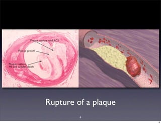
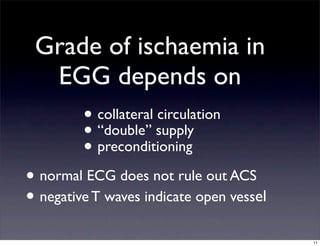
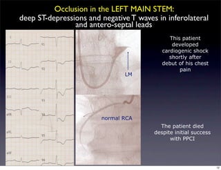
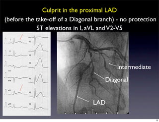
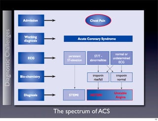
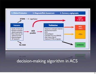
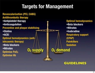
Chest pain of recent
onset and ACS
few highlights
GP - meeting at NNUH
13 September 2011
Toomas Särev
Consultant Cardiologist
NNUH-JPUH
1
2
Challenges - Chest pain + ECG & Lab
3
Concept of Supply - Demand
O2 supply O2 demand
Coronary anatomy
Diastolic BP
Heart Rate
Characteristics of
blood
O2-extraction
•Hb
•PaO2
Heart Rate
Preload
Afterload
Contractility
4
Pathophysiology Clinical Diagnosis
UNSTABLEANGINA
ASYMPTOMATIC/
SYMPTOMATIC
CHRONIC
ACUTESTEMI
Markers of myocardial injury (TnI, CK-Mb)
ECG
RISK
PLAQUE RUPTURE
INTRACORONARY
THROMBUS
DECREASED FLOW
MYOCARDIAL HYPOXIA
ISCHAEMIA IN MYOCYTES
→
5
6
Rupture of a plaque
6
The spectrum of ACS
DiagnosticChallenges
7
diagnostic challenges?
• risk assessment
• individuals without clear symptoms or ECG
features
• atypical presentations (dyspnea, syncope,
abdominal pain)
• older patients (> 75 y)
• women
• diabetes, chronic renal failure, dementia
8
How to identify high risk patients?
9
ECG - when
should you
be
concerned?
10
• collateral circulation
• “double” supply
• preconditioning
Grade of ischaemia in
EGG depends on
• normal ECG does not rule out ACS
• negative T waves indicate open vessel
11
This patient
developed
cardiogenic shock
shortly after
debut of his chest
pain
LM
normal RCA
The patient died
despite initial success
with PPCI
Occlusion in the LEFT MAIN STEM:
deep ST-depressions and negative T waves in inferolateral
and antero-septal leads
12
Culprit in the proximal LAD
(before the take-off of a Diagonal branch) - no protection
LAD
Diagonal
Intermediate
ST elevations in I, aVL andV2-V5
13
RCAa 100%
RCA POST-PTCA
Occlusion in the proximal RCA:
ST-elevaton in in II, III, aVF +V1 & V4R
14
The spectrum of ACS
DiagnosticChallenges
15
Abnormal Troponin
possible causes
• chronic or acute renal dysfunction
• severe congestive heart failure - acute and
chronic
• hypertensive crisis
• tachy- or bradyarrhythmias
• pulmonary embolism, PAH
• myocarditis
• acute neurological disease, stroke, SAH
16
ACS in the
elderly
• clinical presentation might
be different (dyspnea)
• more extensive and severe
CAD,
• more comorbidities, level of
frailty very individual
• worse prognosis
• different benefit/risk ratio
with usual therapies
• higher rate of secondary
effects and complications
© Gary Larson 2002
17
How to manage?
When to refer?
© Gary Larson 2002
18
decision-making algorithm in ACS
19
Targets for Management
O2 supply O2 demand
Revascularisation (PCI, CABG)
Antithrombotic therapy
•Antiplatelet therapy
•Anticoagulation
Preventive and plaque stabilising
•Statins
•ACEi
Optimal hemodynamics (anti-
ishcaemic therapy)
•Beta blockers
•Nitrates
Optimise PaO2
Optimise Hb
Optimal hemodynamics
•Beta blockers
•Nitrates
•Ivabradine
Respiratory support
(CPAP)
Painkillers
Sedation
GUIDELINES
20
Revascularisation
• medical therapy if no critical coronary
lesions if no options for revascularisation
• PCI with stenting of the cuprit lesion
• individualised decision in multivessel disease
• staged PCI or all at once
• PCI at first and then CABG
• CABG
21
new guidelines summary
22
Thank You!
this presentation can be
downloaded from:
www.slideshare.net/
kardiostar
comments:
kardostar@mac.com
© Gary Larson 2002
23

Acs gpmeeting-110912161748-phpapp02

  • 1.
    Chest pain ofrecent onset and ACS few highlights GP - meeting at NNUH 13 September 2011 Toomas Särev Consultant Cardiologist NNUH-JPUH 1
  • 2.
  • 3.
    Challenges - Chestpain + ECG & Lab 3
  • 4.
    Concept of Supply- Demand O2 supply O2 demand Coronary anatomy Diastolic BP Heart Rate Characteristics of blood O2-extraction •Hb •PaO2 Heart Rate Preload Afterload Contractility 4
  • 5.
    Pathophysiology Clinical Diagnosis UNSTABLEANGINA ASYMPTOMATIC/ SYMPTOMATIC CHRONIC ACUTESTEMI Markersof myocardial injury (TnI, CK-Mb) ECG RISK PLAQUE RUPTURE INTRACORONARY THROMBUS DECREASED FLOW MYOCARDIAL HYPOXIA ISCHAEMIA IN MYOCYTES → 5
  • 6.
  • 7.
    The spectrum ofACS DiagnosticChallenges 7
  • 8.
    diagnostic challenges? • riskassessment • individuals without clear symptoms or ECG features • atypical presentations (dyspnea, syncope, abdominal pain) • older patients (> 75 y) • women • diabetes, chronic renal failure, dementia 8
  • 9.
    How to identifyhigh risk patients? 9
  • 10.
    ECG - when shouldyou be concerned? 10
  • 11.
    • collateral circulation •“double” supply • preconditioning Grade of ischaemia in EGG depends on • normal ECG does not rule out ACS • negative T waves indicate open vessel 11
  • 12.
    This patient developed cardiogenic shock shortlyafter debut of his chest pain LM normal RCA The patient died despite initial success with PPCI Occlusion in the LEFT MAIN STEM: deep ST-depressions and negative T waves in inferolateral and antero-septal leads 12
  • 13.
    Culprit in theproximal LAD (before the take-off of a Diagonal branch) - no protection LAD Diagonal Intermediate ST elevations in I, aVL andV2-V5 13
  • 14.
    RCAa 100% RCA POST-PTCA Occlusionin the proximal RCA: ST-elevaton in in II, III, aVF +V1 & V4R 14
  • 15.
    The spectrum ofACS DiagnosticChallenges 15
  • 16.
    Abnormal Troponin possible causes •chronic or acute renal dysfunction • severe congestive heart failure - acute and chronic • hypertensive crisis • tachy- or bradyarrhythmias • pulmonary embolism, PAH • myocarditis • acute neurological disease, stroke, SAH 16
  • 17.
    ACS in the elderly •clinical presentation might be different (dyspnea) • more extensive and severe CAD, • more comorbidities, level of frailty very individual • worse prognosis • different benefit/risk ratio with usual therapies • higher rate of secondary effects and complications © Gary Larson 2002 17
  • 18.
    How to manage? Whento refer? © Gary Larson 2002 18
  • 19.
  • 20.
    Targets for Management O2supply O2 demand Revascularisation (PCI, CABG) Antithrombotic therapy •Antiplatelet therapy •Anticoagulation Preventive and plaque stabilising •Statins •ACEi Optimal hemodynamics (anti- ishcaemic therapy) •Beta blockers •Nitrates Optimise PaO2 Optimise Hb Optimal hemodynamics •Beta blockers •Nitrates •Ivabradine Respiratory support (CPAP) Painkillers Sedation GUIDELINES 20
  • 21.
    Revascularisation • medical therapyif no critical coronary lesions if no options for revascularisation • PCI with stenting of the cuprit lesion • individualised decision in multivessel disease • staged PCI or all at once • PCI at first and then CABG • CABG 21
  • 22.
  • 23.
    Thank You! this presentationcan be downloaded from: www.slideshare.net/ kardiostar comments: kardostar@mac.com © Gary Larson 2002 23