Crimson Publishers- Maternal Profile, Assisted Reproductive Technology, and P...CrimsonPublishers-PRM
Crimson Publishers- Maternal Profile, Assisted Reproductive Technology, and Perinatal Health Indicators by José Manuel Terán*in Perceptions in Reproductive Medicine
Crimson Publishers- Maternal Profile, Assisted Reproductive Technology, and P...CrimsonPublishers-PRM
Crimson Publishers- Maternal Profile, Assisted Reproductive Technology, and Perinatal Health Indicators by José Manuel Terán*in Perceptions in Reproductive Medicine
stillbirth based on Williams Obstetrics, 25th Edition,2019 is 35th chapter of this book
this slide is so useful for medical student$resident of gynecology in all of the world
for new slide from every part of medical please contact with me
this slide has all of figure and important text from williams book
Exposure to Toxic Environmental Agents - Resources for Healthy Children www.scribd.com/doc/254613619 - For more information, Please see Organic Edible Schoolyards & Gardening with Children www.scribd.com/doc/254613963 - Gardening with Volcanic Rock Dust www.scribd.com/doc/254613846 - Double Food Production from your School Garden with Organic Tech www.scribd.com/doc/254613765 - Free School Gardening Art Posters www.scribd.com/doc/254613694 - Increase Food Production with Companion Planting in your School Garden www.scribd.com/doc/254609890 - Healthy Foods Dramatically Improves Student Academic Success www.scribd.com/doc/254613619 - City Chickens for your Organic School Garden www.scribd.com/doc/254613553 - Huerto Ecológico, Tecnologías Sostenibles, Agricultura Organica www.scribd.com/doc/254613494 - Simple Square Foot Gardening for Schools - Teacher Guide www.scribd.com/doc/254613410 - Free Organic Gardening Publications www.scribd.com/doc/254609890 ~
Risk Factors and Pregnancy Outcome of Preterm Laboriosrjce
IOSR Journal of Nursing and health Science is ambitious to disseminate information and experience in education, practice and investigation between medicine, nursing and all the sciences involved in health care. Nursing & Health Sciences focuses on the international exchange of knowledge in nursing and health sciences. The journal publishes peer-reviewed papers on original research, education and clinical practice.
By encouraging scholars from around the world to share their knowledge and expertise, the journal aims to provide the reader with a deeper understanding of the lived experience of nursing and health sciences and the opportunity to enrich their own area of practice. The journal publishes original papers, reviews, special and general articles, case management etc.
Running head MATERNAL, INFANT AND CHILD HEALTH .docxcowinhelen
Running head: MATERNAL, INFANT AND CHILD HEALTH 1
MATERNAL, INFANT AND CHILD HEALTH 9
Maternal, infant and child health
Name
Institution
Abstract
Maternal, infant health is very essential for the progress of any country since they form the pillar of our future generations. United States has made significant strides towards securing the maternal and child health through various initiatives and programs within the country and around the globe. Despite the existence of health care initiatives to promote maternal, infant and child health, maternal and infant mortalities are still recorded on a daily basis in the U.S. Risk factors to maternal, infant and child mortalities include poor and a lack of a antenatal care attendance, unskilled birth attendants,ce and childhood illnesses. More than a quarter of every single maternal mortality is because of postpartum hemorrhaginge, for the most part after labor.
Infant mortality is another prevalent case that contributes to the worsening situation in child and maternal health, because of untimely births represent more than a quarter of infant mortalities, trailed by mortalities during births and neonatal sepsis. Maternal and child health (MCH) programs concentrate on medical problems concerning related to mothers, children, and families – such as , for example, access to suitable pre-natal and child welfare services, baby mortality mitigation initiatives, emergency medical services, prevention of injuries, infant screening, and administrations to kidschildren children with unique health care needs. The United States is working to prevent maternal deaths, infant mortalities, and child mortalities, and to reduce the prevalence of these incidences. It calls for a multidisciplinary approach in order to eliminate this issue affecting the mothers and children. Reinforcing referral systems and linkages between various levels of hospital-based patient care, and between healthcare organizations providers and the general population, must be a top needpriority.
1- (the things in red is the corrections, if its underline means this is the correct world and if its cross off means you have to delete it)
2- ( the things in yellow you have to delete it and write the topic and the purpose of the paper and I will write it for you at the end of the first paragraph).
3- Change anything about child health and just focus on mortality maternal unless there is something related to the child health so then you can mention that.
4- Scoop of the problem
5- Associated factors
6- solutions
Maternal child and infant health
Enhancing the prosperity of mothers, newborn children, and young children is a vital public health objective for the United States and the entire globe. Their prosperity dictates the strength of the people in the future and can anticipate future public wellbeing challenges for fam ...
Hospital based study on perinatal mortality in RIMS,Manipuriosrjce
IOSR Journal of Dental and Medical Sciences is one of the speciality Journal in Dental Science and Medical Science published by International Organization of Scientific Research (IOSR). The Journal publishes papers of the highest scientific merit and widest possible scope work in all areas related to medical and dental science. The Journal welcome review articles, leading medical and clinical research articles, technical notes, case reports and others.
Title: Sense of Taste
Presenter: Dr. Faiza, Assistant Professor of Physiology
Qualifications:
MBBS (Best Graduate, AIMC Lahore)
FCPS Physiology
ICMT, CHPE, DHPE (STMU)
MPH (GC University, Faisalabad)
MBA (Virtual University of Pakistan)
Learning Objectives:
Describe the structure and function of taste buds.
Describe the relationship between the taste threshold and taste index of common substances.
Explain the chemical basis and signal transduction of taste perception for each type of primary taste sensation.
Recognize different abnormalities of taste perception and their causes.
Key Topics:
Significance of Taste Sensation:
Differentiation between pleasant and harmful food
Influence on behavior
Selection of food based on metabolic needs
Receptors of Taste:
Taste buds on the tongue
Influence of sense of smell, texture of food, and pain stimulation (e.g., by pepper)
Primary and Secondary Taste Sensations:
Primary taste sensations: Sweet, Sour, Salty, Bitter, Umami
Chemical basis and signal transduction mechanisms for each taste
Taste Threshold and Index:
Taste threshold values for Sweet (sucrose), Salty (NaCl), Sour (HCl), and Bitter (Quinine)
Taste index relationship: Inversely proportional to taste threshold
Taste Blindness:
Inability to taste certain substances, particularly thiourea compounds
Example: Phenylthiocarbamide
Structure and Function of Taste Buds:
Composition: Epithelial cells, Sustentacular/Supporting cells, Taste cells, Basal cells
Features: Taste pores, Taste hairs/microvilli, and Taste nerve fibers
Location of Taste Buds:
Found in papillae of the tongue (Fungiform, Circumvallate, Foliate)
Also present on the palate, tonsillar pillars, epiglottis, and proximal esophagus
Mechanism of Taste Stimulation:
Interaction of taste substances with receptors on microvilli
Signal transduction pathways for Umami, Sweet, Bitter, Sour, and Salty tastes
Taste Sensitivity and Adaptation:
Decrease in sensitivity with age
Rapid adaptation of taste sensation
Role of Saliva in Taste:
Dissolution of tastants to reach receptors
Washing away the stimulus
Taste Preferences and Aversions:
Mechanisms behind taste preference and aversion
Influence of receptors and neural pathways
Impact of Sensory Nerve Damage:
Degeneration of taste buds if the sensory nerve fiber is cut
Abnormalities of Taste Detection:
Conditions: Ageusia, Hypogeusia, Dysgeusia (parageusia)
Causes: Nerve damage, neurological disorders, infections, poor oral hygiene, adverse drug effects, deficiencies, aging, tobacco use, altered neurotransmitter levels
Neurotransmitters and Taste Threshold:
Effects of serotonin (5-HT) and norepinephrine (NE) on taste sensitivity
Supertasters:
25% of the population with heightened sensitivity to taste, especially bitterness
Increased number of fungiform papillae
Acute scrotum is a general term referring to an emergency condition affecting the contents or the wall of the scrotum.
There are a number of conditions that present acutely, predominantly with pain and/or swelling
A careful and detailed history and examination, and in some cases, investigations allow differentiation between these diagnoses. A prompt diagnosis is essential as the patient may require urgent surgical intervention
Testicular torsion refers to twisting of the spermatic cord, causing ischaemia of the testicle.
Testicular torsion results from inadequate fixation of the testis to the tunica vaginalis producing ischemia from reduced arterial inflow and venous outflow obstruction.
The prevalence of testicular torsion in adult patients hospitalized with acute scrotal pain is approximately 25 to 50 percent
- Video recording of this lecture in English language: https://youtu.be/lK81BzxMqdo
- Video recording of this lecture in Arabic language: https://youtu.be/Ve4P0COk9OI
- Link to download the book free: https://nephrotube.blogspot.com/p/nephrotube-nephrology-books.html
- Link to NephroTube website: www.NephroTube.com
- Link to NephroTube social media accounts: https://nephrotube.blogspot.com/p/join-nephrotube-on-social-media.html
Couples presenting to the infertility clinic- Do they really have infertility...Sujoy Dasgupta
Dr Sujoy Dasgupta presented the study on "Couples presenting to the infertility clinic- Do they really have infertility? – The unexplored stories of non-consummation" in the 13th Congress of the Asia Pacific Initiative on Reproduction (ASPIRE 2024) at Manila on 24 May, 2024.
More Related Content
Similar to ACOG Management of Stillbirth 2015 OBGYN
stillbirth based on Williams Obstetrics, 25th Edition,2019 is 35th chapter of this book
this slide is so useful for medical student$resident of gynecology in all of the world
for new slide from every part of medical please contact with me
this slide has all of figure and important text from williams book
Exposure to Toxic Environmental Agents - Resources for Healthy Children www.scribd.com/doc/254613619 - For more information, Please see Organic Edible Schoolyards & Gardening with Children www.scribd.com/doc/254613963 - Gardening with Volcanic Rock Dust www.scribd.com/doc/254613846 - Double Food Production from your School Garden with Organic Tech www.scribd.com/doc/254613765 - Free School Gardening Art Posters www.scribd.com/doc/254613694 - Increase Food Production with Companion Planting in your School Garden www.scribd.com/doc/254609890 - Healthy Foods Dramatically Improves Student Academic Success www.scribd.com/doc/254613619 - City Chickens for your Organic School Garden www.scribd.com/doc/254613553 - Huerto Ecológico, Tecnologías Sostenibles, Agricultura Organica www.scribd.com/doc/254613494 - Simple Square Foot Gardening for Schools - Teacher Guide www.scribd.com/doc/254613410 - Free Organic Gardening Publications www.scribd.com/doc/254609890 ~
Risk Factors and Pregnancy Outcome of Preterm Laboriosrjce
IOSR Journal of Nursing and health Science is ambitious to disseminate information and experience in education, practice and investigation between medicine, nursing and all the sciences involved in health care. Nursing & Health Sciences focuses on the international exchange of knowledge in nursing and health sciences. The journal publishes peer-reviewed papers on original research, education and clinical practice.
By encouraging scholars from around the world to share their knowledge and expertise, the journal aims to provide the reader with a deeper understanding of the lived experience of nursing and health sciences and the opportunity to enrich their own area of practice. The journal publishes original papers, reviews, special and general articles, case management etc.
Running head MATERNAL, INFANT AND CHILD HEALTH .docxcowinhelen
Running head: MATERNAL, INFANT AND CHILD HEALTH 1
MATERNAL, INFANT AND CHILD HEALTH 9
Maternal, infant and child health
Name
Institution
Abstract
Maternal, infant health is very essential for the progress of any country since they form the pillar of our future generations. United States has made significant strides towards securing the maternal and child health through various initiatives and programs within the country and around the globe. Despite the existence of health care initiatives to promote maternal, infant and child health, maternal and infant mortalities are still recorded on a daily basis in the U.S. Risk factors to maternal, infant and child mortalities include poor and a lack of a antenatal care attendance, unskilled birth attendants,ce and childhood illnesses. More than a quarter of every single maternal mortality is because of postpartum hemorrhaginge, for the most part after labor.
Infant mortality is another prevalent case that contributes to the worsening situation in child and maternal health, because of untimely births represent more than a quarter of infant mortalities, trailed by mortalities during births and neonatal sepsis. Maternal and child health (MCH) programs concentrate on medical problems concerning related to mothers, children, and families – such as , for example, access to suitable pre-natal and child welfare services, baby mortality mitigation initiatives, emergency medical services, prevention of injuries, infant screening, and administrations to kidschildren children with unique health care needs. The United States is working to prevent maternal deaths, infant mortalities, and child mortalities, and to reduce the prevalence of these incidences. It calls for a multidisciplinary approach in order to eliminate this issue affecting the mothers and children. Reinforcing referral systems and linkages between various levels of hospital-based patient care, and between healthcare organizations providers and the general population, must be a top needpriority.
1- (the things in red is the corrections, if its underline means this is the correct world and if its cross off means you have to delete it)
2- ( the things in yellow you have to delete it and write the topic and the purpose of the paper and I will write it for you at the end of the first paragraph).
3- Change anything about child health and just focus on mortality maternal unless there is something related to the child health so then you can mention that.
4- Scoop of the problem
5- Associated factors
6- solutions
Maternal child and infant health
Enhancing the prosperity of mothers, newborn children, and young children is a vital public health objective for the United States and the entire globe. Their prosperity dictates the strength of the people in the future and can anticipate future public wellbeing challenges for fam ...
Hospital based study on perinatal mortality in RIMS,Manipuriosrjce
IOSR Journal of Dental and Medical Sciences is one of the speciality Journal in Dental Science and Medical Science published by International Organization of Scientific Research (IOSR). The Journal publishes papers of the highest scientific merit and widest possible scope work in all areas related to medical and dental science. The Journal welcome review articles, leading medical and clinical research articles, technical notes, case reports and others.
Similar to ACOG Management of Stillbirth 2015 OBGYN (18)
Title: Sense of Taste
Presenter: Dr. Faiza, Assistant Professor of Physiology
Qualifications:
MBBS (Best Graduate, AIMC Lahore)
FCPS Physiology
ICMT, CHPE, DHPE (STMU)
MPH (GC University, Faisalabad)
MBA (Virtual University of Pakistan)
Learning Objectives:
Describe the structure and function of taste buds.
Describe the relationship between the taste threshold and taste index of common substances.
Explain the chemical basis and signal transduction of taste perception for each type of primary taste sensation.
Recognize different abnormalities of taste perception and their causes.
Key Topics:
Significance of Taste Sensation:
Differentiation between pleasant and harmful food
Influence on behavior
Selection of food based on metabolic needs
Receptors of Taste:
Taste buds on the tongue
Influence of sense of smell, texture of food, and pain stimulation (e.g., by pepper)
Primary and Secondary Taste Sensations:
Primary taste sensations: Sweet, Sour, Salty, Bitter, Umami
Chemical basis and signal transduction mechanisms for each taste
Taste Threshold and Index:
Taste threshold values for Sweet (sucrose), Salty (NaCl), Sour (HCl), and Bitter (Quinine)
Taste index relationship: Inversely proportional to taste threshold
Taste Blindness:
Inability to taste certain substances, particularly thiourea compounds
Example: Phenylthiocarbamide
Structure and Function of Taste Buds:
Composition: Epithelial cells, Sustentacular/Supporting cells, Taste cells, Basal cells
Features: Taste pores, Taste hairs/microvilli, and Taste nerve fibers
Location of Taste Buds:
Found in papillae of the tongue (Fungiform, Circumvallate, Foliate)
Also present on the palate, tonsillar pillars, epiglottis, and proximal esophagus
Mechanism of Taste Stimulation:
Interaction of taste substances with receptors on microvilli
Signal transduction pathways for Umami, Sweet, Bitter, Sour, and Salty tastes
Taste Sensitivity and Adaptation:
Decrease in sensitivity with age
Rapid adaptation of taste sensation
Role of Saliva in Taste:
Dissolution of tastants to reach receptors
Washing away the stimulus
Taste Preferences and Aversions:
Mechanisms behind taste preference and aversion
Influence of receptors and neural pathways
Impact of Sensory Nerve Damage:
Degeneration of taste buds if the sensory nerve fiber is cut
Abnormalities of Taste Detection:
Conditions: Ageusia, Hypogeusia, Dysgeusia (parageusia)
Causes: Nerve damage, neurological disorders, infections, poor oral hygiene, adverse drug effects, deficiencies, aging, tobacco use, altered neurotransmitter levels
Neurotransmitters and Taste Threshold:
Effects of serotonin (5-HT) and norepinephrine (NE) on taste sensitivity
Supertasters:
25% of the population with heightened sensitivity to taste, especially bitterness
Increased number of fungiform papillae
Acute scrotum is a general term referring to an emergency condition affecting the contents or the wall of the scrotum.
There are a number of conditions that present acutely, predominantly with pain and/or swelling
A careful and detailed history and examination, and in some cases, investigations allow differentiation between these diagnoses. A prompt diagnosis is essential as the patient may require urgent surgical intervention
Testicular torsion refers to twisting of the spermatic cord, causing ischaemia of the testicle.
Testicular torsion results from inadequate fixation of the testis to the tunica vaginalis producing ischemia from reduced arterial inflow and venous outflow obstruction.
The prevalence of testicular torsion in adult patients hospitalized with acute scrotal pain is approximately 25 to 50 percent
- Video recording of this lecture in English language: https://youtu.be/lK81BzxMqdo
- Video recording of this lecture in Arabic language: https://youtu.be/Ve4P0COk9OI
- Link to download the book free: https://nephrotube.blogspot.com/p/nephrotube-nephrology-books.html
- Link to NephroTube website: www.NephroTube.com
- Link to NephroTube social media accounts: https://nephrotube.blogspot.com/p/join-nephrotube-on-social-media.html
Couples presenting to the infertility clinic- Do they really have infertility...Sujoy Dasgupta
Dr Sujoy Dasgupta presented the study on "Couples presenting to the infertility clinic- Do they really have infertility? – The unexplored stories of non-consummation" in the 13th Congress of the Asia Pacific Initiative on Reproduction (ASPIRE 2024) at Manila on 24 May, 2024.
NVBDCP.pptx Nation vector borne disease control programSapna Thakur
NVBDCP was launched in 2003-2004 . Vector-Borne Disease: Disease that results from an infection transmitted to humans and other animals by blood-feeding arthropods, such as mosquitoes, ticks, and fleas. Examples of vector-borne diseases include Dengue fever, West Nile Virus, Lyme disease, and malaria.
Lung Cancer: Artificial Intelligence, Synergetics, Complex System Analysis, S...Oleg Kshivets
RESULTS: Overall life span (LS) was 2252.1±1742.5 days and cumulative 5-year survival (5YS) reached 73.2%, 10 years – 64.8%, 20 years – 42.5%. 513 LCP lived more than 5 years (LS=3124.6±1525.6 days), 148 LCP – more than 10 years (LS=5054.4±1504.1 days).199 LCP died because of LC (LS=562.7±374.5 days). 5YS of LCP after bi/lobectomies was significantly superior in comparison with LCP after pneumonectomies (78.1% vs.63.7%, P=0.00001 by log-rank test). AT significantly improved 5YS (66.3% vs. 34.8%) (P=0.00000 by log-rank test) only for LCP with N1-2. Cox modeling displayed that 5YS of LCP significantly depended on: phase transition (PT) early-invasive LC in terms of synergetics, PT N0—N12, cell ratio factors (ratio between cancer cells- CC and blood cells subpopulations), G1-3, histology, glucose, AT, blood cell circuit, prothrombin index, heparin tolerance, recalcification time (P=0.000-0.038). Neural networks, genetic algorithm selection and bootstrap simulation revealed relationships between 5YS and PT early-invasive LC (rank=1), PT N0—N12 (rank=2), thrombocytes/CC (3), erythrocytes/CC (4), eosinophils/CC (5), healthy cells/CC (6), lymphocytes/CC (7), segmented neutrophils/CC (8), stick neutrophils/CC (9), monocytes/CC (10); leucocytes/CC (11). Correct prediction of 5YS was 100% by neural networks computing (area under ROC curve=1.0; error=0.0).
CONCLUSIONS: 5YS of LCP after radical procedures significantly depended on: 1) PT early-invasive cancer; 2) PT N0--N12; 3) cell ratio factors; 4) blood cell circuit; 5) biochemical factors; 6) hemostasis system; 7) AT; 8) LC characteristics; 9) LC cell dynamics; 10) surgery type: lobectomy/pneumonectomy; 11) anthropometric data. Optimal diagnosis and treatment strategies for LC are: 1) screening and early detection of LC; 2) availability of experienced thoracic surgeons because of complexity of radical procedures; 3) aggressive en block surgery and adequate lymph node dissection for completeness; 4) precise prediction; 5) adjuvant chemoimmunoradiotherapy for LCP with unfavorable prognosis.
263778731218 Abortion Clinic /Pills In Harare ,sisternakatoto
263778731218 Abortion Clinic /Pills In Harare ,ABORTION WOMEN’S CLINIC +27730423979 IN women clinic we believe that every woman should be able to make choices in her pregnancy. Our job is to provide compassionate care, safety,affordable and confidential services. That’s why we have won the trust from all generations of women all over the world. we use non surgical method(Abortion pills) to terminate…Dr.LISA +27730423979women Clinic is committed to providing the highest quality of obstetrical and gynecological care to women of all ages. Our dedicated staff aim to treat each patient and her health concerns with compassion and respect.Our dedicated group ABORTION WOMEN’S CLINIC +27730423979 IN women clinic we believe that every woman should be able to make choices in her pregnancy. Our job is to provide compassionate care, safety,affordable and confidential services. That’s why we have won the trust from all generations of women all over the world. we use non surgical method(Abortion pills) to terminate…Dr.LISA +27730423979women Clinic is committed to providing the highest quality of obstetrical and gynecological care to women of all ages. Our dedicated staff aim to treat each patient and her health concerns with compassion and respect.Our dedicated group of receptionists, nurses, and physicians have worked together as a teamof receptionists, nurses, and physicians have worked together as a team wwww.lisywomensclinic.co.za/
TEST BANK for Operations Management, 14th Edition by William J. Stevenson, Ve...kevinkariuki227
TEST BANK for Operations Management, 14th Edition by William J. Stevenson, Verified Chapters 1 - 19, Complete Newest Version.pdf
TEST BANK for Operations Management, 14th Edition by William J. Stevenson, Verified Chapters 1 - 19, Complete Newest Version.pdf
Recomendações da OMS sobre cuidados maternos e neonatais para uma experiência pós-natal positiva.
Em consonância com os ODS – Objetivos do Desenvolvimento Sustentável e a Estratégia Global para a Saúde das Mulheres, Crianças e Adolescentes, e aplicando uma abordagem baseada nos direitos humanos, os esforços de cuidados pós-natais devem expandir-se para além da cobertura e da simples sobrevivência, de modo a incluir cuidados de qualidade.
Estas diretrizes visam melhorar a qualidade dos cuidados pós-natais essenciais e de rotina prestados às mulheres e aos recém-nascidos, com o objetivo final de melhorar a saúde e o bem-estar materno e neonatal.
Uma “experiência pós-natal positiva” é um resultado importante para todas as mulheres que dão à luz e para os seus recém-nascidos, estabelecendo as bases para a melhoria da saúde e do bem-estar a curto e longo prazo. Uma experiência pós-natal positiva é definida como aquela em que as mulheres, pessoas que gestam, os recém-nascidos, os casais, os pais, os cuidadores e as famílias recebem informação consistente, garantia e apoio de profissionais de saúde motivados; e onde um sistema de saúde flexível e com recursos reconheça as necessidades das mulheres e dos bebês e respeite o seu contexto cultural.
Estas diretrizes consolidadas apresentam algumas recomendações novas e já bem fundamentadas sobre cuidados pós-natais de rotina para mulheres e neonatos que recebem cuidados no pós-parto em unidades de saúde ou na comunidade, independentemente dos recursos disponíveis.
É fornecido um conjunto abrangente de recomendações para cuidados durante o período puerperal, com ênfase nos cuidados essenciais que todas as mulheres e recém-nascidos devem receber, e com a devida atenção à qualidade dos cuidados; isto é, a entrega e a experiência do cuidado recebido. Estas diretrizes atualizam e ampliam as recomendações da OMS de 2014 sobre cuidados pós-natais da mãe e do recém-nascido e complementam as atuais diretrizes da OMS sobre a gestão de complicações pós-natais.
O estabelecimento da amamentação e o manejo das principais intercorrências é contemplada.
Recomendamos muito.
Vamos discutir essas recomendações no nosso curso de pós-graduação em Aleitamento no Instituto Ciclos.
Esta publicação só está disponível em inglês até o momento.
Prof. Marcus Renato de Carvalho
www.agostodourado.com
New Drug Discovery and Development .....NEHA GUPTA
The "New Drug Discovery and Development" process involves the identification, design, testing, and manufacturing of novel pharmaceutical compounds with the aim of introducing new and improved treatments for various medical conditions. This comprehensive endeavor encompasses various stages, including target identification, preclinical studies, clinical trials, regulatory approval, and post-market surveillance. It involves multidisciplinary collaboration among scientists, researchers, clinicians, regulatory experts, and pharmaceutical companies to bring innovative therapies to market and address unmet medical needs.
New Directions in Targeted Therapeutic Approaches for Older Adults With Mantl...i3 Health
i3 Health is pleased to make the speaker slides from this activity available for use as a non-accredited self-study or teaching resource.
This slide deck presented by Dr. Kami Maddocks, Professor-Clinical in the Division of Hematology and
Associate Division Director for Ambulatory Operations
The Ohio State University Comprehensive Cancer Center, will provide insight into new directions in targeted therapeutic approaches for older adults with mantle cell lymphoma.
STATEMENT OF NEED
Mantle cell lymphoma (MCL) is a rare, aggressive B-cell non-Hodgkin lymphoma (NHL) accounting for 5% to 7% of all lymphomas. Its prognosis ranges from indolent disease that does not require treatment for years to very aggressive disease, which is associated with poor survival (Silkenstedt et al, 2021). Typically, MCL is diagnosed at advanced stage and in older patients who cannot tolerate intensive therapy (NCCN, 2022). Although recent advances have slightly increased remission rates, recurrence and relapse remain very common, leading to a median overall survival between 3 and 6 years (LLS, 2021). Though there are several effective options, progress is still needed towards establishing an accepted frontline approach for MCL (Castellino et al, 2022). Treatment selection and management of MCL are complicated by the heterogeneity of prognosis, advanced age and comorbidities of patients, and lack of an established standard approach for treatment, making it vital that clinicians be familiar with the latest research and advances in this area. In this activity chaired by Michael Wang, MD, Professor in the Department of Lymphoma & Myeloma at MD Anderson Cancer Center, expert faculty will discuss prognostic factors informing treatment, the promising results of recent trials in new therapeutic approaches, and the implications of treatment resistance in therapeutic selection for MCL.
Target Audience
Hematology/oncology fellows, attending faculty, and other health care professionals involved in the treatment of patients with mantle cell lymphoma (MCL).
Learning Objectives
1.) Identify clinical and biological prognostic factors that can guide treatment decision making for older adults with MCL
2.) Evaluate emerging data on targeted therapeutic approaches for treatment-naive and relapsed/refractory MCL and their applicability to older adults
3.) Assess mechanisms of resistance to targeted therapies for MCL and their implications for treatment selection
Title: Sense of Smell
Presenter: Dr. Faiza, Assistant Professor of Physiology
Qualifications:
MBBS (Best Graduate, AIMC Lahore)
FCPS Physiology
ICMT, CHPE, DHPE (STMU)
MPH (GC University, Faisalabad)
MBA (Virtual University of Pakistan)
Learning Objectives:
Describe the primary categories of smells and the concept of odor blindness.
Explain the structure and location of the olfactory membrane and mucosa, including the types and roles of cells involved in olfaction.
Describe the pathway and mechanisms of olfactory signal transmission from the olfactory receptors to the brain.
Illustrate the biochemical cascade triggered by odorant binding to olfactory receptors, including the role of G-proteins and second messengers in generating an action potential.
Identify different types of olfactory disorders such as anosmia, hyposmia, hyperosmia, and dysosmia, including their potential causes.
Key Topics:
Olfactory Genes:
3% of the human genome accounts for olfactory genes.
400 genes for odorant receptors.
Olfactory Membrane:
Located in the superior part of the nasal cavity.
Medially: Folds downward along the superior septum.
Laterally: Folds over the superior turbinate and upper surface of the middle turbinate.
Total surface area: 5-10 square centimeters.
Olfactory Mucosa:
Olfactory Cells: Bipolar nerve cells derived from the CNS (100 million), with 4-25 olfactory cilia per cell.
Sustentacular Cells: Produce mucus and maintain ionic and molecular environment.
Basal Cells: Replace worn-out olfactory cells with an average lifespan of 1-2 months.
Bowman’s Gland: Secretes mucus.
Stimulation of Olfactory Cells:
Odorant dissolves in mucus and attaches to receptors on olfactory cilia.
Involves a cascade effect through G-proteins and second messengers, leading to depolarization and action potential generation in the olfactory nerve.
Quality of a Good Odorant:
Small (3-20 Carbon atoms), volatile, water-soluble, and lipid-soluble.
Facilitated by odorant-binding proteins in mucus.
Membrane Potential and Action Potential:
Resting membrane potential: -55mV.
Action potential frequency in the olfactory nerve increases with odorant strength.
Adaptation Towards the Sense of Smell:
Rapid adaptation within the first second, with further slow adaptation.
Psychological adaptation greater than receptor adaptation, involving feedback inhibition from the central nervous system.
Primary Sensations of Smell:
Camphoraceous, Musky, Floral, Pepperminty, Ethereal, Pungent, Putrid.
Odor Detection Threshold:
Examples: Hydrogen sulfide (0.0005 ppm), Methyl-mercaptan (0.002 ppm).
Some toxic substances are odorless at lethal concentrations.
Characteristics of Smell:
Odor blindness for single substances due to lack of appropriate receptor protein.
Behavioral and emotional influences of smell.
Transmission of Olfactory Signals:
From olfactory cells to glomeruli in the olfactory bulb, involving lateral inhibition.
Primitive, less old, and new olfactory systems with different path
Pulmonary Thromboembolism - etilogy, types, medical- Surgical and nursing man...VarunMahajani
Disruption of blood supply to lung alveoli due to blockage of one or more pulmonary blood vessels is called as Pulmonary thromboembolism. In this presentation we will discuss its causes, types and its management in depth.
Pulmonary Thromboembolism - etilogy, types, medical- Surgical and nursing man...
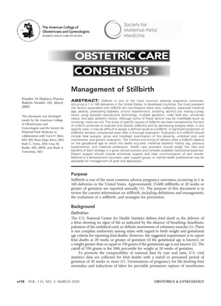
ACOG Management of Stillbirth 2015 OBGYN
1. Number 10 (Replaces Practice
Bulletin Number 102, March
2009)
This document was developed
jointly by the American College
of Obstetricians and
Gynecologists and the Society for
Maternal-Fetal Medicine in
collaboration with Torri D. Metz,
MD, MS; Rana Snipe Berry, MD;
Ruth C. Fretts, MD; Uma M.
Reddy, MD, MPH; and Mark A.
Turrentine, MD.
Management of Stillbirth
ABSTRACT: Stillbirth is one of the most common adverse pregnancy outcomes,
occurring in 1 in 160 deliveries in the United States. In developed countries, the most prevalent
risk factors associated with stillbirth are non-Hispanic black race, nulliparity, advanced maternal
age, obesity, preexisting diabetes, chronic hypertension, smoking, alcohol use, having a preg-
nancy using assisted reproductive technology, multiple gestation, male fetal sex, unmarried
status, and past obstetric history. Although some of these factors may be modifiable (such as
smoking), many are not. The study of specific causes of stillbirth has been hampered by the lack
of uniform protocols to evaluate and classify stillbirths and by decreasing autopsy rates. In any
specific case, it may be difficult to assign a definite cause to a stillbirth. A significant proportion of
stillbirths remains unexplained even after a thorough evaluation. Evaluation of a stillbirth should
include fetal autopsy; gross and histologic examination of the placenta, umbilical cord, and
membranes; and genetic evaluation. The method and timing of delivery after a stillbirth depend
on the gestational age at which the death occurred, maternal obstetric history (eg, previous
hysterotomy), and maternal preference. Health care providers should weigh the risks and
benefits of each strategy in a given clinical scenario and consider available institutional expertise.
Patient support should include emotional support and clear communication of test results.
Referral to a bereavement counselor, peer support group, or mental health professional may be
advisable for management of grief and depression.
Purpose
Stillbirth is one of the most common adverse pregnancy outcomes, occurring in 1 in
160 deliveries in the United States. Approximately 23,600 stillbirths at 20 weeks or
greater of gestation are reported annually (1). The purpose of this document is to
review the current information on stillbirth, including definitions and management,
the evaluation of a stillbirth, and strategies for prevention.
Background
Definition
The U.S. National Center for Health Statistics defines fetal death as the delivery of
a fetus showing no signs of life as indicated by the absence of breathing, heartbeats,
pulsation of the umbilical cord, or definite movements of voluntary muscles (1). There
is not complete uniformity among states with regard to birth weight and gestational
age criteria for reporting fetal deaths. However, the suggested requirement is to report
fetal deaths at 20 weeks or greater of gestation (if the gestational age is known), or
a weight greater than or equal to 350 grams if the gestational age is not known (2). The
cutoff of 350 grams is the 50th percentile for weight at 20 weeks of gestation.
To promote the comparability of national data by year and state, U.S. vital
statistics data are collected for fetal deaths with a stated or presumed period of
gestation of 20 weeks or more (1). Terminations of pregnancy for life-limiting fetal
anomalies and inductions of labor for previable premature rupture of membranes
e110 VOL. 135, NO. 3, MARCH 2020 OBSTETRICS & GYNECOLOGY
2. are specifically excluded from the stillbirth statistics and
are classified as terminations of pregnancy (1).
The term stillbirth is preferred among parent
groups, and more recent research efforts have begun
using this term in place of fetal death. Therefore, in this
document, the term stillbirth is used.
Frequency of Occurrence
In 2013, the stillbirth rate in the United States was 5.96
per 1,000 live births, a decrease from 6.61 in 2006 and
6.05 per 1,000 births in 2012 (1). Between 2006 and 2012,
the rate of early stillbirth (20–27 weeks) remained essen-
tially unchanged, but between 2012 and 2013, the rate
decreased from 3.11 to 3.01 per 1,000 births. The rate of
late stillbirth (28 weeks or greater) has been relatively
stable since 2006 and did not change significantly
between 2012 and 2013 at 2.96 and 2.97 per 1,000 births,
respectively (1). There is ongoing discussion regarding
the most useful calculation for analysis of stillbirth oc-
currences. Currently, fetal mortality rates are widely cal-
culated using a birth-based approach: the number of
stillbirths per 1,000 live births and stillbirths (1).
There may be some utility in changing the denom-
inator to better capture the population at risk, that is, all
women who are still pregnant at a given gestational age.
Using a denominator of women who are still pregnant at
a given gestational age allows for calculation of a pro-
spective fetal mortality rate defined as the number of
stillbirths at a given gestational age (in single weeks)
per 1,000 live births and stillbirths at that gestational
age or greater (3). This approach produces the prospec-
tive risk of stillbirth, which can be clinically valuable to
make predictions for individual pregnancies and to help
health care providers balance the risks of expectant man-
agement with those of intervention (1) (Fig. 1).
Risk Factors
In developed countries, the most prevalent risk factors
associated with stillbirth are non-Hispanic black race,
nulliparity, advanced maternal age, obesity, preexisting
diabetes, chronic hypertension, smoking, alcohol use, having
a pregnancy using assisted reproductive technology, multi-
ple gestation, male fetal sex, unmarried status, and past
obstetric history (4, 5). Although some of these factors may
be modifiable (such as smoking), many are not.
Social Demographic Factors Affecting Stillbirth
Race. Non-Hispanic black women have a stillbirth rate
that is more than twice the rate of other racial groups
(10.53 deaths per 1,000 livebirths and stillbirths) (1). In
the United States, the stillbirth rates for other groups
were 4.88 for non-Hispanic white women, 5.22 for His-
panic women, 6.22 for American Indian or Alaska
Native, and 4.68 for Asian or Pacific Islanders (1).
The reason for this health care disparity in stillbirth
rates is multifactorial and the subject of ongoing research
(6). Higher rates of stillbirth persist among non-Hispanic
black women with adequate prenatal care; this has been
attributed to higher rates of diabetes mellitus, hyperten-
sion, placental abruption, and premature rupture of
membranes (7, 8). The educational level for Hispanic
and non-Hispanic black women does not appear to be
protective as compared with white women, with the wid-
est disparities observed between white and non-Hispanic
Figure 1. Prospective fetal mortality rate, by single week of gestation: United States, 2013. Note: The prospective fetal mortality rate is the number
of stillbirths at a given gestational age per 1,000 live births and stillbirths at that gestational age or greater. (MacDorman MF, Gregory ECW. Fetal and
perinatal mortality: United States, 2013. National vital statistics reports; vol. 64 no. 8. Hyattsville, MD: National Center for Health Statistics. 2015.)
VOL. 135, NO. 3, MARCH 2020 Obstetric Care Consensus Management of Stillbirth e111
3. black stillbirths at 20–27 weeks of gestation, regardless of
educational attainment (9). Implicit and explicit bias and
racism are implicated in many health disparities includ-
ing perinatal morbidity and mortality (10). It remains to
be better characterized how biologic and modifiable risk
factors, including care disparities and environmental
stressors, biases, and racism further contribute to the risk
for non-Hispanic black women (11).
Multiple Gestations
The stillbirth rate among twin pregnancies is approximately
2.5 times higher than that of singletons (14.07 versus 5.65
per 1,000 live births and stillbirths) (1). The risk of stillbirth
increases in all twins with advancing gestational age, and it
is significantly greater in monochorionic as compared with
dichorionic twins (12). The stillbirth rate for triplet preg-
nancies and higher order multiples is reported as 30.53 per
1,000 live births and stillbirths. Higher rates are due to
complications specific to multiple gestation (such as
twin–twin transfusion syndrome), as well as to increased
risks of common complications such as aneuploidy, con-
genital anomalies, and growth restriction (1, 13).
Past Obstetric History
Women with a previous stillbirth are at increased risk of
recurrence. Compared with women with no history of
stillbirth, women who had a stillbirth in an index pregnancy
had an increased risk in subsequent pregnancies (pooled
odds ratio, 4.83; 95% CI, 3.77–6.18), which remained signif-
icant after adjustment for confounding factors (14).
Women with previous adverse pregnancy outcomes,
such as preterm delivery, growth restriction, or pre-
eclampsia, are at increased risk of stillbirth in subsequent
pregnancies (15). The relationship between previous
adverse pregnancy outcomes and stillbirth is strongest
in the case of explained stillbirth. However, there re-
mains a persistent 1.7-fold to 2-fold increase in unex-
plained stillbirth associated with a history of adverse
pregnancy outcomes. In a study that examined previous
preterm and small for gestational age (SGA) births and
the risk of stillbirth in a subsequent pregnancy, the risk
of stillbirth was increased in the setting of a prior SGA
infant; the highest risk was for a prior SGA infant born at
less than 32 weeks (OR, 8.0; 95% CI, 4.7–13.7) (16).
The relationship between previous cesarean delivery
and subsequent stillbirth remains controversial. In two
large studies from the United Kingdom, previous cesarean
delivery was associated with an increased rate of explained
(17) and unexplained stillbirth (15) with an adjusted hazard
ratio ranging from 2.08 (95% CI, 1.00–4.31) and 1.75 (95%
CI, 1.30–2.37), respectively, for all causes of subsequent
stillbirth. A Danish analysis showed a slight increase in
the rate of stillbirth after cesarean in explained and unex-
plained stillbirths, but neither reached statistical significance
(18). In addition, three large observational studies from the
United States (19–21) and one from Canada (22) found no
association between history of cesarean and stillbirth. In the
largest of these studies, the unexplained stillbirth rates at
term for women with and without a previous cesarean
delivery were 0.8 and 0.7 per 1,000 births, respectively (rel-
ative risk [RR] 0.90; 95% CI, 0.76–1.06) (20).
The extremes of parity have also been associated
with stillbirth. Higher rates of stillbirth are observed in
nulliparous women as well as multiparas women with
greater than three previous pregnancies when compared
to women with one or two previous births (23).
Male sex
Male sex of the fetus has been observed as a risk factor
for stillbirth. In a recent review of data from more than
30 million births, in a wide range of high-income and
low-income countries, the crude mean rate (stillbirths per
1,000 total births) was 6.23 for males and 5.74 for females.
The pooled RR was 1.10 (95% CI, 1.07–1.13), which in-
dicates that a male fetus has approximately a 10% higher
risk for stillbirth (24). Although this meta-analysis identi-
fies fetal sex as an important risk factor for stillbirth, the
reason why males are at higher risk is unknown.
Younger and Older Maternal Age
Maternal age at either end of the reproductive age spectrum
(less than 15 years and greater than 35 years) is an
independent risk factor for stillbirth. Maternal age greater
than or equal to 35 years of age is associated with an
increased risk of stillbirth in nulliparous and multiparous
women (25, 26). A significant proportion of perinatal deaths
seen in older women are related to lethal congenital and
chromosomal anomalies. The introduction of population-
based screening for chromosomal abnormalities has con-
tributed to lower rates of explained stillbirth or neonatal
death resulting from chromosomal abnormalities (27). Large
observational studies demonstrate that advanced maternal
age is an independent risk factor for stillbirth even after
controlling for risk factors such as hypertension, diabetes,
placenta previa, and multiple gestation (26, 28, 29). In addi-
tion, there appears to be an interaction between first birth
and increasing maternal age that places nulliparous older
women at higher risk (27). Based on one study, the esti-
mated risk of stillbirth is 1 in 116 in a 40-year-old nullipa-
rous woman after 37 weeks of gestation, compared with 1 in
304 in a multiparous woman of the same age (27).
The stillbirth rate for teenagers younger than 15
years of age is 15.88 per 1,000 live births. This is nearly
three times the rate of the lowest risk group, aged 25–29
years, with a rate of 5.34 per 1,000 live births. The rate
for teenagers aged 15–17 years was 7.03 per 1,000, and
the rate for 18–19-year olds was 6.52 per 1,000 live
births. These were 32% and 22% higher than the lowest
risk age group (1). This bimodal peak at extremes of
reproductive age has been observed in several studies
as well as confirmed in a large population-based cohort
study using the Centers for Disease Control and Preven-
tion’s “Linked Birth-Infant Death” and “Fetal Death”
data files of 37,504,230 births (30).
e112 Obstetric Care Consensus Management of Stillbirth OBSTETRICS & GYNECOLOGY
4. Comorbid Medical Conditions
Many maternal medical conditions are associated with
an increased risk of stillbirth (Table 1). Hypertension
and diabetes are two of the most common comorbid
pregnancy conditions (4, 31). Population-based studies
demonstrated almost a twofold to fivefold increase in the
risk of stillbirth among women with pregestational dia-
betes and gestational diabetes (4, 32–34). There appears
to be a joint effect of pregestational diabetes and obesity
that is stronger than the individual effects of each risk
factor (35). However, with prepregnancy strict glycemic
control aiming for HgbA1C values less than 7% and
maintenance of maternal euglycemia during pregnancy,
the risk of stillbirth may be reduced (36, 37). The peri-
natal mortality rate reported with maternal chronic
hypertension is 2–4 times higher than that of the general
population (38), and the increased risk of stillbirth or
neonatal death appears to be independent of other pos-
sible contributors such as superimposed preeclampsia or
fetal growth restriction. The precise blood pressure level
at which antihypertensive therapy is indicated during
pregnancy in women with chronic hypertension contin-
ues to be debated; similarly, it is unknown if strict blood
pressure control reduces the risk of stillbirth (38). There
also appears to be interaction between chronic hyperten-
sion and pregestational diabetes on having a stillbirth
and in women with both comorbidities, an even higher
risk has been reported (39).
Numerous other medical conditions including sys-
temic lupus erythematosus, renal disease, uncontrolled
thyroid disease, and cholestasis of pregnancy have been
associated with stillbirth (Table 1). For guidance regard-
ing antenatal fetal surveillance based on anticipated risk
of stillbirth, refer to ACOG Practice Bulletin No. 145,
Antepartum Fetal Surveillance.
Acquired and Inherited Thrombophilias
Antiphospholipid syndrome (APS) is an acquired throm-
bophilia that has been associated with stillbirth. The
diagnosis of APS depends on women meeting laboratory
and clinical criteria for the disorder. One of the clinical
criteria for APS is history of stillbirth. As such, women
with a stillbirth are typically tested for APS (see ACOG
Practice Bulletin No. 132, Antiphospholipid Syndrome,
for details of testing and management). In contrast, in-
herited thrombophilias have not been associated with
stillbirth, and testing for them as part of a stillbirth eval-
uation is not recommended (40) (Table 2).
Obesity and Gestational Weight Gain
Obesity is defined as a prepregnancy BMI (defined as
weight in kilograms divided by height in meters squared)
of 30 or greater and is the fastest growing health problem
in the United States (41). Obesity in pregnancy is asso-
ciated with an increased risk of early fetal loss and
Table 1. Estimated Rate of Stillbirth With Maternal or
Fetal Conditions
Condition
Estimated Rate of
Stillbirth*
All pregnancies 6.4/1000
Diabetes
Treated with diet (A1) 6–10/1000
Treated with insulin 6–35/1000
Hypertensive disorder
Chronic hypertension 6–25/1000
Preeclampsia
without severe features 9–51/1000
with severe features 12–29/1000
Growth restricted fetus 10–47/1000
Multiple gestation
Twins 12/1000
Triplets 34/1000
Oligohydramnios 14/1000
Late term pregnancy (greater than 41 weeks) 14–40/1000†
Previous stillbirth 9–20/1000
Decreased fetal movement 13/1000
Systemic lupus erythematosus 40–150/1000
Renal disease 15–200/1000
Cholestasis of pregnancy 12–30/1000
Advanced maternal age
35–39 years 11–14/1000
40 years or greater 11–21/1000
Black maternal race 12–14/1000
Maternal age less than 20 years 7–13/1000
Assisted reproductive technology 12/1000
Obesity (prepregnancy)
BMI equal to or greater than 30 kg/m2 13–18/1000
Smoking greater than 10 cigarettes per day 10–15/1000
*Rate per 1,000 live births and stillbirths
†
Data from Rosenstein MG, Snowden JM, Cheng YW, Caughey AB. The mortality
risk of expectant management compared with delivery stratified by gestational age
and race and ethnicity. Am J Obstet Gynecol 2014;211:660.e1–8.
Adapted from Signore C, Freeman RK, Spong CY. Antenatal testing—a reevaluation:
executive summary of a Eunice Kennedy Shriver National Institute of Child Health
and Human Development workshop. Obstet Gynecol 2009;113:687–701 and Fretts
RC. Etiology and prevention of stillbirth. Am J Obstet Gynecol 2005;193:1923–35.
VOL. 135, NO. 3, MARCH 2020 Obstetric Care Consensus Management of Stillbirth e113
5. stillbirth (42). A comprehensive study of five high-
income countries found that maternal overweight and
obesity (BMI greater than 25) was the most common
modifiable risk factor for stillbirth (43). A meta-
analysis of 38 studies that included 16,274 stillbirths
found that even small increases in maternal BMI were
associated with an increased risk of stillbirth. For BMI
levels of 20, 25, and 30, absolute risks per 1,000 preg-
nancies were 4.0 (reference standard), 4.8 (95% CI, 46–
51), and 5.9 (95% CI, 55–63), respectively (44). Further,
Table 2. Stillbirth Recommendations
Recommendation Grade of Recommendation
Inherited thrombophilias have not been associated with stillbirth, and testing for
them as part of a stillbirth evaluation is not recommended.
1C
Strong recommendation, low-quality evidence
In women who decline invasive testing, a portion of the placenta, an umbilical
cord segment, or internal fetal tissue can be sent for genetic analysis.
1B
Strong recommendation, moderate-quality evidence
Microarray analysis, incorporated into the stillbirth workup, improves the test
success rate and the detection of genetic anomalies compared with conventional
karyotyping.
1A
Strong recommendation, high-quality evidence
Genetic evaluation for specific abnormalities should be guided by the clinical
history and detected fetal abnormalities.
1C
Strong recommendation, low-quality evidence
Evaluation of a stillbirth should include fetal autopsy; gross and histologic
examination of the placenta, umbilical cord, and membranes; and genetic
evaluation.
1A
Strong recommendation, high-quality evidence
Gross and microscopic examination of the placenta, umbilical cord, and fetal
membranes by a trained pathologist is the single most useful aspect of the
evaluation of stillbirth and is an essential component of the evaluation.
1A
Strong recommendation, high-quality evidence
The general examination of the stillborn fetus should be done promptly, noting
any dysmorphic features and obtaining measurements of weight, length, and
head circumference.
1C
Strong recommendation, low-quality evidence
Fetal autopsy should be offered because it is one of the most useful diagnostic
tests in determining the cause of death.
1A
Strong recommendation, high-quality evidence
Genetic analyses are of sufficient yield that they should be performed in all
cases of stillbirth after appropriate parental permission is obtained.
1A
Strong recommendation, high-quality evidence
Appropriate history and physical findings should be included in the requisition
sent to the laboratory to assist the laboratory personnel to interpret cytogenetic tests.
Best practice
A thorough maternal history should be taken to look for known conditions or
symptoms suggestive of those that have been associated with stillbirth.
Best practice
Health care providers should weigh the risks and benefits of each strategy in
a given clinical scenario and consider available institutional expertise. Shared
decision-making plays an important role in determining the optimal method for
delivery in the setting of fetal demise.
Best practice
The results of the autopsy, placental examination, laboratory tests, and
cytogenetic studies should be communicated to the involved clinicians and to the
family of the deceased infant in a timely manner.
Best practice
Bereavement care should be individualized to recognize bereaved parents’
personal, cultural or religious needs.
Best practice
For patients with a previous stillbirth at or after 32 0/7 weeks, once or twice weekly
antenatal surveillance is recommended at 32 0/7 weeks or starting at 1–2 weeks
before the gestational age of the previous stillbirth. For stillbirth that occurred before
32 0/7 weeks of gestation, individualized timing of antenatal surveillance may be
considered.
2C
Weak recommendation, low-quality evidence
e114 Obstetric Care Consensus Management of Stillbirth OBSTETRICS & GYNECOLOGY
6. excessive weight gain was associated with higher risk of
stillbirth among obese and morbidly obese women (45).
There is some evidence that the obesity-related stillbirth
risk increases with gestational age. In one study, the
hazard ratio for stillbirth increased from 2.1 at 28–36
weeks to 4.6 at 40 weeks of gestation (46). The reason
for this association is likely multifactorial, but obesity is
associated with a fivefold increased risk of stillbirth re-
sulting from placental dysfunction. Obesity remains an
independent risk factor for stillbirth even after control-
ling for smoking, gestational diabetes, and preeclampsia
(47–49); however, the optimal BMI to minimize stillbirth
risk remains unknown (44).
Substance Use
Maternal cocaine, methamphetamine, other illicit drug
use, and smoking tobacco, are all significant contributors
to abruption and stillbirth (50–54). In a secondary anal-
ysis of a case–control study from the Eunice Kennedy
Shriver National Institute of Child Health and Human
Development Stillbirth Collaborative Research Network,
any illicit drug use as detected by biological sampling of
the umbilical cord homogenate was associated with an
increased risk of stillbirth (OR, 1.94; 95% CI, 1.16–3.27)
(55). Smoking is a particularly common risk factor,
especially and increasingly in high-income countries. In
a recent large systematic review, smoking during preg-
nancy was significantly associated with a 47% increase in
the odds of stillbirth (OR, 1.47; 95% CI, 1.37–1.57,
P,.0001) (56). The causal relationship of smoking and
stillbirth has been established through many studies that
demonstrated differential effects based on timing and
amount of tobacco exposure.
Exposure to secondhand smoke also increases risk.
Women with exposure to secondhand smoke were also
at higher risk of stillbirth than never smokers with
lower or no secondhand exposure and had comparable
risks to some active smokers (57). Timing of exposure
is also relevant; smoking during the first trimester is
associated with increased risk of stillbirth (adjusted
hazard ratio, 2.4; 95% CI, 1.2–4.9) (58). There is also
a clear dose-response effect of maternal smoking in
pregnancy on risk of stillbirth. Smoking one to nine
cigarettes per day was associated with a 9% increased
odds of having a stillbirth compared with women who
do not smoke in pregnancy (OR, 1.09, 95% CI, 1.09–
1.24, P5.55, six studies), and smoking 10 or more
cigarettes per day was associated with a 52% increase
in odds of stillbirth (OR, 1.52; 95% CI, 1.30–1.78,
P,.0001, seven studies) (56). Quitting smoking
between pregnancies is protective. Women who
smoked during the first pregnancy but not during
the second do not have an increased risk of recurrent
stillbirth (OR, 1.02; 95% CI, 0.79–1.30), compared
with woman who did not smoke in either pregnancy.
The risk among women who smoked during both
pregnancies was 1.35 (95% CI, 1.15–1.58) (59).
Assisted Reproductive Technology
Pregnancies achieved by in vitro fertilization (IVF)
appear to be associated with an elevated risk (twofold
to threefold increase) of stillbirth even after controlling
for age, parity, and multifetal gestations (60–63). A more
recent study from California for the years 2009–2011
confirms that the stillbirth risk is elevated at 5.5 per
1,000 (64). Whether this is related to the procedures
themselves or to unmeasured confounding variables
associated with underlying causes of infertility is less
clear. Couples with a waiting time to pregnancy of 1 year
or more and women who became pregnant after non-
IVF assisted reproductive technology had a risk for still-
birth similar to that of fertile couples and a lower risk
than women who became pregnant after IVF or intra-
cytoplasmic sperm injection, which indicates that the
increased rate of stillbirth risk may be a result of the
IVF or intracytoplasmic sperm injection and not the
underlying infertility (62).
Late-Term and Postterm Pregnancies
In a Cochrane review of 30 RCTs of 12,479 women that
compared expectant management with induction of
labor in term and postterm pregnancies, induction of
labor was associated with a decreased risk of perinatal
death and cesarean delivery (65). Based on these and
other observational data, induction of labor for an indi-
cation of late-term and postterm pregnancy is recom-
mended after 42 0/7 weeks of gestation and can be
considered at or after 41 weeks 0/7 days of gestation
(66). Estimates of the risk of stillbirth after 41 weeks
differ by race and ethnicity and range from 14–40 per
1,000 live births (67). For the California population
overall from 1997–2006, mortality risks of stillbirth
and neonatal death were equivalent at 38 weeks of ges-
tation, but at later gestational ages the mortality risk of
expectant management exceeded that of delivery with
a mortality risk of 17.6 per 10,000 compared with 10.8
per 10,000 ongoing pregnancies at 42 weeks of gestation
(68). The RR of stillbirth in this cohort was 2.9 (95% CI,
2.6–3.2) at 41 weeks and 5.1 (95% CI, 4.4–6.0) at 42
weeks, when compared with a referent stillbirth rate at
37 weeks (68).
Potential Causes of Stillbirth
The study of specific causes of stillbirth has been
hampered by the lack of uniform protocols to evaluate
and classify stillbirths and by decreasing autopsy rates. In
most cases, stillbirth certificates are filled out before a full
postnatal investigation has been completed and amended
death certificates are rarely filed when additional infor-
mation from the stillbirth evaluation emerges. In any
specific case, it may be difficult to assign a definite cause
to a stillbirth. A significant proportion of stillbirths
remains unexplained even after a thorough evaluation
(69).
VOL. 135, NO. 3, MARCH 2020 Obstetric Care Consensus Management of Stillbirth e115
7. Fetal Growth Restriction
Fetal growth restriction is associated with a significant
increase in the risk of stillbirth. The most severely
affected fetuses (weight less than the 2.5th percentile)
are at greatest risk (70, 71). The cumulative risk of still-
birth is approximately 1.5% at fetal weights less than the
10th percentile, and the risk increases to 2.5% at less than
the 5th percentile for gestational age (72, 73). Similarly,
using data from all births in the United States, investi-
gators demonstrated increased risk of stillbirth with
increasing severity of growth restriction. The risk of still-
birth was highest among fetuses estimated to be less than
the 3rd percentile for growth (58.0 per 10,000 at risk),
decreased for those less than the 5th percentile (43.9 per
10,000 at risk) and was the lowest for those less than the
10th percentile (26.3 per 10,000 at risk) (71). Fetal
growth restriction is associated with some fetal aneuploi-
dies, fetal infection, maternal smoking, hypertension,
autoimmune disease, obesity, and diabetes, which also
modify the risk of stillbirth.
Placental Abruption
Placental abruption is identified as the cause of stillbirth
in 5–10% of cases (69). Maternal cocaine and other
illicit drug use, and smoking tobacco, are all significant
contributors to abruption and stillbirth (50–53). If
abruption occurs in the preterm fetus or involves
a larger surface area of the placenta (74), it is more
likely to cause stillbirth. The rates of abruption appear
to be increasing (75). Hemodynamically significant fe-
tomaternal hemorrhage in the absence of placental
abruption is a rare cause of stillbirth and occurs mainly
in unusual scenarios, such as chorioangioma or chorio-
carcinoma (76, 77).
Chromosomal and Genetic Abnormalities
An abnormal karyotype can be found in approximately
6–13% of stillbirths (69, 78–80). The rate of karyotypic
abnormalities exceeds 20% in fetuses with anatomic
abnormalities or in those with growth restriction, but
the rate of chromosomal anomalies found in normally
formed fetuses was found to be 4.6% in one large series
(80). If an abnormal karyotype is found in association
with stillbirth, the most common abnormalities are tri-
somy 21 (31%), monosomy X (22%), trisomy 18 (22%),
and trisomy 13 (8%) (80).
Karyotypic analysis underestimates the contribution
of genetic abnormalities to stillbirth because in up to 50%
of karyotype attempts, cell culture is unsuccessful (79).
One strategy to increase the yield of cell culture is to
perform chorionic villi sampling or amniocentesis before
the delivery. In a large study in the Netherlands, invasive
testing had a much greater tissue culture rate (85%) than
fetal tissue sampling after birth (28%) (80). In women
who decline invasive testing, a portion of the placenta, an
umbilical cord segment, or internal fetal tissue can be
sent for genetic analysis (Fig. 2).
Microarray analysis not only detects aneuploidy but
also detects copy number variants (smaller deletions and
duplications) that are not detectable by karyotype. As
compared to karyotype analysis, microarray analysis
increased the diagnosis of a genetic cause to 41.9% in
all stillbirths, 34.5% in antepartum stillbirths, and 53.8%
in stillbirths with anomalies (81). Microarray analysis
was more likely than karyotype analysis to provide
a genetic diagnosis, primarily because of its success with
nonviable tissue, and it was especially valuable in analy-
ses of stillbirths with congenital anomalies or when kar-
yotype results could not be obtained. Thus, microarray
analysis, incorporated into the stillbirth workup, im-
proves the test success rate and the detection of genetic
anomalies compared with conventional karyotyping (82).
Microarrray is the preferred method of evaluation for
these reasons but, due to cost and logistic concerns, kar-
yotype may be the only method readily available for
some patients. In the future, whole exome sequencing
or whole genome sequencing may be part of the stillbirth
workup, but it is not currently part of the standard
evaluation.
Confined placental mosaicism in which the karyo-
type of the fetus is euploid despite an abnormal cell line
in the placenta also has been associated with an
increased risk of stillbirth, but currently it is not part
of standard testing (83). Autosomal dominant disorders
caused by spontaneous mutations (eg, skeletal dyspla-
sias) or inherited parental mutations leading to long QT
syndrome may contribute to stillbirth (84, 85). How-
ever, routine assessments for single gene defects and
microdeletions currently are limited because it is
unlikely that any single gene defect will be responsible
for a significant proportion of stillbirths. Genetic eval-
uation for specific abnormalities should be guided by
the clinical history and detected fetal abnormalities.
Approximately 20% of stillborn fetuses have dysmor-
phic features or skeletal abnormalities and 15–20% have
a major malformation (78, 86).
Infection
Infection is associated with approximately 10–20% of
stillbirths in developed countries and a greater percent-
age in developing countries (69, 87). In developed coun-
tries, infection accounts for a greater percentage of
preterm stillbirths than of term stillbirths (69, 88). Infec-
tious pathogens may result in stillbirth by producing
direct fetal infection, placental dysfunction, severe mater-
nal illness, or by stimulating spontaneous preterm birth.
Placental and fetal infections originate from either
ascending (eg, group B streptococcus or Escherichia coli)
or hematogenous spread of agents such as Listeria mono-
cytogenes or syphilis. Viral infections associated with
stillbirth include cytomegalovirus, parvovirus, and Zika.
Serology for toxoplasmosis, rubella, cytomegalovirus,
and herpes simplex virus are not included because they
are of unproven benefit and not recommended (89).
e116 Obstetric Care Consensus Management of Stillbirth OBSTETRICS & GYNECOLOGY
8. Umbilical Cord Events
Umbilical cord abnormalities account for approximately
10% of stillbirths but this diagnosis should be made with
caution (69). In a cohort–control study of almost 14,000
deliveries, single nuchal cords were present at birth in
23.6% of deliveries and multiple nuchal cords in 3.7%.
Single or multiple nuchal cords were not associated with
an increased risk of stillbirth in this cohort (90). The
criteria for considering a cord abnormality to be a cause
of death were rigorous in the Stillbirth Collaborative
Research Network and included vasa previa, cord
entrapment, and evidence of occlusion and fetal hypoxia,
prolapse, or stricture with thrombi (69). Nuchal cord
alone was not considered a cause of death. In addition,
other causes of stillbirth should be excluded.
Clinical Considerations and Management
► What are the essential components of a stillbirth
evaluation?
Evaluation of a stillbirth should include fetal autopsy;
gross and histologic examination of the placenta, umbil-
ical cord, and membranes; and genetic evaluation (91).
An algorithm for evaluation is provided in Figure 2. Spe-
cific aspects of the evaluation are outlined as follows and
in Figure 3.
Figure 2. Fetal and placental evaluation
VOL. 135, NO. 3, MARCH 2020 Obstetric Care Consensus Management of Stillbirth e117
9. Examination of the Placenta
Gross and microscopic examination of the placenta,
umbilical cord, and fetal membranes by a trained
pathologist is the single most useful aspect of the
evaluation of stillbirth and is an essential component of
the evaluation (91, 92). Gross evaluation may reveal con-
ditions such as abruption, umbilical cord thrombosis,
velamentous cord insertion, and vasa previa. Placental
evaluation may also provide information regarding infec-
tion, genetic abnormalities, and anemia. Examination of
the placental vasculature and membranes can be partic-
ularly revealing in stillbirths that occur as part of a multi-
fetal gestation. Chorionicity should be established and
vascular anastomoses identified.
Umbilical cord knots or tangling should be noted
but interpreted with caution, as cord entanglement
occurs in approximately 25% of normal pregnancies
and most true knots are found after live births.
Corroborating evidence should be sought before con-
cluding that a cord accident is the likely cause of death
(eg, evidence of cord occlusion and hypoxia on perinatal
postmortem examination and histologic examination of
the placenta and umbilical cord). The minimal histologic
criteria for considering a diagnosis of cord accident
should include vascular ectasia and thrombosis in the
umbilical cord, chorionic plate, and stem villi. In
addition to the previous findings, for a probable diag-
nosis, a regional distribution of avascular villi or villi
showing stromal karyorrhexis is suggested (93).
Examination of the Stillborn Fetus
The general examination of the stillborn fetus should be
done promptly, noting any dysmorphic features and
obtaining measurements of weight, length, and head
circumference (94–96). Foot length may be especially
useful before 23 weeks of gestation to ascertain gesta-
tional age. Photographs of the whole body (unclothed);
frontal and profile views of the face, extremities, and
palms; and close-up photographs of specific abnormali-
ties are vital for subsequent review and consultation with
a specialist, particularly if no geneticist is available at the
institution.
Fetal autopsy should be offered because it is one of
the most useful diagnostic tests in determining the cause
of death. The yield is increased when dysmorphic
features, inconsistent growth measurements, anomalies,
hydrops, or growth restriction are present. If families are
uncomfortable with a complete autopsy, other options
such as partial autopsy, gross examination by a trained
pathologist, ultrasonography and especially magnetic
resonance imaging are particularly useful. Parents should
be given the opportunity to hold the baby and perform
cultural or religious activities before the autopsy. Whole-
body X-ray with anterior–posterior and lateral views may
reveal an unrecognized skeletal abnormality or further
define a grossly apparent deformity.
When a full autopsy is performed, it should follow
published guidelines and protocols for perinatal autopsy
(97, 98). These include measurements to establish
Figure 3. Evaluation of stillbirth based on test utility in a variety of clinical scenarios. (Adapted from Page JM, Christiansen-Lindquist L, Thorsten
V, Parker CB, Reddy UM, Dudley DJ, et al. Diagnostic Tests for Evaluation of Stillbirth: Results From the Stillbirth Collaborative Research
Network. Obstet Gynecol 2017;129:699–706.)
e118 Obstetric Care Consensus Management of Stillbirth OBSTETRICS & GYNECOLOGY
10. gestational age, such as foot length and body weight.
Recommendations also include an estimation of the
interval between death and delivery, identification of
intrinsic abnormalities and developmental disorders,
and investigation for evidence of infection. It is prefera-
ble to use a pathologist who is experienced in perinatal
autopsy and to have a physician who is experienced in
genetics and dysmorphology examine the fetus. The cli-
nician should communicate the obstetric and pertinent
medical history to the pathology team and request any
tissue collection that may be needed for additional
analysis.
Fetal Laboratory Studies
Genetic analyses are of sufficient yield that they should
be performed in all cases of stillbirth after appropriate
parental permission is obtained (80). Karyotype or mi-
croarray are of higher yield if the fetus displays dysmor-
phic features, inconsistent growth measurements,
anomalies, hydrops, or growth restriction (81). Compar-
ative genomic hybridization or single nucleotide probe
and copy number probe microarrays provide almost the
same information as karyotype plus they detect abnor-
malities in smaller regions of chromosomes that are
missed by traditional karyotyping. Single nucleotide
probe arrays also can detect uniparental disomy and
consanguinity. Fetal karyotype is important if a parent
carries a balanced chromosomal rearrangement (eg,
translocation or inversion) or has a mosaic karyotype.
Acceptable cytogenetic specimens include amniotic
fluid and a placental block taken from below the cord
insertion site that includes the chorionic plate, an
umbilical cord segment, or an internal fetal tissue
specimen that thrives under low-oxygen tension such
as costochondral or patellar tissue. Fetal skin is sub-
optimal (see Fig. 1) (99–101). Amniocentesis for fetal
karyotyping has the highest yield and is particularly valu-
able if delivery is not expected imminently (80). Appro-
priate history and physical findings should be included in
the requisition sent to the laboratory to assist the labo-
ratory personnel to interpret cytogenetic tests. Cost of
various genetic analyses may affect patient decision mak-
ing at the time of stillbirth evaluation, and efforts should
be made to communicate information about anticipated
cost whenever possible.
Maternal Evaluation
A thorough maternal history should be taken to look for
known conditions or symptoms suggestive of those that
have been associated with stillbirth (Table 3). In addition
to the medical and obstetric history, including exposures
(eg, medications and viral infections), a family history
with a three-generation pedigree including stillborn in-
fants should be reviewed. Any pertinent information in
the maternal or paternal pedigree should be documented
and investigated further. Recurrent pregnancy losses and
the presence of live born individuals with developmental
delay or structural anomalies may be clues to single-gene
disorders. Consanguinity should be identified because of
the increased possibility of severe autosomal recessive
disorders. A detailed history of arrhythmias and sudden
death (including sudden infant death syndrome) should
be ascertained, because prolonged QT syndrome may be
associated with stillbirth.
Relevant original medical records and documenta-
tion should be obtained whenever possible. The gesta-
tional age by last menstrual period, maternal
examinations, laboratory data, and ultrasound examina-
tion should be recorded for correlation with the physical
examination of the neonate. Possible nongenetic causes,
such as infection, placental abruption, and umbilical cord
abnormality also should be considered.
Although fetomaternal hemorrhage is an uncom-
mon cause of stillbirth, Kleihaur-Betke testing could be
falsely elevated after delivery; therefore, testing for
significant fetomaternal hemorrhage either with a Klei-
haur-Betke or flow cytometry test should be conducted
as soon as possible after the diagnosis of stillbirth (102).
Antiphospholipid syndrome testing is recommended
in many stillbirths, especially when accompanied by fetal
growth restriction, severe preeclampsia, or other evidence
of placental insufficiency. Laboratory testing is performed
by testing for lupus anticoagulant as well as immuno-
globulin G and immunoglobulin M for both anticardio-
lipin and b2-glycoprotein antibodies. A moderate to high
immunoglobulin G phospholipid or immunoglobulin M
phospholipid titer (greater than 40 immunoglobulin M
phospholipid or immunoglobulin G phospholipid, or
greater than 99th percentile) is considered positive but
must be confirmed with repeat testing after 12 weeks.
Elevated levels of anticardiolipin and anti-b2-glycopro-
tein-I antibodies are associated with a threefold to fivefold
increased odds of stillbirth, which supports testing for
antiphospholipid antibodies in cases of otherwise unex-
plained stillbirth (103). However, testing for inherited
thrombophilias is not recommended (40).
The percentage of cases in which the various
components of the stillbirth evaluation were considered
useful to establish a cause of stillbirth in the Stillbirth
Collaborative Research Network study of 512 stillbirths
that underwent a complete evaluation was as follows:
64.6% placental pathology (95% CI, 57.9–72.0), 42.4%
fetal autopsy (95% CI, 36.9–48.4), 11.9% genetic testing
by karyotype or microarray (95% CI, 9.1–15.3), 11.1%
testing for antiphospholipid antibodies (95% CI, 8.4–
14.4), 6.4% fetal–maternal hemorrhage (95% CI, 4.4–
9.1), 1.6% glucose screen (95% CI, 0.7–3.1), 0.4% par-
vovirus (95% CI, 0.0–1.4), and 0.2% syphilis (95% CI,
0.0–1.1). The utility of the tests varied by clinical pre-
sentation, which suggests a customized approach for
each patient. The most useful tests were placental
pathology and fetal autopsy followed by genetic testing
and testing for antiphospholipid antibodies. Further
testing is indicated based on the results of the
VOL. 135, NO. 3, MARCH 2020 Obstetric Care Consensus Management of Stillbirth e119
11. Table 3. Elements of a Stillbirth Evaluation
Key Components Details Comments
Patient History Family history
Recurrent spontaneous abortions
Venous thromboembolism
Congenital anomaly or chromosomal abnormalities
Hereditary condition or syndrome
Developmental delay
Consanguinity
Maternal history
Previous venous thromboembolism
Diabetes mellitus
Chronic hypertension
Thrombophilia
Systemic lupus erythematosus
Autoimmune disease
Epilepsy
Severe anemia
Heart disease
Tobacco, alcohol, drug or medication use
Obstetric history
Recurrent miscarriages
Previous child with anomaly, hereditary condition,
or growth restriction
Previous gestational hypertension or preeclampsia
Previous gestational diabetes mellitus
Previous placental abruption
Previous fetal demise
Current pregnancy
Maternal age
Gestational age at stillbirth
Medical conditions complicating pregnancy
B Cholestasis
Pregnancy weight gain and body mass index
(continued)
e120 Obstetric Care Consensus Management of Stillbirth OBSTETRICS GYNECOLOGY
12. Table 3. Elements of a Stillbirth Evaluation (continued)
Key Components Details Comments
Complications of multifetal gestation, such as twin–twin
transfusion syndrome, twin reversed arterial perfusion
syndrome, and discordant growth
Placental abruption
Abdominal trauma
Preterm labor or rupture of membranes
Gestational age at onset of prenatal care
Abnormalities seen on an ultrasound image
Infections or chorioamnionitis
Fetal autopsy If patient declines, external evaluation by a trained perinatal
pathologist. Other options include photographs, X-ray imaging,
ultrasonography, magnetic resonance imaging, and sampling of
tissues, such as blood or skin.
Provides important
information in
approximately 30% of cases
Placental examination Includes evaluation for signs of viral or bacterial infection. Discuss
available tests with pathologist.
Provides additional
information in 30% of
cases. Infection is more
common in preterm
stillbirth (19% vs. 2% at
term)
Fetal karyotype/microarray Amniocentesis before delivery provides the greatest yield. Umbilical
cord proximal to placenta if amniocentesis declined
Abnormalities found in
approximately 8% of
cases
Maternal evaluation at time of
demise
Fetal–maternal hemorrhage screen: Kleihauer-Betke test or flow cy-
tometry for fetal cells in maternal circulation
Routine testing for
inherited thrombophilias
is not recommended.
Consider in cases with
a personal or family
history of
thromboembolic disease.
Syphilis
Lupus anticoagulant
Anticardiolipin antibodies
b2 glycoprotein antibodies
In selected cases Indirect Coombs If not performed
previously in pregnancy.
Glucose screening (oral glucose tolerance test, hemoglobin A1C) In the large for
gestational age baby
Toxicology screen In cases of placental
abruption or when drug
use is suspected
VOL. 135, NO. 3, MARCH 2020 Obstetric Care Consensus Management of Stillbirth e121
13. postmortem examination and placental histology, as well
as the clinical circumstances accompanying the stillbirth
(91) (see Fig. 3, Evaluation of Stillbirth).
► What are the options for management of the current
pregnancy after confirmation of a diagnosis of
stillbirth?
Methods of Delivery
The method and timing of delivery after a stillbirth
depend on the gestational age at which the death
occurred, maternal obstetric history (eg, previous hys-
terotomy), and maternal preference. Although most
patients desire prompt delivery, the timing of delivery
is not critical; coagulopathies associated with prolonged
fetal retention are uncommon.
Options for delivery of the stillborn fetus typically
include dilation and evacuation or induction of labor. In
the second trimester, dilation and evacuation can be
offered if an experienced health care provider is available,
although patients should be counseled that dilation and
evacuation may limit efficacy of autopsy for the detection
of macroscopic fetal abnormalities, and often precludes
seeing or holding the fetus after removal. On the other
hand, women undergoing induction of labor, especially
early in the second trimester, are at high risk of requiring
a dilation and curettage for removal of the placenta after
delivery of the fetus. In addition, induction of labor for
pregnancies with a fetal demise between 14 weeks and 24
weeks of gestation has been associated with an increased
risk of maternal morbidity (predominantly infection
morbidity that requires intravenous antibiotics) when
compared with surgical uterine evacuation (104). Induc-
tion of labor has also been demonstrated to be less effec-
tive and to have higher complication rates than dilation
and evacuation between 13 weeks and 24 weeks of ges-
tation with an adjusted risk ratio of 8.5 (95% CI, 3.7–
19.8) (105). Health care providers should weigh the risks
and benefits of each strategy in a given clinical scenario
and consider available institutional expertise. Shared
decision making plays an important role in determining
the optimal method for delivery in the setting of fetal
demise.
Appropriate methods for labor induction vary
based on gestational age at the time of fetal demise.
Much of the data for management of fetal demise are
extrapolated from randomized trials that evaluated
optimal methods for second trimester pregnancy
termination. Before 28 weeks of gestation, vaginal
misoprostol appears to be the most efficient method
of induction, regardless of cervical Bishop score (106,
107), although high-dose oxytocin infusion also is an
acceptable choice (108–111). A meta-analysis of 14
randomized controlled trials that evaluated methods
of induction for second and third trimester stillbirth
demonstrated that both vaginal and oral misoprostol
regimens were 100% effective in achieving uterine
evacuation within 48 hours (112). Dose regimens
and frequency of administration differed in the
included trials, which makes direct comparisons of
dose strategy challenging. Based on limited evidence,
before 28 weeks of gestation, typical dosages for miso-
prostol are 400–600 micrograms vaginally every 3–6
hours. Doses less than 400 micrograms have decreased
efficacy (113). After 28 weeks of gestation, induction
of labor should be managed according to usual obstet-
ric protocols.
There is high-quality evidence to support the use of
mifepristone plus misoprostol for management of
pregnancy loss before 20 weeks when compared to
misoprostol alone (114). Data regarding the use of mif-
epristone as an adjunct to misoprostol for pregnancy
loss from 24–28 weeks are more limited (115–117).
Mifepristone (either 200 or 600 mg orally) can be used
as an adjunct to misoprostol for induction of labor in
the setting of stillbirth and reduces the time to delivery
when compared with misoprostol alone. However, it
does not appear to increase overall efficacy of induction
(115). When available, mifepristone can be adminis-
tered 24–48 hours before initiation of induction with
misoprostol.
Both induction of labor and dilation and evacuation
remain options for women with a previous hysterotomy.
In a population-based case–control study of 611 still-
births, induction of labor resulted in vaginal delivery for
91% (41 of 45) of women with a history of cesarean
delivery with two cases of uterine rupture (118).
Although induction of labor is preferred rather than
cesarean delivery in the setting of fetal demise, the
presence of a previous hysterotomy modifies manage-
ment. Several studies have evaluated the use of miso-
prostol at a dosage of 400 micrograms every 6 hours in
women with a stillbirth between 24 and 28 weeks of
gestation and a previous uterine scar (119, 120). Avail-
able evidence from randomized trials supports the use of
vaginal misoprostol as a medical treatment to terminate
nonviable pregnancies before 24 weeks of gestation
(110). Further research is required to assess effectiveness
and safety, optimal route of administration, and dose,
especially in women between 24 weeks and 28 weeks of
gestation in whom lower doses of misoprostol (200 mi-
crograms per dose) may be preferred (113). Women with
a previous hysterotomy and fetal demise after 28 weeks
of gestation should undergo induction of labor per
standard obstetric protocols for trial of labor after
cesarean (see ACOG Practice Bulletin No. 205, Vaginal
Birth After Cesarean Delivery) rather than misoprostol
administration.
In patients after 28 weeks of gestation with a pre-
vious hysterotomy, cervical ripening with a transcer-
vical Foley catheter has been associated with uterine
rupture rates comparable to spontaneous labor (121),
and this may be a helpful adjunct in patients with an
unfavorable cervical examination. Therefore, based on
e122 Obstetric Care Consensus Management of Stillbirth OBSTETRICS GYNECOLOGY
14. limited data in patients with one previous low trans-
verse cesarean delivery, trial of labor remains a favor-
able option. There are limited data to guide clinical
practice in a patient with a previous classical cesarean
delivery or multiple previous cesarean deliveries, and
the delivery plan should be individualized based on
individual circumstances and patient preference. In
general, cesarean delivery for fetal demise should be
reserved for unusual circumstances because it is asso-
ciated with potential maternal morbidity without any
fetal benefit. In women with an increased risk of uter-
ine rupture (eg, history of classical hysterotomy or
transfundal surgery), repeat cesarean delivery is a rea-
sonable option. Women with an increased risk of uter-
ine rupture who opt for induction of labor should do
so with an understanding of the increased risk, and
health care providers need to be attuned to signs and
symptoms of uterine rupture throughout the labor
course.
► What support services and clinical counseling should
be offered to the patient with a stillbirth?
Patient support should include emotional support and
clear communication of test results. Bereavement care
should be individualized to recognize bereaved pa-
rents’ personal, cultural, or religious needs. Other
components of bereavement care after a stillbirth
include good communication; shared decision making;
Table 4. Principles of Bereavement Care
Individualized
bereavement care
Bereavement care should be individualized to recognize bereaved parents’ personal, cultural, or religious
needs. Time needs to be spent with bereaved parents to gain an understanding of their wishes.
Good communication Communication with bereaved parents should be clear and honest. The term “your baby or babies”
should be used in conversation; terms such as fetus, embryo, or spontaneous abortion should be
avoided.
Shared decision making Parents should be provided with full information into any important decisions to be made regarding
themselves or their baby (babies). Parents should be given adequate time to consider all options
available to them.
Recognition of parenthood Recognition of parenthood and the role of memory making is vitally important as it is thought to assist
with the actualization of grief and the slow transition of the parents’ relationship with their baby from
one of presence to one of memory. One of the greatest regrets that bereaved parents have reported is the
lack of memories of their baby.
Acknowledging a partner’s and
families’ grief
Recognition that a partner’s and family’s grief can be as profound as that of the mother and that their
need for support should be considered and met. Support services should be made available and
resources given to the parents and their families.
Acknowledging that grief is
individual
Recognition of the grief journey and that all bereaved parents will handle and react differently to grief.
The intensity and duration of grief will be different. Health professionals should be made aware that
different grief responses are normal and that there is no perfect way to grieve.
Awareness of burials,
cremation, and funerals
All babies, no matter what gestation, should be treated with respect at all times. Options for burial,
cremation, taking baby home, home funerals, and conventional funerals should be discussed before
the baby is born, if possible, to give as much time to organize, consider, and for all options to remain
open. Health professionals should be aware of burial, cremation, and funeral options available in their
local area.
Ongoing emotional and
practical support
Bereaved parents should be provided with information and referrals to both professional support and
peer-to-peer support services such as First Candle. The concept of seeking support (professional or
peer) should be normalized for bereaved parents and encouraged. Bereaved parents who access
support services report that they feel their grief was heard, understood, and validated have greater
prospects of hope for the future.
Health professionals trained in
bereavement care
All health care professionals who interact with bereaved parents should aim to attend
professional development opportunities and to be familiar with the principles of bereavement care.
Health professionals with
access to self-care
It is ok not to be ok after the death of a baby. All staff who care for bereaved parents before, during,
and after the death of a baby will be affected emotionally. Health professionals are in the “helping”
profession and when they cannot help this can bring up difficult emotions. Staff should have good
access to information about effective self-care.
Modified from Sands Australian Principles of Bereavement Care: Miscarriage, Stillbirth and Newborn Death, 1st edition, May 2018.
VOL. 135, NO. 3, MARCH 2020 Obstetric Care Consensus Management of Stillbirth e123
15. recognition of parenthood; acknowledgement of a part-
ners’ and families’ grief; acknowledgement that grief is
individual; awareness of burials, cremation, and funer-
als; ongoing emotional and practical support; health
professionals trained in bereavement care; and health
professionals with access to self-care (122) (Table 4).
Referral to a bereavement counselor, peer support
group, or mental health professional may be advisable
for management of grief and depression. Feelings of
guilt or anger in parents who have experienced a still-
birth are common and may be magnified when there is
an abnormal child or a genetic defect. However, some
parents may welcome discussion and find relief in
autopsy results. The results of the tests are important
even when no specific diagnosis is identified (123).
The results of the autopsy, placental examination, lab-
oratory tests, and cytogenetic studies should be com-
municated to the involved clinicians and to the family
of the deceased infant in a timely manner. If there was
no growth of the fetal chromosomes (or these were not
obtained), further consultation with a genetic or
maternal–fetal medicine subspecialist is advised to
discuss the need for parental chromosomal testing. A
copy of the results of the tests and a list of diagnoses
excluded should be provided to the patients if desired.
► For the patient with a history of an unexplained
stillbirth in a previous pregnancy, how should clin-
ical management be altered in subsequent
pregnancies?
Data on management of pregnancies after an unex-
plained stillbirth are scarce. Women should be encour-
aged to minimize the risk of stillbirth attributable to
modifiable risk factors (eg, optimize glycemic control in
the setting of diabetes). However, a 2018 Cochrane
review found insufficient evidence to inform clinical
practice regarding effective interventions to improve care
for women with a history of stillbirth (124).
Risk of Stillbirth Recurrence Counseling
The evidence surrounding the recurrence risk of stillbirth
remains controversial and limited (14). Counseling can be
hampered by insufficient information regarding the etiol-
ogy of the previous stillbirth. In many cases, the previous
stillbirth may be unexplained despite a thorough evalua-
tion. In a systematic review and meta-analysis of 13 cohort
and three case–control studies, increased risk of stillbirth
was found among women with a history of any stillbirth
(2.5%) compared with those with a history of live birth
(0.4%) (pooled OR, 4.83; 95% CI, 3.77–6.18). In this meta-
analysis, the authors were unable to pool the studies that
specifically evaluated the risk of stillbirth in the setting of
previous unexplained stillbirth. Two studies included in
the systematic review reported adjusted risks for stillbirth
in a subsequent pregnancy after previous unexplained
stillbirth of 3.11 (95% CI, 0.72–13.50) and 1.00 (95% CI,
0.23–4.30) (125, 126). A retrospective analysis reported
adjusted risks for unexplained stillbirth after one previous
stillbirth of 4.18 (95% CI, 1.36–12.89) (127).
When specific risks for stillbirth are identified, the
risk of recurrence may be better quantified (Table 1).
Rates of recurrent fetal loss are higher in women with
medical complications such as diabetes or hypertension
or in those with obstetric problems with a significant
recurrence risk, such as placental abruption. Despite re-
assurances, the patient is likely to be anxious and to
require extra support (128).
Antepartum Surveillance
There are little data to guide the treating clinician in the
antepartum surveillance of a patient who had a previous
unexplained stillbirth. Compared with women whose
first infant was live born, those with a previous stillborn
infant are 2.5 times (95% CI, 1.4–4.7) more likely to have
a subsequent stillbirth (16). The risk of recurrent still-
birth may be increased as high as 10-fold depending on
maternal race and characteristics of the previous still-
birth, such as etiology, gestational age, and presence of
fetal growth restriction (129). Using maternal linked
cohort data, stillbirth occurred in 22.7 per 1,000 women
with a previous stillbirth compared with 4.7 per 1,000 for
those without such a history (130). The etiology of a pre-
vious stillbirth also affects the ability of antenatal testing
to prevent recurrences. However, for many cases of still-
birth the etiology is unknown (131).
For stillbirths associated with specific conditions,
such as hypertension or diabetes, the fetal surveillance
should be part of the recommended management guide-
lines for such conditions. For patients with a previous
stillbirth at or after 32 0/7 weeks, once or twice weekly
antenatal surveillance is recommended at 32 0/7 weeks
or starting at 1–2 weeks before the gestational age of the
previous stillbirth. For prior stillbirth that occurred
before 32 0/7 weeks of gestation, individualized timing
of antenatal surveillance may be considered. However,
this approach is associated with potential morbidity
and cost: rates of delivery for abnormal or equivocal
testing were 16.3% at or before 39 weeks of gestation
and 1% before 36 weeks of gestation. Similarly, the au-
thors of one study estimate that antenatal testing before
37 weeks of gestation results in a 1.5% rate of iatrogenic
prematurity for intervention based on false-positive test
results (132). The excess risk of infant mortality because
of late preterm birth is 8.8 per 1,000 live births at 32–33
weeks of gestation and 3 per 1,000 at 34–36 weeks of
gestation (133), and this must be considered in any strat-
egy that may lead to iatrogenic late preterm birth.
Fetal Kick Counting for Women with History of
Unexplained Stillbirth
Multiple studies have demonstrated that women who
report decreased fetal movement are at increased risk for
adverse perinatal outcomes (134). Although fetal kick
counting is an inexpensive test of fetal well-being,
e124 Obstetric Care Consensus Management of Stillbirth OBSTETRICS GYNECOLOGY
16. evidence of its effectiveness in preventing stillbirth re-
mains uncertain (135, 136). One study demonstrated
that a combination of providing uniform information
to patients and improving standardized guidelines for
health care providers in the management of decreased
fetal movement was associated with a reduction in still-
birth rates (137). However, a large randomized study of
fetal movement awareness with a primary outcome of
stillbirth did not demonstrate a reduction in stillbirth
rates, and there was an observed increase in interven-
tions such as inductions and hospital admissions (138).
There are insufficient data to make specific recommen-
dations regarding fetal kick counts. Best practices
regarding fetal kick counting seems to involve encour-
agement of awareness of fetal movement patterns, being
attentive to the complaint of reduced fetal movements,
addressing the complaint in a systematic way, and the
use of shared decision making to employ interventions
safely (139).
Timing of Delivery
The decision to proceed with early delivery to prevent
stillbirth must incorporate an understanding of the
increased risks of maternal and neonatal complications
compared with the potential benefits. Risks of pregnancy
continuation will be variable and largely dependent on
underlying maternal and fetal comorbidities in the current
pregnancy. Deliveries before 39 weeks of gestation are
associated with an increased risk of admission to neonatal
special care units for respiratory complications and other
neonatal morbidities; however, maternal anxiety with
a history of stillbirth should be considered and may warrant
an early term delivery (37 0/7 weeks to 38 6/7 weeks) in
women who are educated regarding, and accept, the
Box 1.
Management of Subsequent Pregnancy After Stillbirth
Prepregnancy or Initial Prenatal Visit
Detailed medical and obstetric history
Evaluation and workup of previous stillbirth
Determination of recurrence risk
Smoking cessation
Weight loss in obese women (prepregnancy only)
Genetic counseling if family genetic condition exists
Diabetes screen
Acquired thrombophilia testing: lupus anticoagulant as well as IgG and IgM for both anticardiolipin and b2-glycoprotein
antibodies
Support and reassurance
First Trimester
Dating ultrasonography
First-trimester screen: pregnancy-associated plasma protein A, human chorionic gonadotropin, and nuchal translucency* or
cell-free fetal DNA testing
Support and reassurance
Second Trimester
Fetal sonographic anatomic survey at 18–20 weeks
Offer genetic screening if not performed in the first trimester or single marker alpha fetoprotein if first trimester screening
already performed
Support and reassurance
Third Trimester
Sonographic screening for fetal growth restriction after 28 weeks
Antepartum fetal surveillance starting at 32 weeks of gestation or 1–2 weeks earlier than previous stillbirth
Support and reassurance
Delivery
Planned delivery at 39 0/7 weeks of gestation or as dictated by other maternal or fetal comorbid conditions. In cases of severe
patient anxiety, where there is a preference to proceed with early term delivery (37 0/7 weeks to 38 6/7 weeks) to prevent
recurrent stillbirth, such decisions must incorporate the understanding of the increased risks of neonatal complications with
early term delivery compared with the potential benefit.
*Provides risk modification but does not alter management.
(Adapted from Reddy UM. Prediction and prevention of recurrent stillbirth. Obstet Gynecol 2007;110:1151–64.)
VOL. 135, NO. 3, MARCH 2020 Obstetric Care Consensus Management of Stillbirth e125
17. associated neonatal risks. Ultimately, actual care and
delivery interventions should be based on all potential
aspects of the maternal and fetal conditions, as well as the
risks and benefits associated with the suggested timing of
delivery. When the suggested timing of delivery occurs
during early term periods, timing of delivery must balance
the maternal and newborn risk of early term delivery with
the risks of further continuation of pregnancy. Amniocen-
tesis for the determination of fetal lung maturity generally
should not be used to guide the timing of delivery. Details of
pregnancy management recommendations for women with
a previous stillbirth are listed in Box 1.
References
1. MacDorman MF, Gregory EC. Fetal and perinatal mor-
tality: United States, 2013. Natl Vital Stat Rep 2015;64:1–
24.
2. National Center for Health Statistics. Model state vital
statistics act and regulations. Atlanta, GA: Centers for
Disease Control and Prevention; 1992. Available at:
https://www.cdc.gov/nchs/data/misc/mvsact92b.pdf. Re-
trieved September 16, 2019.
3. Yudkin PL, Wood L, Redman CW. Risk of unexplained
stillbirth at different gestational ages. Lancet 1987;1:1192–4.
4. Reddy UM, Laughon SK, Sun L, Troendle J, Willinger M,
Zhang J. Prepregnancy risk factors for antepartum still-
birth in the United States. Obstet Gynecol 2010;116:1119–
26.
5. Fretts R. Stillbirth epidemiology, risk factors, and oppor-
tunities for stillbirth prevention. Clin Obstet Gynecol
2010;53:588–96.
6. Rowland Hogue CJ, Silver RM. Racial and ethnic dispar-
ities in United States: stillbirth rates: trends, risk factors,
and research needs. Semin Perinatol 2011;35:221–33.
7. Healy AJ, Malone FD, Sullivan LM, Porter TF, Luthy DA,
Comstock CH, et al. Early access to prenatal care: impli-
cations for racial disparity in perinatal mortality. FASTER
Trial Research Consortium. Obstet Gynecol 2006;107:
625–31.
8. Vintzileos AM, Ananth CV, Smulian JC, Scorza WE,
Knuppel RA. Prenatal care and black-white fetal death
disparity in the United States: heterogeneity by high-risk
conditions. Obstet Gynecol 2002;99:483–9.
9. Willinger M, Ko CW, Reddy UM. Racial disparities in
stillbirth risk across gestation in the United States. Am J
Obstet Gynecol 2009;201:469.e1–8.
10. Alhusen JL, Bower KM, Epstein E, Sharps P. Racial dis-
crimination and adverse birth outcomes: an integrative
review. J Midwifery Womens Health 2016;61:707–20.
11. Fretts RC. The study of stillbirth. Am J Obstet Gynecol
2009;201:429–30.
12. Cheong-See F, Schuit E, Arroyo-Manzano D, Khalil A,
Barrett J, Joseph KS, et al. Prospective risk of stillbirth
and neonatal complications in twin pregnancies: system-
atic review and meta-analysis. Global Obstetrics Network
(GONet) Collaboration. BMJ 2016;354:i4353.
13. Bell R, Glinianaia SV, Rankin J, Wright C, Pearce MS,
Parker L. Changing patterns of perinatal death, 1982-
2000: a retrospective cohort study. Arch Dis Child Fetal
Neonatal Ed 2004;89:F531–6.
14. Lamont K, Scott NW, Jones GT, Bhattacharya S. Risk of
recurrent stillbirth: systematic review and meta-analysis.
BMJ 2015;350:h3080.
15. Smith GC, Shah I, White IR, Pell JP, Dobbie R. Previous
preeclampsia, preterm delivery, and delivery of a small for
gestational age infant and the risk of unexplained stillbirth
in the second pregnancy: a retrospective cohort study,
Scotland, 1992-2001. Am J Epidemiol 2007;165:194–202.
16. Surkan PJ, Stephansson O, Dickman PW, Cnattingius S.
Previous preterm and small-for-gestational-age births and
the subsequent risk of stillbirth. N Engl J Med 2004;350:
777–85.
17. Gray R, Quigley MA, Hockley C, Kurinczuk JJ, Goldacre
M, Brocklehurst P. Caesarean delivery and risk of still-
birth in subsequent pregnancy: a retrospective cohort
study in an English population. BJOG 2007;114:264–70.
18. O’Neill SM, Agerbo E, Kenny LC, Henriksen TB, Kearney
PM, Greene RA, et al. Cesarean section and rate of sub-
sequent stillbirth, miscarriage, and ectopic pregnancy:
a Danish register-based cohort study. PLoS Med 2014;
11:e1001670.
19. Salihu HM, Sharma PP, Kristensen S, Blot C, Alio AP,
Ananth CV, et al. Risk of stillbirth following a cesarean
delivery: black-white disparity. Obstet Gynecol 2006;107:
383–90.
20. Bahtiyar MO, Julien S, Robinson JN, Lumey L, Zybert P,
Copel JA, et al. Prior cesarean delivery is not associated
with an increased risk of stillbirth in a subsequent preg-
nancy: analysis of U.S. perinatal mortality data, 1995-
1997. Am J Obstet Gynecol 2006;195:1373–8.
21. Landon MB, Hauth JC, Leveno KJ, Spong CY, Leindecker
S, Varner MW, et al. Maternal and perinatal outcomes
associated with a trial of labor after prior cesarean deliv-
ery. National Institute of Child Health and Human Devel-
opment Maternal-Fetal Medicine Units Network. N Engl J
Med 2004;351:2581–9.
22. Wood S, Ross S, Sauve R. Cesarean section and subse-
quent stillbirth, is confounding by indication responsible
for the apparent association? An updated cohort analysis
of a large perinatal database. PLoS One 2015;10:e0136272.
23. Gardosi J, Madurasinghe V, Williams M, Malik A, Francis
A. Maternal and fetal risk factors for stillbirth: population
based study. BMJ 2013;346:f108.
24. Mondal D, Galloway TS, Bailey TC, Mathews F. Elevated
risk of stillbirth in males: systematic review and meta-
analysis of more than 30 million births. BMC Med
2014;12:22–4.
25. Fretts RC, Schmittdiel J, McLean FH, Usher RH, Gold-
man MB. Increased maternal age and the risk of fetal
death. N Engl J Med 1995;333:953–7.
26. Reddy UM, Ko CW, Willinger M. Maternal age and the
risk of stillbirth throughout pregnancy in the United
States. Am J Obstet Gynecol 2006;195:764–70.
e126 Obstetric Care Consensus Management of Stillbirth OBSTETRICS GYNECOLOGY
18. 27. Liu S, Joseph KS, Kramer MS, Allen AC, Sauve R, Rusen
ID, et al. Relationship of prenatal diagnosis and preg-
nancy termination to overall infant mortality in Canada.
Fetal and Infant Health Study Group of the Canadian
Perinatal Surveillance System. JAMA 2002;287:1561–7.
28. Froen JF, Arnestad M, Frey K, Vege A, Saugstad OD,
Stray-Pedersen B. Risk factors for sudden intrauterine
unexplained death: epidemiologic characteristics of single-
ton cases in Oslo, Norway, 1986-1995. Am J Obstet Gy-
necol 2001;184:694–702.
29. Huang DY, Usher RH, Kramer MS, Yang H, Morin L,
Fretts RC. Determinants of unexplained antepartum fetal
deaths. Obstet Gynecol 2000;95:215–21.
30. Balayla J, Azoulay L, Assayag J, Benjamin A, Abenhaim
HA. Effect of maternal age on the risk of stillbirth: a pop-
ulation-based cohort study on 37 million births in the
United States. Am J Perinatol 2011;28:643–50.
31. Martin JA, Hamilton BE, Osterman MJ, Driscoll AK,
Drake P. Births: final data for 2017. Natl Vital Stat Rep
2018;67(8):1–49.
32. Casson IF, Clarke CA, Howard CV, McKendrick O, Pen-
nycook S, Pharoah PO, et al. Outcomes of pregnancy in
insulin dependent diabetic women: results of a five year
population cohort study. BMJ 1997;315:275–8.
33. Dunne F, Brydon P, Smith K, Gee H. Pregnancy in
women with Type 2 diabetes: 12 years outcome data
1990-2002. Diabet Med 2003;20:734–8.
34. Rosenstein MG, Cheng YW, Snowden JM, Nicholson JM,
Doss AE, Caughey AB. The risk of stillbirth and infant
death stratified by gestational age in women with gesta-
tional diabetes. Am J Obstet Gynecol 2012;206:309.e1–7.
35. Browne K, Park BY, Goetzinger KR, Caughey AB, Yao R.
The joint effects of obesity and pregestational diabetes on
the risk of stillbirth. J Matern Fetal Neonatal Med 2019.
DOI: 10.1080/14767058.2019.1607287.
36. Starikov R, Dudley D, Reddy UM. Stillbirth in the preg-
nancy complicated by diabetes. Curr Diab Rep 2015;15:11.
37. Tennant PW, Glinianaia SV, Bilous RW, Rankin J, Bell R.
Pre-existing diabetes, maternal glycated haemoglobin, and
the risks of fetal and infant death: a population-based
study. Diabetologia 2014;57:285–94.
38. Chronic hypertension in pregnancy. ACOG Practice Bul-
letin No. 203. American College of Obstetricians and Gy-
necologists. Obstet Gynecol 2019;133:e26–50.
39. Yanit KE, Snowden JM, Cheng YW, Caughey AB. The
impact of chronic hypertension and pregestational diabe-
tes on pregnancy outcomes. Am J Obstet Gynecol 2012;
207:333.e1–6.
40. Inherited thrombophilias in pregnancy. ACOG Practice
Bulletin No. 197. American College of Obstetricians and
Gynecologists [published erratum appears in Obstet Gy-
necol 2018;132:1069]. Obstet Gynecol 2018;132:e18–34.
41. Hales CM, Carroll MD, Fryar CD, Ogden CL. Prevalence
of obesity among adults and youth: United States, 2015-
2016. NCHS Data Brief 2017(288):1–8.
42. Catalano PM. Management of obesity in pregnancy. Ob-
stet Gynecol 2007;109:419–33.
43. Flenady V, Koopmans L, Middleton P, Froen JF, Smith
GC, Gibbons K, et al. Major risk factors for stillbirth in
high-income countries: a systematic review and meta-
analysis. Lancet 2011;377:1331–40.
44. Aune D, Saugstad OD, Henriksen T, Tonstad S. Maternal
body mass index and the risk of fetal death, stillbirth, and
infant death: a systematic review and meta-analysis. JA-
MA 2014;311:1536–46.
45. Yao R, Park BY, Foster SE, Caughey AB. The association
between gestational weight gain and risk of stillbirth: a popu-
lation-based cohort study. Ann Epidemiol 2017;27:638–44.e1.
46. Nohr EA, Bech BH, Davies MJ, Frydenberg M, Henriksen
TB, Olsen J. Prepregnancy obesity and fetal death: a study
within the Danish National Birth Cohort. Obstet Gynecol
2005;106:250–9.
47. Cnattingius S, Bergstrom R, Lipworth L, Kramer MS. Pre-
pregnancy weight and the risk of adverse pregnancy out-
comes. N Engl J Med 1998;338:147–52.
48. Cnattingius S, Lambe M. Trends in smoking and over-
weight during pregnancy: prevalence, risks of pregnancy
complications, and adverse pregnancy outcomes. Semin
Perinatol 2002;26:286–95.
49. Stephansson O, Dickman PW, Johansson A, Cnattingius S.
Maternal weight, pregnancy weight gain, and the risk of
antepartum stillbirth. Am J Obstet Gynecol 2001;184:463–9.
50. Hoskins IA, Friedman DM, Frieden FJ, Ordorica SA,
Young BK. Relationship between antepartum cocaine
abuse, abnormal umbilical artery Doppler velocimetry,
and placental abruption. Obstet Gynecol 1991;78:279–82.
51. Hulse GK, Milne E, English DR, Holman CD. Assessing
the relationship between maternal cocaine use and
abruptio placentae. Addiction 1997;92:1547–51.
52. Kramer MS, Usher RH, Pollack R, Boyd M, Usher S.
Etiologic determinants of abruptio placentae. Obstet Gy-
necol 1997;89:221–6.
53. Ananth CV, Oyelese Y, Yeo L, Pradhan A, Vintzileos AM.
Placental abruption in the United States, 1979 through
2001: temporal trends and potential determinants. Am J
Obstet Gynecol 2005;192:191–8.
54. Gorman MC, Orme KS, Nguyen NT, Kent EJ III, Caugh-
ey AB. Outcomes in pregnancies complicated by metham-
phetamine use. Am J Obstet Gynecol 2014;211:429.e1–7.
55. Varner MW, Silver RM, Rowland Hogue CJ, Willinger M,
Parker CB, Thorsten VR, et al. Association between still-
birth and illicit drug use and smoking during pregnancy.
Eunice Kennedy Shriver National Institute of Child
Health and Human Development Stillbirth Collaborative
Research Network. Obstet Gynecol 2014;123:113–25.
56. Marufu TC, Ahankari A, Coleman T, Lewis S. Maternal
smoking and the risk of still birth: systematic review and
meta-analysis. BMC Public Health 2015;15:23–5.
57. Hyland A, Piazza KM, Hovey KM, Ockene JK, Andrews
CA, Rivard C, et al. Associations of lifetime active and
passive smoking with spontaneous abortion, stillbirth and
tubal ectopic pregnancy: a cross-sectional analysis of his-
torical data from the Women’s Health Initiative. Tob
Control 2015;24:328–35.
VOL. 135, NO. 3, MARCH 2020 Obstetric Care Consensus Management of Stillbirth e127
19. 58. Dodds L, King WD, Fell DB, Armson BA, Allen A, Nim-
rod C. Stillbirth risk factors according to timing of expo-
sure. Ann Epidemiol 2006;16:607–13.
59. Högberg L, Cnattingius S. The influence of maternal
smoking habits on the risk of subsequent stillbirth: is
there a causal relation? BJOG 2007;114:699–704.
60. Jackson RA, Gibson KA, Wu YW, Croughan MS. Perina-
tal outcomes in singletons following in vitro fertilization:
a meta-analysis. Obstet Gynecol 2004;103:551–63. (Meta-
Analysis)
61. Marino JL, Moore VM, Willson KJ, Rumbold A, Whitrow
MJ, Giles LC, et al. Perinatal outcomes by mode of assis-
ted conception and sub-fertility in an Australian data
linkage cohort. PLoS One 2014;9:e80398.
62. Wisborg K, Ingerslev HJ, Henriksen TB. IVF and still-
birth: a prospective follow-up study. Hum Reprod 2010;
25:1312–6.
63. Bay B, Lyngso J, Hohwu L, Kesmodel US. Childhood
growth of singletons conceived following in vitro fertilisa-
tion or intracytoplasmic sperm injection: a systematic
review and meta-analysis. BJOG 2019;126:158–66.
64. Merritt TA, Goldstein M, Philips R, Peverini R, Iwakoshi
J, Rodriguez A, et al. Impact of ART on pregnancies in
California: an analysis of maternity outcomes and insights
into the added burden of neonatal intensive care.
J Perinatol 2014;34:345–50.
65. Middleton P, Shepherd E, Crowther CA. Induction of
labour for improving birth outcomes for women at or
beyond term. Cochrane Database of Systematic Reviews
2018, Issue 5. Art. No.: CD004945. DOI: 10.
1002/14651858.CD004945.pub4.
66. Management of late-term and postterm pregnancies.
Practice Bulletin No. 146. American College of Obstetri-
cians and Gynecologists. Obstet Gynecol 2014;124:390–6.
67. Rosenstein MG, Snowden JM, Cheng YW, Caughey AB.
The mortality risk of expectant management compared
with delivery stratified by gestational age and race and
ethnicity. Am J Obstet Gynecol 2014;211:660.e1–8.
68. Rosenstein MG, Cheng YW, Snowden JM, Nicholson JM,
Caughey AB. Risk of stillbirth and infant death stratified
by gestational age. Obstet Gynecol 2012;120:76–82. (2)
69. Causes of death among stillbirths. Stillbirth Collaborative
Research Network Writing Group. JAMA 2011;306:2459–
68.
70. Clausson B, Gardosi J, Francis A, Cnattingius S. Perinatal
outcome in SGA births defined by customised versus
population-based birthweight standards. BJOG 2001;108:
830–4.
71. Pilliod RA, Cheng YW, Snowden JM, Doss AE, Caughey
AB. The risk of intrauterine fetal death in the small-for-
gestational-age fetus. Am J Obstet Gynecol 2012;207:318.
e1–6.
72. Getahun D, Ananth CV, Kinzler WL. Risk factors for
antepartum and intrapartum stillbirth: a population-
based study. Am J Obstet Gynecol 2007;196:499–507.
73. Ego A, Subtil D, Grange G, Thiebaugeorges O, Senat MV,
Vayssiere C, et al. Customized versus population-based
birth weight standards for identifying growth restricted
infants: a French multicenter study. Am J Obstet Gynecol
2006;194:1042–9.
74. Downes KL, Grantz KL, Shenassa ED. Maternal, labor,
delivery, and perinatal outcomes associated with placental
abruption: a systematic review. Am J Perinatol 2017;34:
935–57. (Systematic Review)
75. Ananth CV, Smulian JC, Demissie K, Vintzileos AM,
Knuppel RA. Placental abruption among singleton and
twin births in the United States: risk factor profiles. Am
J Epidemiol 2001;153:771–8.
76. Kawano R, Takemoto S, Shimamatsu K, Hori D, Kamura
T. Fetomaternal hemorrhage with intraplacental cho-
rioangioma. J Obstet Gynaecol Res 2013;39:583–7.
77. She Q, Cheng Z, El-Chaar D, Luo F, Guo X, Wen SW.
Intraplacental choriocarcinoma coexisting with fetomater-
nal hemorrhage: case report, chemotherapy management,
and literature review. Medicine (Baltimore) 2018;97:e9977.
78. Pauli RM, Reiser CA, Lebovitz RM, Kirkpatrick SJ. Wis-
consin Stillbirth Service Program: I. Establishment and
assessment of a community-based program for etiologic
investigation of intrauterine deaths. Am J Med Genet
1994;50:116–34.
79. Laury A, Sanchez-Lara PA, Pepkowitz S, Graham JM Jr.
A study of 534 fetal pathology cases from prenatal diag-
nosis referrals analyzed from 1989 through 2000. Am J
Med Genet A 2007;143A:3107–20.
80. Korteweg FJ, Bouman K, Erwich JJ, Timmer A, Veeger NJ,
Ravise JM, et al. Cytogenetic analysis after evaluation of
750 fetal deaths: proposal for diagnostic workup. Obstet
Gynecol 2008;111:865–74.
81. Reddy UM, Page GP, Saade GR, Silver RM, Thorsten VR,
Parker CB, et al. Karyotype versus microarray testing for
genetic abnormalities after stillbirth. NICHD Stillbirth
Collaborative Research Network. N Engl J Med 2012;
367:2185–93.
82. Martinez-Portilla RJ, Pauta M, Hawkins-Villarreal A, Rial-
Crestelo M, Paz Y Mino F, Madrigal I, et al. Added value of
chromosomal microarray analysis over conventional karyo-
typing in stillbirth work-up: systematic review and meta-
analysis. Ultrasound Obstet Gynecol 2019;53:590–7.
83. Kalousek DK. Pathogenesis of chromosomal mosaicism
and its effect on early human development. Am J Med
Genet 2000;91:39–45.
84. Nelson DB, Dashe JS, McIntire DD, Twickler DM. Fetal
skeletal dysplasias: sonographic indices associated with
adverse outcomes. J Ultrasound Med 2014;33:1085–90.
85. Crotti L, Tester DJ, White WM, Bartos DC, Insolia R,
Besana A, et al. Long QT syndrome-associated mutations
in intrauterine fetal death. JAMA 2013;309:1473–82.
86. Pauli RM, Reiser CA. Wisconsin Stillbirth Service Program:
II. Analysis of diagnoses and diagnostic categories in the
first 1,000 referrals. Am J Med Genet 1994;50:135–53.
87. Petersson K, Bremme K, Bottinga R, Hofsjo A, Hulthen-
Varli I, Kublickas M, et al. Diagnostic evaluation of intra-
uterine fetal deaths in Stockholm 1998-99. Acta Obstet
Gynecol Scand 2002;81:284–92.
e128 Obstetric Care Consensus Management of Stillbirth OBSTETRICS GYNECOLOGY
20. 88. Copper RL, Goldenberg RL, DuBard MB, Davis RO. Risk
factors for fetal death in white, black, and Hispanic
women. Collaborative Group on Preterm Birth Preven-
tion. Obstet Gynecol 1994;84:490–5.
89. Incerpi MH, Miller DA, Samadi R, Settlage RH, Goodwin
TM. Stillbirth evaluation: what tests are needed? Am J
Obstet Gynecol 1998;178:1121–50.
90. Carey JC, Rayburn WF. Nuchal cord encirclements and
risk of stillbirth. Int J Gynaecol Obstet 2000;69:173–4.
91. Page JM, Christiansen-Lindquist L, Thorsten V, Parker CB,
Reddy UM, Dudley DJ, et al. Diagnostic tests for evaluation
of stillbirth: results from the Stillbirth Collaborative
Research Network. Obstet Gynecol 2017;129:699–706.
92. Korteweg FJ, Erwich JJ, Timmer A, van der Meer J, Ravise
JM, Veeger NJ, et al. Evaluation of 1025 fetal deaths: proposed
diagnostic workup. Am J Obstet Gynecol 2012;206:3.e1–12.
93. Parast MM, Crum CP, Boyd TK. Placental histologic cri-
teria for umbilical blood flow restriction in unexplained
stillbirth. Hum Pathol 2008;39:948–53.
94. Reed GB, Claireaux AE, Cockburn F, editors. Diseaseas of
the fetus and newborn: pathology, imaging, genetics and
management. 2nd ed. London, UK: Chapman Hall
Medical; 1995.
95. Stocker JT, Dehner LP, editors. Pediatric pathology. 2nd ed.
Philadelphia, PA: Lippincott Williams Wilkins; 2001.
96. Naeye RL. Disorders of the placenta, fetus, and neonate:
diagnosis and clinical significance. St. Louis, MO: Mosby
Year Book; 1992.
97. Valdes-Dapena M, Huff DS. Perinatal autopsy manual.
Washington, DC: Armed Forces Institute of Pathology; 1983.
98. Pinar H, Koch MA, Hawkins H, Heim-Hall J, Abramow-
sky CR, Thorsten VR, et al. The stillbirth collaborative
research network postmortem examination protocol. Still-
birth Collaborative Research Network. Am J Perinatol
2012;29:187–202.
99. Smith A, Bannatyne P, Russell P, Ellwood D, den Dulk G.
Cytogenetic studies in perinatal death. Aust N Z J Obstet
Gynaecol 1990;30:206–10.
100. Baena N, Guitart M, Ferreres JC, Gabau E, Corona M,
Mellado F, et al. Fetal and placenta chromosome consti-
tution in 237 pregnancy losses. Ann Genet 2001;44:83–8.
101. Gelman-Kohan Z, Rosensaft J, Ben-Hur H, Haber A,
Chemke J. Cytogenetic analysis of fetal chondrocytes:
a comparative study. Prenat Diagn 1996;16:165–8.
102. Biankin SA, Arbuckle SM, Graf NS. Autopsy findings in
a series of five cases of fetomaternal haemorrhages.
Pathology 2003;35:319–24.
103. Silver RM, Parker CB, Reddy UM, Goldenberg R, Coustan
D, Dudley DJ, et al. Antiphospholipid antibodies in still-
birth. Obstet Gynecol 2013;122:641–57.
104. Edlow AG, Hou MY, Maurer R, Benson C, Delli-Bovi L,
Goldberg AB. Uterine evacuation for second-trimester
fetal death and maternal morbidity. Obstet Gynecol
2011;117:307–16.
105. Bryant AG, Grimes DA, Garrett JM, Stuart GS. Second-
trimester abortion for fetal anomalies or fetal death: labor
induction compared with dilation and evacuation. Obstet
Gynecol 2011;117:788–92.
106. Dickinson JE, Evans SF. The optimization of intravaginal
misoprostol dosing schedules in second-trimester preg-
nancy termination [published erratum appears in Am J
Obstet Gynecol 2005;193:597]. Am J Obstet Gynecol
2002;186:470–4.
107. Tang OS, Lau WN, Chan CC, Ho PC. A prospective
randomised comparison of sublingual and vaginal miso-
prostol in second trimester termination of pregnancy.
BJOG 2004;111:1001–5.
108. Toaff R, Ayalon D, Gogol G. Clinical use of high concen-
tration oxytocin drip. Obstet Gynecol 1971;37:112–20.
109. Winkler CL, Gray SE, Hauth JC, Owen J, Tucker JM. Mid-
second-trimester labor induction: concentrated oxytocin
compared with prostaglandin E2 vaginal suppositories.
Obstet Gynecol 1991;77:297–300.
110. Lemmers M, Verschoor MA, Kim BV, Hickey M, Vazquez
JC, Mol BW, et al. Medical treatment for early fetal death
(less than 24 weeks). Cochrane Database of Systematic
Reviews 2019, Issue 6. Art. No.: CD002253. DOI: 10.
1002/14651858.CD002253.pub4.
111. Nuthalapaty FS, Ramsey PS, Biggio JR, Owen J. High-dose
vaginal misoprostol versus concentrated oxytocin plus
low-dose vaginal misoprostol for midtrimester labor
induction: a randomized trial. Am J Obstet Gynecol
2005;193:1065–70.
112. Gomez Ponce de Leon R, Wing DA. Misoprostol for ter-
mination of pregnancy with intrauterine fetal demise in
the second and third trimester of pregnancy - a systematic
review. Contraception 2009;79:259–71.
113. Borgatta L, Kapp N. Clinical guidelines. Labor induction
abortion in the second trimester. Society of Family Plan-
ning. Contraception 2011;84:4–18.
114. Early pregnancy loss. ACOG Practice Bulletin No. 200.
American College of Obstetricians and Gynecologists.
Obstet Gynecol 2018;132:e197–207.
115. Perritt JB, Burke A, Edelman AB. Interruption of non-
viable pregnancies of 24-28 weeks’ gestation using medical
methods: release date June 2013 SFP guideline #20133.
Contraception 2013;88:341–9.
116. Fyfe R, Murray H. Comparison of induction of labour
regimes for termination of pregnancy, with and without
mifepristone, from 20 to 41 weeks gestation. Aust N Z J
Obstet Gynaecol 2017;57:604–8.
117. Muglu J, Rather H, Arroyo-Manzano D, Bhattacharya S,
Balchin I, Khalil A, et al. Risks of stillbirth and neonatal
death with advancing gestation at term: a systematic
review and meta-analysis of cohort studies of 15 million
pregnancies. PLoS Med 2019;16:e1002838.
118. Boyle A, Preslar JP, Hogue CJ, Silver RM, Reddy UM,
Goldenberg RL, et al. Route of delivery in women with
stillbirth: results from the Stillbirth Collaborative Research
Network. Obstet Gynecol 2017;129:693–8.
119. Dickinson JE. Misoprostol for second-trimester preg-
nancy termination in women with a prior cesarean deliv-
ery. Obstet Gynecol 2005;105:352–6.
VOL. 135, NO. 3, MARCH 2020 Obstetric Care Consensus Management of Stillbirth e129
21. 120. Daskalakis GJ, Mesogitis SA, Papantoniou NE, Moulo-
poulos GG, Papapanagiotou AA, Antsaklis AJ. Misopros-
tol for second trimester pregnancy termination in women
with prior caesarean section. BJOG 2005;112:97–9.
121. Bujold E, Blackwell SC, Gauthier RJ. Cervical ripening
with transcervical Foley catheter and the risk of uterine
rupture. Obstet Gynecol 2004;103:18–23.
122. Sands Australian principles of bereavement care: miscar-
riage, stillbirth and newborn death. Box Hill, Australia:
Sands; 2018. Available at: https://www.sands.org.au/
bereavement-care. Retrieved September 16, 2019.
123. Laing IA. Clinical aspects of neonatal death and autopsy.
Semin Neonatol 2004;9:247–54.
124. Wojcieszek AM, Shepherd E, Middleton P, Lassi ZS, Wil-
son T, Murphy MM, et al. Care prior to and during sub-
sequent pregnancies following stillbirth for improving
outcomes. Cochrane Database of Systematic Reviews
2018, Issue 12. Art. No.: CD012203. DOI: 10.
1002/14651858.CD012203.pub2.
125. Gordon A, Raynes-Greenow C, McGeechan K, Morris J,
Jeffery H. Stillbirth risk in a second pregnancy. Obstet
Gynecol 2012;119:509–17.
126. Robson S, Chan A, Keane RJ, Luke CG. Subsequent birth
outcomes after an unexplained stillbirth: preliminary
population-based retrospective cohort study. Aust N Z J
Obstet Gynaecol 2001;41:29–35.
127. Measey MA, Tursan d’Espaignet E, Charles A, Douglass
C. Unexplained fetal death: are women with a history of
fetal loss at higher risk? Aust N Z J Obstet Gynaecol 2009;
49:151–7.
128. Wojcieszek AM, Boyle FM, Belizan JM, Cassidy J, Cassidy
P, Erwich J, et al. Care in subsequent pregnancies follow-
ing stillbirth: an international survey of parents. BJOG
2018;125:193–201.
129. Reddy UM. Prediction and prevention of recurrent still-
birth. Obstet Gynecol 2007;110:1151–64.
130. Sharma PP, Salihu HM, Oyelese Y, Ananth CV, Kirby RS.
Is race a determinant of stillbirth recurrence? Obstet Gy-
necol 2006;107:391–7.
131. Reddy UM, Goldenberg R, Silver R, Smith GC, Pauli RM,
Wapner RJ, et al. Stillbirth classification—developing an
international consensus for research: executive summary
of a National Institute of Child Health and Human Devel-
opment workshop [published erratum appears in Obstet
Gynecol 2010;115:191]. Obstet Gynecol 2009;114:901–14.
132. Miller DA, Rabello YA, Paul RH. The modified biophys-
ical profile: antepartum testing in the 1990s. Am J Obstet
Gynecol 1996;174:812–7.
133. Kramer MS, Demissie K, Yang H, Platt RW, Sauve R,
Liston R. The contribution of mild and moderate preterm
birth to infant mortality. Fetal and Infant Health Study
Group of the Canadian Perinatal Surveillance System. JA-
MA 2000;284:843–9.
134. Froen JF. A kick from within—fetal movement counting
and the cancelled progress in antenatal care. J Perinat
Med 2004;32:13–24.
135. Grant A, Elbourne D, Valentin L, Alexander S. Routine
formal fetal movement counting and risk of antepartum
late death in normally formed singletons. Lancet 1989;2:
345–9.
136. Mangesi L, Hofmeyr GJ, Smith V, Smyth RM. Fetal move-
ment counting for assessment of fetal wellbeing. Cochrane
Database of Systematic Reviews 2015, Issue 10. Art. No.:
CD004909. DOI: 10.1002/14651858.CD004909.pub3.
137. Tveit JV, Saastad E, Stray-Pedersen B, Bordahl PE, Fle-
nady V, Fretts R, et al. Reduction of late stillbirth with the
introduction of fetal movement information and guide-
lines—a clinical quality improvement. BMC Pregnancy
Childbirth 2009;9:32.
138. Norman JE, Heazell AEP, Rodriguez A, Weir CJ, Stock
SJE, Calderwood CJ, et al. Awareness of fetal movements
and care package to reduce fetal mortality (AFFIRM):
a stepped wedge, cluster-randomised trial. AFFIRM inves-
tigators. Lancet 2018;392:1629–38.
139. Flenady V, Ellwood D, Bradford B, Coory M, Middleton
P, Gardener G, et al. Beyond the headlines: fetal move-
ment awareness is an important stillbirth prevention
strategy. Women Birth 2019;32:1–2.
e130 Obstetric Care Consensus Management of Stillbirth OBSTETRICS GYNECOLOGY
22. VOL. 135, NO. 3, MARCH 2020 Obstetric Care Consensus Management of Stillbirth e131
23. Published online on February 20, 2020.
Full-text document published concurrently in the March 2020 issue of the American Journal of Obstetrics and Gynecology.
Copyright 2020 by the American College of Obstetricians and Gynecologists. All rights reserved. No part of this publication may be reproduced,
stored in a retrieval system, posted on the Internet, or transmitted, in any form or by any means, electronic, mechanical, photocopying, recording,
or otherwise, without prior written permission from the publisher.
American College of Obstetricians and Gynecologists
409 12th Street SW, Washington, DC 20024-2188
Management of stillbirth. ACOG Obstetric Care Consensus No. 10. American College of Obstetricians and Gynecologists. Obstet Gynecol
2020;135:e110–32.
This information is designed as an educational resource to aid clinicians in providing obstetric and gynecologic care, and use of this information is
voluntary. This information should not be considered as inclusive of all proper treatments or methods of care or as a statement of the standard of care. It
is not intended to substitute for the independent professional judgment of the treating clinician. Variations in practice may be warranted when, in the
reasonable judgment of the treating clinician, such course of action is indicated by the condition of the patient, limitations of available resources, or
advances in knowledge or technology. The American College of Obstetricians and Gynecologists reviews its publications regularly; however, its
publications may not reflect the most recent evidence. Any updates to this document can be found on acog.org or by calling the ACOG Resource Center.
While ACOG makes every effort to present accurate and reliable information, this publication is provided as is without any warranty of accuracy,
reliability, or otherwise, either express or implied. ACOG does not guarantee, warrant, or endorse the products or services of any firm, organization, or
person. Neither ACOG nor its officers, directors, members, employees, or agents will be liable for any loss, damage, or claim with respect to any liabilities,
including direct, special, indirect, or consequential damages, incurred in connection with this publication or reliance on the information presented.
All ACOG committee members and authors have submitted a conflict of interest disclosure statement related to this published product. Any potential
conflicts have been considered and managed in accordance with ACOG’s Conflict of Interest Disclosure Policy. The ACOG policies can be found on acog.
org. For products jointly developed with other organizations, conflict of interest disclosures by representatives of the other organizations are addressed by
those organizations. The American College of Obstetricians and Gynecologists has neither solicited nor accepted any commercial involvement in the
development of the content of this published product.
e132 Obstetric Care Consensus Management of Stillbirth OBSTETRICS GYNECOLOGY