A review peanut fatty acids determination using hyper
1. Food Science and Quality Management www.iiste.org
ISSN 2224-6088 (Paper) ISSN 2225-0557 (Online)
Vol.28, 2014
90
A Review: Peanut Fatty Acids Determination Using Hyper
Spectroscopy Imaging and Its Significance on Food Quality and
Safety
Rehema Mzimbiri1
, Ai-Min Shi2
, Hongzhi Liu 3
, Qiang Wang4 *
Institute of Agro-product Processing Science and Technology, Chinese Academy of Agricultural Sciences, Key
Laboratory of Agricultural Product Processing and Quality Control, Ministry of Agriculture, Beijing 100193,
China
*E-mail of the corresponding author: wangqiang06@caas.cn
This review paper is sponsored by Laboratory of Cereals and Oils Processing
Abstract
This paper is a review of determination of peanut fatty acids by using Hyper Spectral Imaging (HSI) methods as
a non-destructive food quality and safety monitoring. The key spectral areas are the visual and near-infrared
wavelengths. Few have been published on determination of peanut fatty acids by using HSI as an efficient and
effective method for evaluating the quality and safety of oil. Providentially, the use of HSI has been observed to
have positive effects on determination of food quality and safety (Smith B. 2012). It has gained a wide
recognition as a non-destructive, fast, quality and safety analysis, and assessment method for a wide range of
food products. Literature shows that, HSI is not commonly and widely used therefore this paper aspires to
emphasize the use of HSI on improving the quality and safety of peanut oil and its products based on the
determination of peanut fatty acids. The authors predicted that even in its current imperfect on the affordability,
maintenance and complexity on finding solutions or model approaches to their food quality problems from
optics, imaging, and spectroscopy, yet HSI is the best method than other current existing methods, and can give
an idea of how to better meet market and consumer needs on high food quality and safety for their better healthy.
Key words: Hyper spectral imaging, Peanut (Arachis hypogaea), oil, Oleic and linoleic fatty acid, Food quality,
food safety,
1.0 Introduction
Peanut (Arachis hypogaea) belong in the legume or "bean" family (Fabaceae). It is known by many other local
names such as earthnuts, ground nuts, goober peas, monkey nuts, pygmy nuts and pig nuts (Seijo et al., 2007).
Peanut seeds contain 44-56% oil and 22-30% protein on a dry seed basis. In addition, they are a good source of
minerals (phosphorus, calcium, magnesium and potassium) and vitamins (E, K and B groups), (Hassan F. and
Ahmed M., 2012). According to Xue, et al, (2012), peanut also contain polyphenols, phytosterols, active
polysaccharides, phospholipids, dietary fiber and other bioactive ingredients. Fatty acids are important diet for
healthy living. They have several functions in the body including helping in transportation of oxygen in the
bloodstream, aiding cell membrane development and function (necessary for strong organs and tissue), keeping
the skin healthy, preventing early aging, and more importantly, preventing cholesterol build up in the arteries
(Dennis et al, 2003). The composition of fatty acids in peanut oil varies both in quality and in relative proportion
(Onemli F. 2012). These variations may be caused by the nutritional quality of the seed which is strongly
influenced by production location, cultivar and season particularly soil moisture and temperature during crop
growth and seed maturation (Hassan F. and Ahmed M., 2012). It is important to know the content of peanut
fatty acids for the better quality and safety of its product and this will be successfully through using efficient
method such as hyper spectral imaging.
1.1 Composition of peanut oil
Peanut oil like other vegetable oil is determined on the ester which is made up of straight chain higher fatty acids
and glycerine. The fatty acids include the unsaturated; palmitic acid and stearic acid, mono unsaturated fatty
acids; such as oleic acid, and polyunsaturated fatty acids such as linoleic acid, linolenic acid; docosahexaenoic
acid (DHA) and eicosapentaenoic acid (EPA) (Xue, et al, 2012). Peanut oil is characterized by 45.2% oleic acid
(18:1) and 32.4% linoleic acid (18:2), palmitic (C16:0), and a trace amount of linolenic fatty acid (C18:3),
(Carrin M.E. and Carelli A.A., 2010; Mondragón M. G.et al, 2009). It also contains some stearic acid, arachidic
acid, arachidonic acid, behenic acid, lignoceric acid and other fatty acids (Anneken et al, 2006).
1.2 Importance of oleic and linoleic fatty acid to oil quality and safety
At present, the fatty acid composition of peanuts has become increasingly important with the realization that the
compositions of Oleic and Linoleic fatty acids have a large and important bearing on the stability, nutritional
2. Food Science and Quality Management www.iiste.org
ISSN 2224-6088 (Paper) ISSN 2225-0557 (Online)
Vol.28, 2014
91
quality, and flavor of peanut oil and its derived products (Chamberlin K.D, 2014; Onemli F, 2012). The choice
of the fatty acid (FA) is a crucial step in obtaining good results, in particular with short-chain and conjugated FA
(Juanéda P., 2007). Measuring and reporting of the fatty acid content of food is an important step that allows
consumers the opportunity to establish a healthy dietary strategy (Buchanan M.D., 2011). In view of the fact
that, the two leading fatty acids in peanut oil are Oleic and Linoleic fatty acids (Onemli F. 2012), high oleic to
linoleic acid ratio characteristic could confer a significant health advantage to the consumer and has the potential
to greatly enhance the marketability of peanuts (Hassan F. and Ahmed M., 2012).
1.3 Common methods used to determine fatty acids
Generally, the methods for food analysis can be classified as chemical or biological analysis (Alander J.T.,
2013). Different methods used for quality evaluation and determination of fatty acids in peanut oil mostly are
slow and destructive (Nicolaï et l., 2007). Some of the methods include Gas Chromatography (GC), Thin Layer
Chromatography (TLC), High performance Liquid Chromatography (HPLC), Capillary Electrophoresis (CE),
Real-Time Polymerase Chain Reaction (RT-PCR), Near-Infrared Reflectance Spectroscopy (NIRS), and HSI.
The most common method used is GC as it has been used traditionally to determine fatty acids in peanut oil
(Chamberlin K.D., 2014). With the exception of NIRS and HSI, other methods are destructive, less efficient and
need preparation of samples, (Scotter, 1997). The well-known, Near-Infrared Reflectance spectroscopy (NIRS)
offers a potential alternative, because it is fast, non-destructive, involves no sample preparation and provides a
safe working environment. Moreover, it is related to overtones and combinations of such chemical bonds as C–
H, O–H, and N–H which has influence on many properties of food and enables both quantitative and qualitative
analysis (Hein M, 1997). However, analysis of spectral measurements is often not easy and requires expertise.
The mathematical and statistical models created might not be general and need to be adjusted to new conditions
and products (Alander J.T., 2013).
Hyper Spectral Imaging (HSI) such as Sichuchema-NIR, Specimen QY, Finland, also known as Chemical or
Spectroscopic Imaging, is an emerging technique that integrates conventional imaging and spectroscopy to attain
both spatial and spectral information from an object (Gowen A.A. et al, 2008). It is the fastest chemical imaging
solution, acquiring spectral images in just a few seconds. The primary advantage of hyper spectral imaging
system is that the operator needs no prior knowledge of the sample because an entire spectrum is acquired at
each point. It can also take advantage of the spatial relationships among the different spectra in a neighborhood,
allowing more elaborate spectral-spatial models for a more accurate segmentation and classification of the image
(Ghita, 2009). Therefore, the purpose of this paper is to emphasize the use HSI as the advanced technology for
determination of peanut fatty acids particularly oleic and linoleic acids and its significance on food quality and
safety.
2.0 Hyper spectral imaging
2.1 Detection of fatty acids by Hyper Spectral Imaging
Hyper spectral imaging is the method used to obtain spectrum for each pixel in the image of a scene that is
invisible to the human eye for the purpose of finding oobjects, identifying materials, or detecting processes. Sisu
CHEMA, like other spectral imaging, collects and processes information from across the electromagnetic
spectrum. Like the human eye sees visible light in three bands (red, green, and blue), spectral imaging extend the
electromagnetic spectrum beyond visible light (400 and 1700 nanometers (Ghita, 2009)) and divides the
spectrum into many more bands. Hyper spectral sensors collect information as a set of 'images', each image
represents a spectral band. These 'images' are then combined and form a three-dimensional hyper spectral data
cube for processing and analysis (Ghita, 2009).
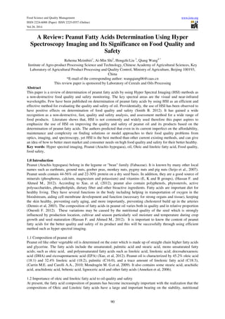
2.2 Production of hyper spectral images
A line of light reflected from the sample enters the objective lens and is separated into its component
wavelengths by diffraction optics contained in the spectrograph; a two dimensional image (spatial dimension -
wavelength dimension) is then formed on the camera and saved on the computer. The sample is moved pass
through the objective lens on a motorized stage and the process repeated; two dimensional line images acquired
at adjacent points on the object are stacked to form a three-dimensional hypercube which may be stored on a PC
for further analysis (Gowen, et al., 2007) as shown in figure 1.
3. Food Science and Quality Management www.iiste.org
ISSN 2224-6088 (Paper) ISSN 2225-0557 (Online)
Vol.28, 2014
92
Figure1: Production and storage of hyper spectral image (Gowen, et al., 2007)
2.3 Analysis of hyper spectral images
There are numerous techniques which are used to analyze hyper spectral data, all of which aim to reduce the
dimensionality of the data while retaining important spectral information with the power to classify important
areas of a scene (Gowen et al., 2007). Typical steps in analyzing hyper spectral images include reflectance
calibration, pre-processing, classification and application. Reflectance calibration accounts for the background
spectral response of the instrument and the ‘dark’ camera response.
2.3.1 Reflectance
For reflectance measurements, the background is obtained by collecting a hyper spectral image from a uniform,
high reflectance standard or white ceramic and the dark response is acquired by turning off the light source,
completely covering the lens with its cap and recording the camera response (Gowen et al., 2007). The corrected
reflectance value (R) is then calculated using the following formula: R ¼ (sample - dark) / (background – dark)
(Gowen et al., 2007).
2.3.2 Pre processing
Pre-processing is normally performed to remove non-chemical biases from the spectral information (e.g.,
scattering effects due to surface in homogeneities) and prepare the data for further processing. A number of
spectral preprocessing techniques exist, including polynomial baseline correction, Savitzkye Golay derivative
conversion, mean centering, and unit variance normalization. Other operations usually carried out at the pre-
processing stage include thresholding and masking to remove redundant background information from the hyper
spectral image. Pre- processing must be handled with care to avoid the spectral and spatial variability (Amigo et
al., 2013).
2.3.3 Classification
Hyper spectral image classification enables the identification of regions with similar spectral characteristics. Due
to the large size of hyper spectral images (which can exceed 50 MB, depending on image resolution, spectral
resolution and pixel binning) complex multivariate analytical tools, such as principal component analysis (PCA),
partial least squares (PLS), linear discriminant analysis (LDA), Fishers discriminant analysis (FDA), multi-linear
regression (MLR) and artificial neural networks (ANN), are usually employed for classification image
processing (Gowen et al., 2007).
2.3.4 Application
Application step involve image processing to convert the contrast developed by the classification steps into a
picture depicting component distribution. Grey scale or color mapping with intensity scaling is commonly used
to display compositional contrast between pixels in an image. Image fusion, in which two or more images at
different wavebands are combined to form a new image, is frequently implemented to provide even greater
contrast between distinct regions of a sample (Pohl, 1998). Images may be combined using algorithms based on
straightforward mathematical operations such as addition, subtraction, multiplication and division. One example
is the band ratio method, in which an image at one wavelength is divided by that at another wavelength (Liu et
al., 2007; Park et al., 2006). The distinction between hyper- and multi-spectral images is sometimes based on an
4. Food Science and Quality Management www.iiste.org
ISSN 2224-6088 (Paper) ISSN 2225-0557 (Online)
Vol.28, 2014
93
arbitrary "number of bands" or on the type of measurement, depending on what is appropriate to the purpose. It
deals with imaging narrow spectral bands over a continuous spectral range, and produces the spectra of all pixels
in the scene. So a sensor with only 20 bands can also be hyper spectral when it covers the range from 500 to
700 nm with 20 bands each 10 nm wide. It is noted that a sensor with 20 discrete bands covering the Visible
Infrared Spectroscopy (VIS), Near Infrared Reflectance(NIR), Short Wavelength Infrared (SWIR), Medium
Wavelength Infrared (MWIR) and Long Wavelength Infrared (LWIR) is be considered as multispectral (Ghita,
2009). The hyper spectral image allows for the visualization of biochemical constituents of a sample, separated
into particular areas of the image, since regions of a sample with similar spectral properties have similar
chemical composition.
2.4 Hyper spectral image acquisition compared to other imaging
HSI has proven to be an outstanding tool for analysis of agricultural and food products. Its fast measurement,
with little or no tedious sample preparation, good adaptability and simultaneous determination of different
attributes makes superior to other imaging methods (Nicolaï et al., 2007) as shown in table 1.
Table1. Comparison of RGB imaging, NIR spectroscopy (NIRS), multispectral imaging (MSI) and hyper
spectral imaging (HSI
Feature RGB imaging NIRS MSI HSI
Spatial information √ Limited √ √
Spectral information √ √ Limited √
Multi-constituent information Limited √ Limited √
Sensitive to minor components √ Limited Limited √
Source: (Gowen, 2007)
2.5 The composition of peanut oleic and linoleic fatty acids and its role in the quality of oil
It is the composition of fatty acid which plays an important role in the quality and safety of oil because of their
relationship to the shelf life, nutrition, and flavor of peanut oil and other derived products. As depicted in the
table 2 below, there are thirteen fatty acids present in peanut oil. The composition of the two leading peanut fatty
acids (oleic and linoleic) are with average of 37.7% and 34.21 % respectively (USDA, 2012). According to
Berry S.K. (1982), by Gas Chromatography method revealed the occurrence of palmitic (12.22 to13.30%),
stearic (3.17 to 3.67%), oleic (37.94 to 41.90%) linoleic (34.59 to 37.51%), arachidic (1.63 to 1.85%)
eicosaenoic (0.99 to 1.22%), behenic (3.24 to 4.36%), and lignoceric (1.08 to 1.44%) as the major fatty acids in
peanut oil. In addition, Chamberlin K.D. (2014) revealed that the two fatty acids, oleic and linoleic acid
comprise over 80% of the oil content in peanut, and these fatty acids have a strong effect on the stability of the
oil and its products.
Table 2. Fatty acid composition in crude and refined peanut oil
Fatty acid
Crude composition (%) Refined
Composition (%)
Caprylic acid (C8:0) 0.013346 0.003208
Capric acid (C10:0) 0.008544 0.005542
Lauric acid (C12:0) 0.275816 0.116076
Myristic acid (C14:0) 0.115270 0.1061
Palmitic acid (C16:0) 8.2280 11.7378
Palmitoleic acid (C16:0) 0.1073 0.1296
Stearic acid (C18:0) 2.4581 2.0606
Oleic acid (C18:1) 58.6871 57.6784
Linoleic acid (18:2) 21.7656 21.5413
Linolenic acid (C18:3) 0.3446 0.2810
Arachidic acid (C20:0) 1.8313 1.4804
Behenic acid (C22:0) 3.8852 2.36610
Source: Aluyor, (2009)
5. Food Science and Quality Management www.iiste.org
ISSN 2224-6088 (Paper) ISSN 2225-0557 (Online)
Vol.28, 2014
94
2.6 Summary
Fast computers, sensitive detectors, and large data storage capacities as that of HSI are needed for analyzing
hyper spectral data. All of these factors greatly increase the cost of acquiring and processing hyper spectral data
(Schurmer, 2003). One of the barrier the researchers have to face is finding ways to program hyper spectral
dependency to sort through data on their own and transmit only the most important images, as both transmission
and storage of that much data could prove difficult and costly (Schurmer, 2003). The full potential use of a
relatively new analytical technique of hyper spectral imaging has not yet been recognized, so emphasis is
needed. In the past it was unfeasible to obtain information in all four-dimensions of a hypercube using other
methods (refer Table 1).
3.0 RESULTS
3.1 Significances of HSI in food quality and safety
The use of HSI in food science and technology has recently been widely studied and developed, resulting in
many successful applications and comprehensive assessment in the food industry for quality and safety
evaluation and inspection (Win D.T., 2005). The primary use of the imaging system is to conduct food safety
and quality research. Statistical combination of measurements by several sensors as applied in HSI will increase
the likelihood of predicting overall quality and safety. This is because; sensor testing and calibration of HSI must
include a wide range of conditions important in minimizing the limitations (Abbort J.A, 1998). For instance,
Singh, Jayas, Paliwal, & White, 2010a demonstrated the use of HSI in seed color classification, seed kernel
purity determination, identification of sound or stained grains, and detection of midge-damaged in wheat
kernels. Whereby six image features and ten histogram features were extracted from the most significant
wavelengths determined according to the PCA analysis on hyper spectral images, and were then used to develop
statistical discriminant classifiers (linear, quadratic, and Mahalanobis) or a back propagation. Therefore, it is
vital to emphasize the use of non-invasive, efficient and quick testing method for monitoring food quality and
safety.
3.2 Existing issues for consideration on Quality and Safety of food
Nielsen, (2009) reported several issues for considerations on food quality and safety including legal issue, food
processing food quality, adulteration and lipid oxidation. Government regulations (legal issue) often demand that
the amounts of saturated, unsaturated and polyunsaturated lipids (fatty acids) and the amount of cholesterol
should be specified on food labels. In food processing, the manufacture of many foods relies on knowledge of
the type of lipids present in order to adjust the processing conditions to their optimum values, e.g. temperature.
Moreover, desirable physical characteristics of foods, such as appearance, flavor, taste and texture, depend on
the type of fatty acids present. Foods which contain high concentrations of unsaturated fatty acids are
particularly susceptible to lipid oxidation, which can lead to the formation of undesirable flavors and aromas, as
well as potentially toxic compounds such as cholesterol oxides. By measuring the type of lipids present and
comparing them with the profile expected for an unadulterated sample it is possible to detect the adulteration of
fats and oils. On these bases, it is important for food scientists to either know or be able to specify the
concentration of the different types of fatty acids present in oil Nielsen, (2009). Food process control necessitates
real-time monitoring at critical processing points. Fast and precise analytical methods such as HSI are essential
to ensure product quality and safety (Gowena et al, 2007).
3.3 Peanut fatty acids and health benefits
Owing to the steadily growing trend towards the intake of a healthy and scientifically balanced diet, the
selection of high-quality vegetable oils is constantly rising (Hein M., 1997). Besides physical
(seed mass and shape, integrity of seed testa and blanching efficiency) and sensory (seed color, texture, flavor)
factors, nutritional (oil, protein contents, fatty acid and amino acid composition) factors are important in the food
trade (Hein M., 1997). Besides, WHO (2003) reported that the global burden of chronic diseases is rapidly
increase, in 2001 chronic diseases including cardio vascular diseases contributed approximately 60% of the 56.5
million total reported deaths in the world. It has been observed that two important problems in modern lipid
chemistry are purity control and the identification of oils and fats (Alander J.T., 2013). Evidence about the
impact of lipids in different clinical diseases is still increasing rapidly as the understanding of the role that
dietary lipids can play at all ages in preventing diseases related to lifestyle (ISSFAL, 2014). Peanut oil has a role
in a healthy balanced diet even though they are energy dense and contain a high proportion of fat (McKelvith B,
2005).Oils that have high oleic acid content and food products containing these oils have been shown to have
6. Food Science and Quality Management www.iiste.org
ISSN 2224-6088 (Paper) ISSN 2225-0557 (Online)
Vol.28, 2014
95
nutritional benefits (Chamberlin K.D., 2014). Oleic acid has been shown to be associated with a reduction in
blood pressure and serum lipoprotein levels. High-oleic peanuts have health benefits over conventional peanuts
because the linoleic (polyunsaturated fat) and palmitic (saturated fat) fatty acids have been naturally replaced by
the healthier oleic fatty acid (monounsaturated fat) (Chamberlin K.D., 2014). According to Win D.T. (2005),
high concentrations of oleic acid can lower cholesterol levels in blood thus, lower the risk of heart problems and
block the action of a cancer-causing oncogene, called HER-2/neu, which is found in about 30% of breast cancer
patients, also has effect on Type II diabetes. Linoleic acid plays a role in pro-inflammatory reactions, blood clots
and allergic reactions.
4.0 Conclusion
HSI are essential for effective food (peanut oil) quality and safety control system as it can sufficient detect the
fatty acid contents (quantity and quality) in the seed and its oil, besides selection of peanut seed varieties with
high oleic and linoleic fatty acids also need to be considered for the good quality and safety of peanut oil and its
product as well.
Food processing industries and food control systems are emphasized to use HSI for ensuring the efficient quality
and safety of the product to meet nutritious and health demand of the consumers. This paper is in line with FAO
and WHO, (2002) guideline in assuring food quality and safety by widening information on good methods or
technique in detecting, evaluating and inspecting and building models on food quality and safety specifically
peanut oil and its products.
HSI has recognized as the best in offering the possibility of designing inspection systems for the automatic
grading and nutrition determination of food quality and safety products. Several applications outlined in this
review show the capability of using HSI for sample classification, and grading, defect and disease detection,
distribution visualization of chemical attributes in chemical images, and evaluations of overall quality and safety
of food products.
Therefore, it is predicted that real-time for food quality and safety surveillance and control systems with this
technique can be expected to meet the requirements of the contemporary food (peanut oil) industrial processing
in the near future, hence maintain health of the consumers.
Acknowledgement
The authors gratefully would like to acknowledge Oils and Cereals Laboratory under the Institute of Agro-
product Processing Science and Technology, Chinese Academy of Agricultural Sciences, for the resources
provided including financial support in this publication.
References
Abbott, J. A., (1998). “Quality measurement of fruits and vegetables”. Elsevier-Institute
Postharvest Biology and Technology 15 (1999) 207–225. Beltsville MD 20705, USA.
http://www.scribd.com/doc/117851214/Quality-measurement-of-fruits-and-vegetables
Alander, J.T., (2013). “A Review of Optical Non-destructive Visual and Near-Infrared Methods for Food Quality
and Safety”. International Journal of Spectroscopy Volume 2013 (2013), University of Vaasa, 65101 Vaasa,
Finland. http://dx.doi.org/10.1155/2013/341402.
Aluyor, E., et al., (2009). “Chromatographic analysis of vegetable oils” A review: Scientific Research and Essay
Vol. 4 (4) pp. 191-197. Department of Chemical Engineering, University of Benin, Benin City, Nigeria.
Available online at http://www.academicjournals.org/SRE
Amigo M.J, et al., (2013). "Hyper Spectral Imaging and Chemo metrics: A Perfect Combination for the Analysis
of Food structure, Composition and Quality. Data Handling in Science and Technology, Vol 28. ISBN: 978-0-
444-59528-7. Amsterdam http://www.elsevier.com/locate/permissionusematerial
Anneken, et al., (2006). "Fatty Acids in Ullmann's Encyclopedia of Industrial Chemistry”. Wiley-VCH,
Weinheim.http://www.jofamericanscience.org/journals/am-sci/am0811/018_11805am0811_128_131.pdf
Berry, S.K., (1982). “Fatty Acid Composition of 16 Groundnut (Arachis hypogaea, L.) Cultivars grown under
Malaysian Conditions”. Universiti Pertanian Malaysia, Serdang, Selangor, Malaysia. Pertanika5(1),20-24(1982).
http://psasir.upm.edu.my/2150/1/Fatty_Acid_Composition_of_16_Groundnut_%28AracHSI_hypogaea,_L.%29.
pdf
Buchanan, M.D., (2011)., “ Gas Chromatograph fatty acid analysis” Reporter US Volume 26.4 pg 1-3.
http://www.sigmaaldrich.com/china-mainland/zh/technical-documents/articles/reporter-us/gc-analyses-of-
free.html#sthash.rvGVTPYo.dpuf
7. Food Science and Quality Management www.iiste.org
ISSN 2224-6088 (Paper) ISSN 2225-0557 (Online)
Vol.28, 2014
96
Chamberlin, K.D., (2014). “A Comparison of Methods Used to Determine the Oleic/Linoleic Acid Ratio in
Cultivated" Peanut (Arachis hypogaea L.)”. Agricultural Sciences, 5, 227-237. doi: 10.4236/as.2014.53026.AS>
Vol.5 No.3., United States Department of Agriculture-Agricultural Research Service (USDA-ARS), Still water,
USA.
Carrin, M.E., and Carelli, A.A., (2010). “Peanut Oil Composition data”. European Journal of Lipid Science and
Technology. Vol 112, Issue 7, Pages 693–818. Wiley Online Library. KGaA, Weinheim.
http://onlinelibrary.wiley.com/doi/10.1002/ejlt.201090012/pdf
Dennis, et al, (2003). “Clear answers for common questions”: FAQ Copyright 3003-2014Conjecture
Cooperation. Wise Geek, Faculty Practice at Columbia University Medical Centre.
www.wisegeek.org/technology Retrieved on 10th
March, 2014
FAO, (2012). “FAO Statistical Year book”. World Food and Agriculture, Rome Italy. ISBN 978-92-5-106913-1.
Available on http://www.fao.org/docrep/015/i2490e/i2490e00.htm
FAO and WHO, (2002). “Assuring Food Safety and Quality: Guidelines for Strengthening Food National
Control System”. Joint FAO/WHO Publication http://www.wpro.who.int/foodsafety/documents/docs/
English_Guidelines_
Food_control.pdf
Ghita, O., et al., (2009), “Spectral and Spatial Feature Integration for Classification of Non-ferrous Materials in
Hyper-spectral Data”. IEEE Transactions on Industrial Informatics, Vol. 5, No. 4, November 2009. Wikipedia
cited on 10th
March, 2014
Gowena, A.A., (2007). “Hyper spectral Imaging System an Emerging Process Analytical Tool for Food Quality
and Safety Control: Trends in Food Science and Technology”, Review. Biosystems engineering, School of
Agriculture, Food Science and Veterinary Medicine, Dublin, Ireland.
Gowen, A. A., et al, (2009) “Potential Applications of Hyper spectral imaging for Quality Control in Dairy
Foods”. Image Analysis for Agricultural Products and Processes. Bornimer Agrartechnische Berichte ISSN
0947-7314. School of Agriculture, Food Science and Veterinary Medicine, University College Dublin, Belfield,
Dublin 4, Ireland. www2.atb-potsdam.de/cigr.../images/07_125_%20Gowen
Grunert, G. K., (2005). “Food quality and safety: consumer perception and demand”. European Review of
Agricultural Economics Vol 32 (3) (2005) pp. 369–391, MAPP—Centre for Research on Customer Relations in
the Food Sector, Aarhus School of Business, Haslegaardsvej, Denmark. doi:10.1093/eurrag/jbi011.
Hassan, F., and Ahmed, M., (2012). “Oil and Fatty Acid Composition of Peanut Cultivars Grown in Pakistan”.
Pak. J. Bot., 44(2): 627-630, 2012. PMAS-Arid Agriculture University, Rawalpindi, Pakistan.
http://www.pakbs.org/pjbot/PDFs/44%282%29/22.pdf
Hein, M., (1997). “D e t e r m i n a t i o n o f u n d e r i v a t e d f a t t y a c i d s b y H P L C ” . Springer-
Verlag. H e i n Universita¨t Hohenheim, Institut fu¨r Lebensmitteltechnologie, Garbenstrasse 25, D-70593
Stuttgart, Germany. http://www.scribd.com/doc/140188034/Determination-of-Underivated-Fatty-
Acids-by-HPLC
ISSFAL, (2014). 11th Congress for the International Society. The Study of Fatty Acids and Lipids. Stockholm
Sweden. Available on http://www.issfal.org/conferences/2014-stockholm
Juanéda, P. et al., (2007). “Analytical Methods for Determination of Trans Fatty Acid Content in Food”.
European Journal of Lipid Science and Technology. Special Issue: Tran’s fatty acids Volume 109, Issue 9, pages
901–917.doi: 10.1002/ejlt.200600277.
Kim, M. S., (2001). “Hyperspectral Reflectance and Fluorescence Imaging System for Food Quality and Safety”
Transactions of the ASAE Vol. 44(3): 721–729 2001 American Society of Agricultural Engineers ISSN 0001–
2351 721.USA. http://naldc.nal.usda.gov/download/26654/PDF
Li W., (2012). “Determining the Contents of Protein and Amino Acids in Peanuts using Near-Infrared
Reflectance Spectroscopy”. Proceedings: 14th
ICC Cereals and Bread Congress and Forum on Fats and Oils.
August 6-9, 2012. Beijing, China.
Ministry of Health and Family Welfare (2012). “Manual of Methods Analysis of Foods-Oils and Fats”. Lab.
Manual 2. Food Safety and Standards Authority of India. Government of India, New Delhi.
http://www.fssai.gov.in/Portals/0/Pdf/15Manuals/oils%20and%20fats.pdf visited on 25th
March, 2014.
Mc.Kevith, B., (2005). “A Review: Nutritional aspects of oilseeds”. British Nutrition Foundation. Nutrition
Bulletin, 30, 13–26, London, UK.www.researchgate.net/.Nutritional. oilseeds/./9fcfd5063. visited on19th April,
2014.
Musa O., and Serap S., (2003). “Physicalandchemicalanalysisandfattyacidcompositionofpeanut,peanutoilandpeanutbutter
from ÇOM and NC-7 cultivars”. Grasasy Aceites Vol. 54. Fasc. 1 (2003), 12-18. Department of Food Engineering, Faculty
ofAgriculture,SelcukUniversity.GesasFoodIndustriesKonya,Turkey.
8. Food Science and Quality Management www.iiste.org
ISSN 2224-6088 (Paper) ISSN 2225-0557 (Online)
Vol.28, 2014
97
Nauman, K., (2012). “Quality Evaluation and Safety Assessment of Different Cooking Oils available in
Pakistan”. Vpol.34, No.3, Department of Food Technology, PMAS –and Agriculture University, Rawalpindi
46300, Pakistan.
http://www.academia.edu/1612665/Quality_Evaluation_and_Safety_Assessment_of_Different_Cooking_Oils_a
vailable_in_Pakistan
Nielsen, (2009). “Food Analysis Fourth Edition”. ISBN 978-1-4419-1477-4 DOI 10.1007/978-1-4419-1478-1.
Purdue University, Dept. Food Science 745 Agriculture Mall Dr. West Lafayette in 47907 USA.
https://ag.purdue.edu/foodsci/Pages/Profile.aspx?. nielsens & int Dir DeptI. visited on 17th
March, 2014.
O’Brien, (1998). “Vegetable Oil in Food Technology; Composition, Properties and Uses”. Emeritus University
of St. Andrews, Crop Research Institute Dundes, Blackwell Publishing, CRC Press.
http://health120years.com/cn/pdf/hd_Vegetable.Oils.pdf
Onemli, F., (2012). “Impact of Climate Change on Oil Fatty Acid Composition of Peanut: In Three Market
Classes”. Chilean Journal of Agricultural Sciences. University of Namik Kemal, Faculty of Agriculture, 59030
Tekirdag, Turkey. http://www.bioline.org.br/request?cj12073
Rao, Y., (2013). “Quantitative and Qualitative Determination of Acid Value of Peanut Oil using Near-Infrared
Spectrometry”. Journal of Food Engineering,Volume 93, Issue 2, July 2009, Pharmaceutical University, 210009
Nanjing, PR China. http://dx.doi.org/10.1016/j.jfoodeng.2009.01.023
Sebedio, (1995). “Fatty acids and their Health Implications”. Third Edition edited by Ching K.C. CPC Press,
Taylor and Fransic Group. Food Science and Technology Department. 6000Broken Sound Parkway NW, Suite
300 U.S.A. ISBN10:0-8493-7261-
http://books.google.com.hk/books?id=Hcl0fkcrfbEC&pg=PA539&lpg=PA539&dq=Sebedio+J.L+1995+percent
age+of+fatty+acids+in+peanut&source=bl&ots=Pd
Seijo,G., et al., (2007). "Genomic relationships between the cultivated peanut (Arachis hypogaea, Leguminosae)
and its close relatives revealed by double GISH". Am. J. Bot. 94 (12): 1963–1971. doi:10.3732/ajb.94.12.1963.
PMID 21636391. Retrieved 2014-04-20.
Smith, B.R., (2012). “Introduction to Hyper spectral Imaging”.Available on
http://www.microimages.com/documentation/Tutorials/hyprspec.pdf
Scotter, (1997). “Non-destructive spectroscopic techniques for the measurement of food quality”. Trends on
Food Sci. &Tech., 8, 285-292.
http://grasasyaceites.revistas.csic.es/index.php/grasasyaceites/article/viewFile/289/291
Yadava, D.K., e.t al., (2012). “Breeding Major Oil Crops: Present Status and Future Research Needs”.
Technological Innovations in Major World Oil Crops, 17, Volume 1: Breeding, doi: 10.1007/978-1-4614-0356-
2_2., India. Available on http://www.springer.com/978-1-4614-0355-5
Yalin, X., et al (2012). “Study on Quality Characteristics of Vegetable Oil and Adulteration Test. Academy of
State Administation of Grain, China”. Proceedings: 14th
ICC Cereal and Bread Congress and Forum on Fats and
Oils. August 6-9, 2012. Beijing, China.pg 626.
Win, D. T., (2005). “Oleic Acid: The Anti-Breast Cancer Component in Olive Oil”. Available on
www.journal.au.edu/au_techno/.../vol9num2_article02.pdf, retrieved on 19thApril, 2014.
Zambiazi, et al., (2007). “Fatty acids Composition of Vegetable Oils and Fats”. Departamento de Ciência dos
Alimentos (DCA), Universidade Federal de Pelotas (UFPEL), Pelotas/RS. B.CEPPA, Curitiba, v. 25, n. 1, p.
111-120, jan./jun. 2007.
Corresponding author Biography:
Qiang Wang Professor, Deputy Director and Chief Expert of the Agricultural Science and Technology
Innovation Program (ASTIP)
Institute of Agro-products Processing Science and Technology
Chinese Academy of Agricultural Sciences (CAAS)
Address: No 2 Yuanmingyuan West Road, Haidian District
P.O.Box5109, Beijing, P.R.of China 100193
E-mail: wangqiang06@caas.cn
9. The IISTE is a pioneer in the Open-Access hosting service and academic event
management. The aim of the firm is Accelerating Global Knowledge Sharing.
More information about the firm can be found on the homepage:
http://www.iiste.org
CALL FOR JOURNAL PAPERS
There are more than 30 peer-reviewed academic journals hosted under the hosting
platform.
Prospective authors of journals can find the submission instruction on the
following page: http://www.iiste.org/journals/ All the journals articles are available
online to the readers all over the world without financial, legal, or technical barriers
other than those inseparable from gaining access to the internet itself. Paper version
of the journals is also available upon request of readers and authors.
MORE RESOURCES
Book publication information: http://www.iiste.org/book/
IISTE Knowledge Sharing Partners
EBSCO, Index Copernicus, Ulrich's Periodicals Directory, JournalTOCS, PKP Open
Archives Harvester, Bielefeld Academic Search Engine, Elektronische
Zeitschriftenbibliothek EZB, Open J-Gate, OCLC WorldCat, Universe Digtial
Library , NewJour, Google Scholar