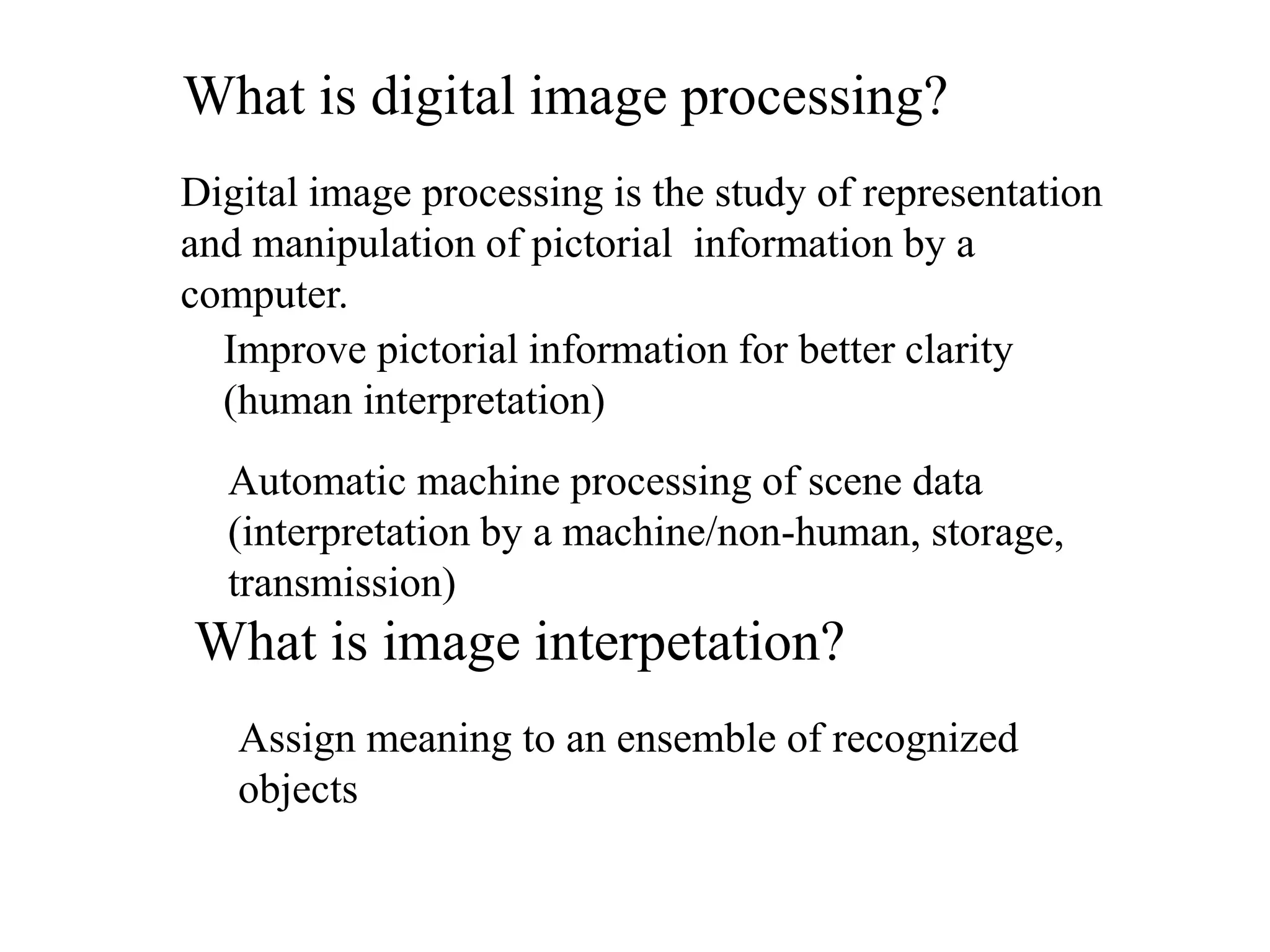
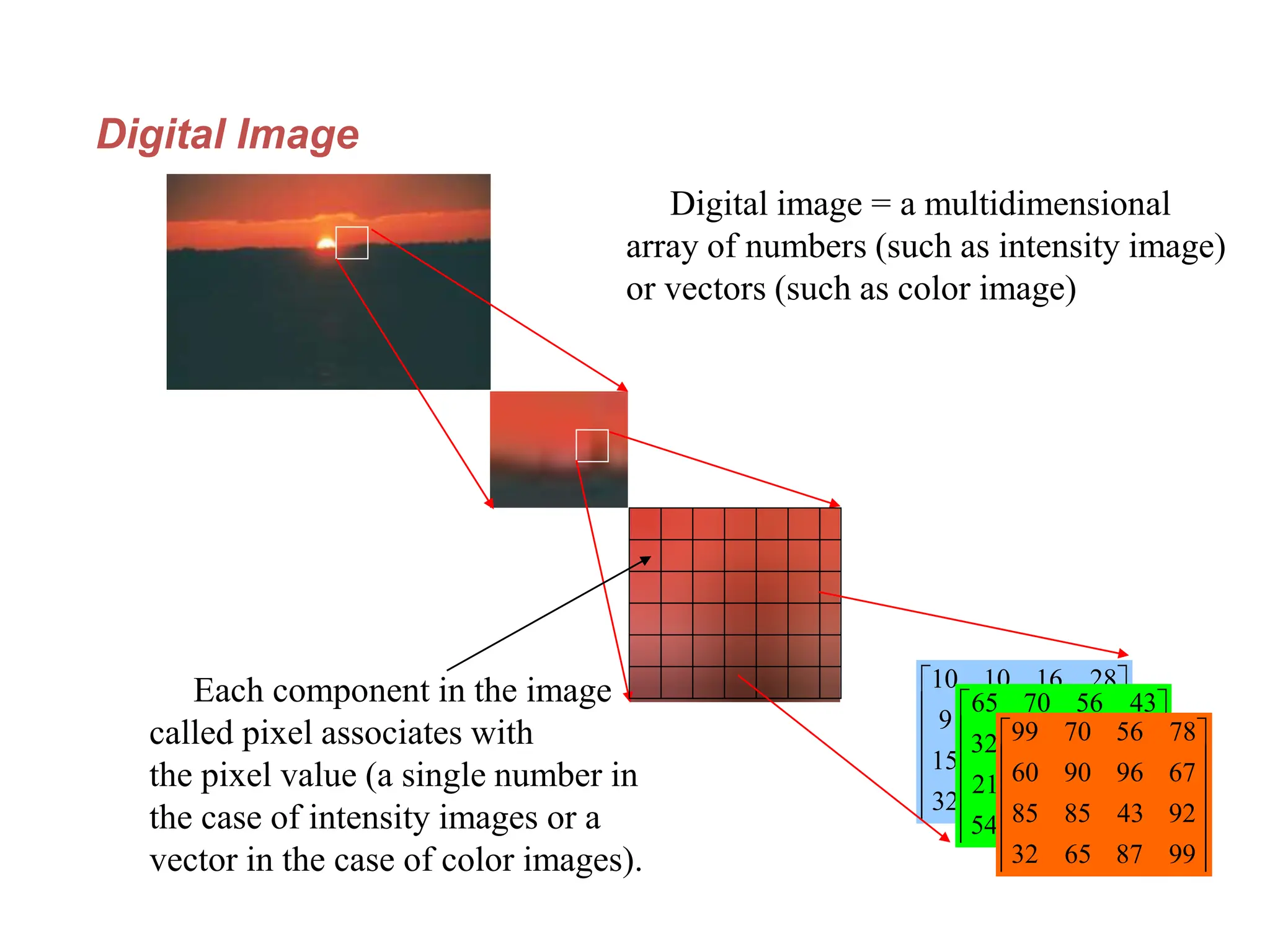
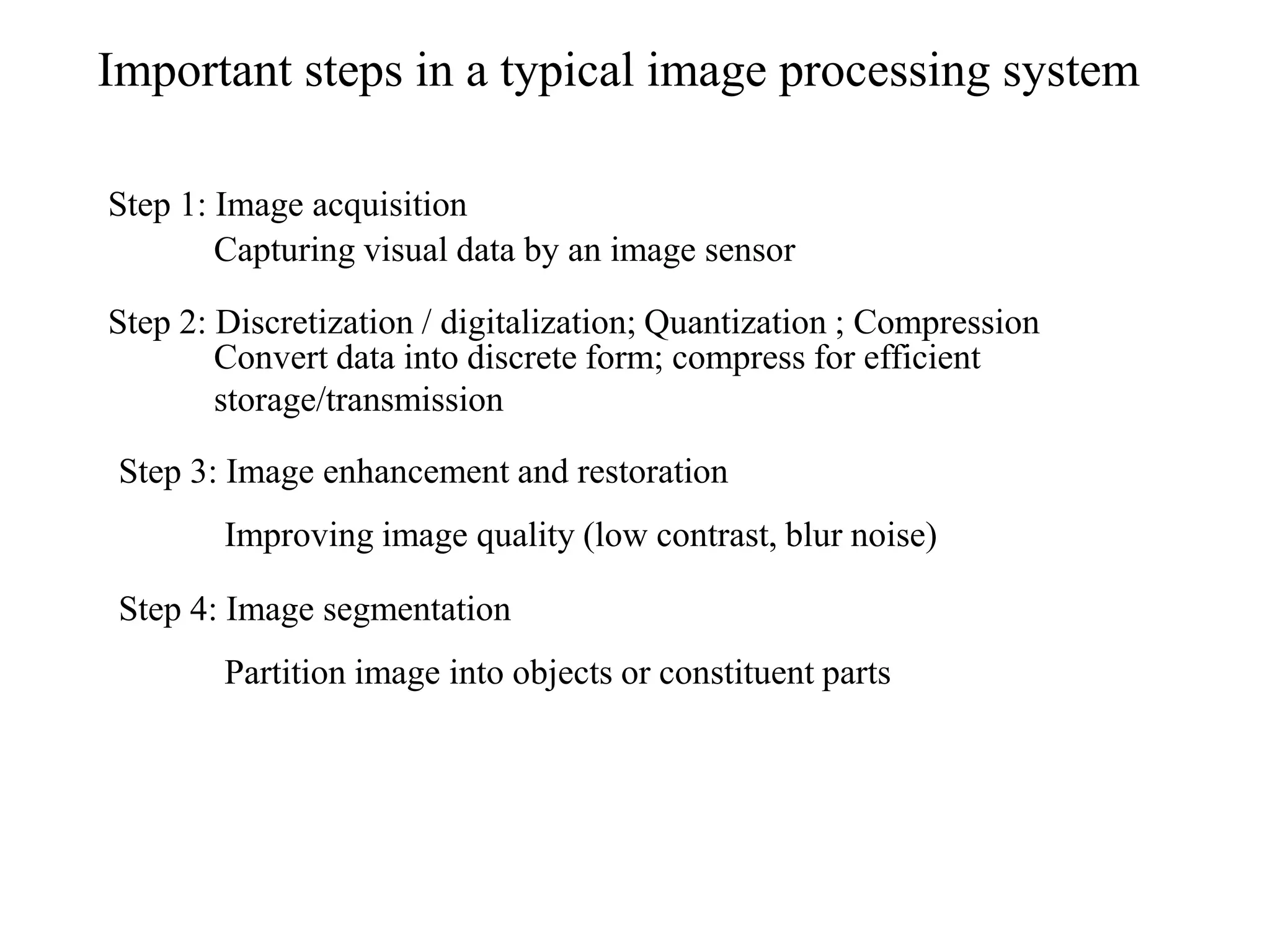
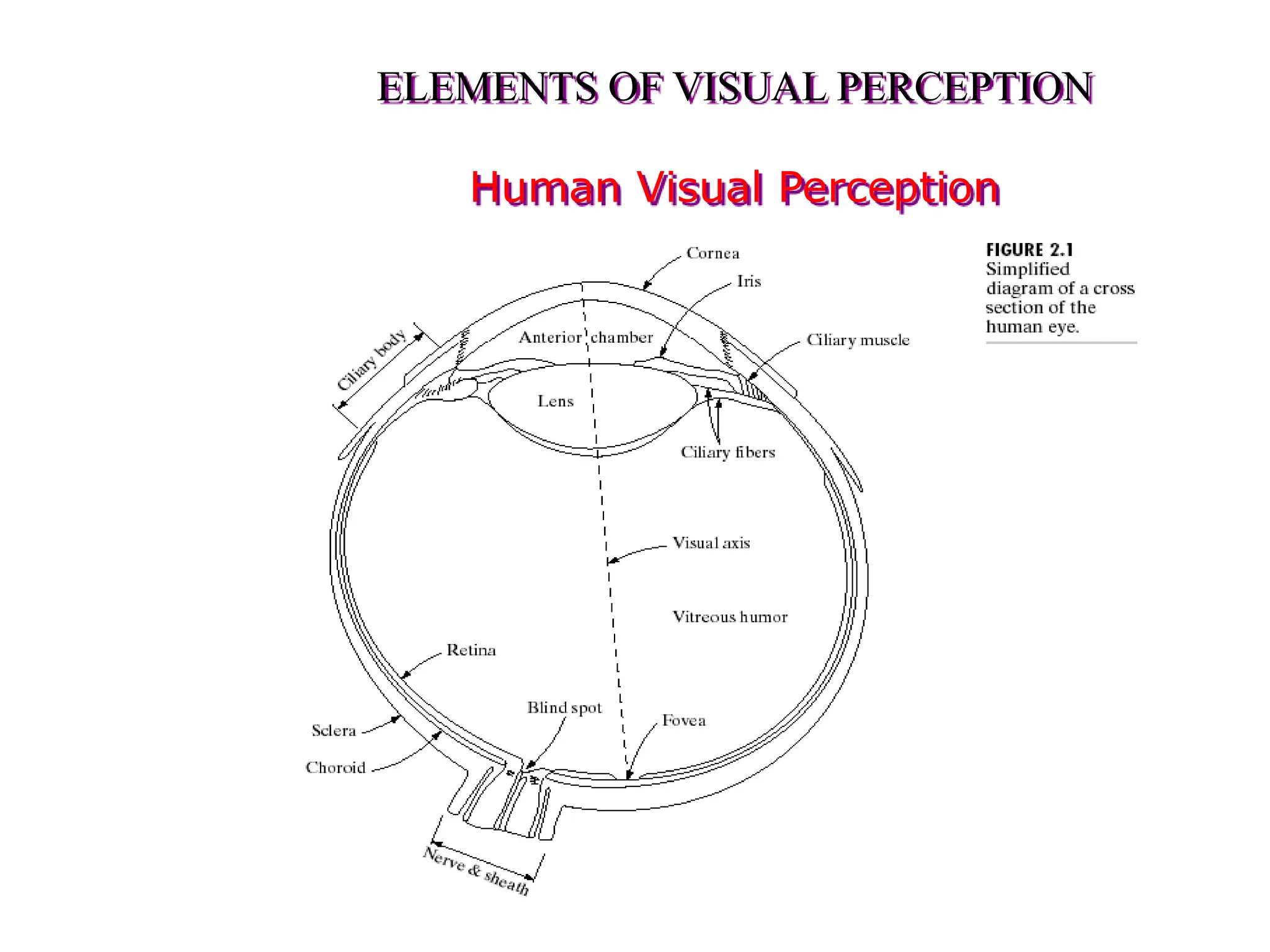
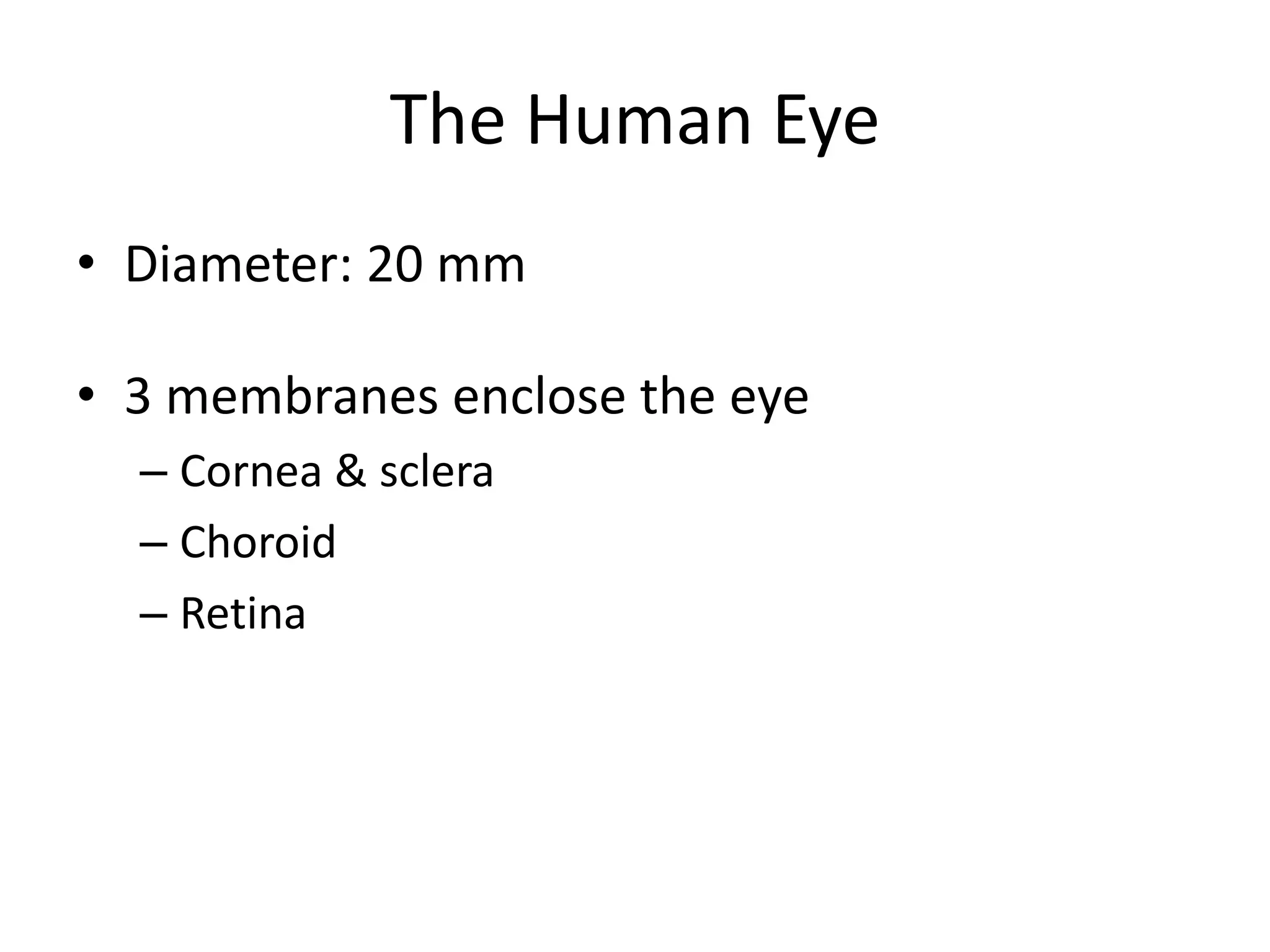
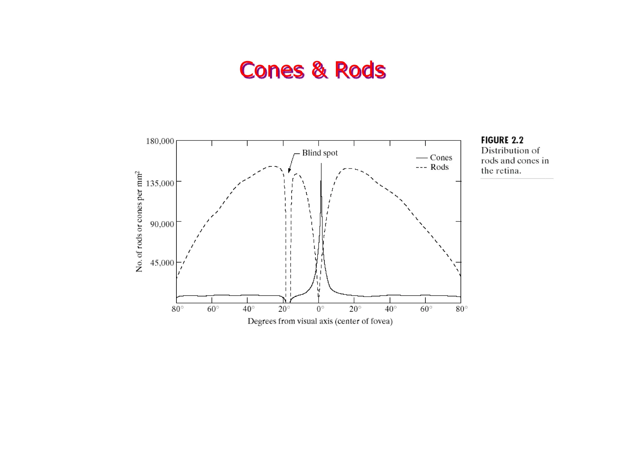
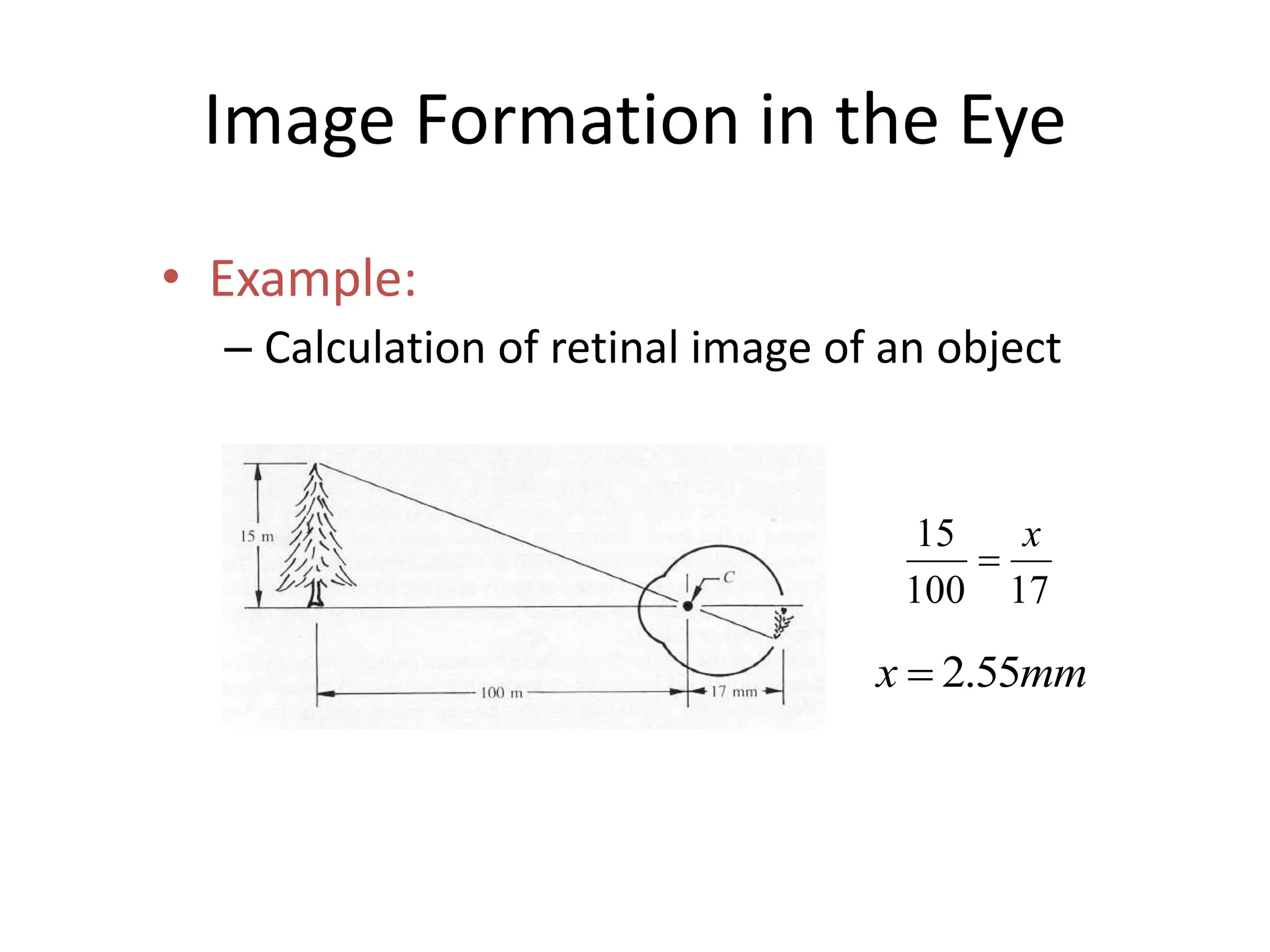
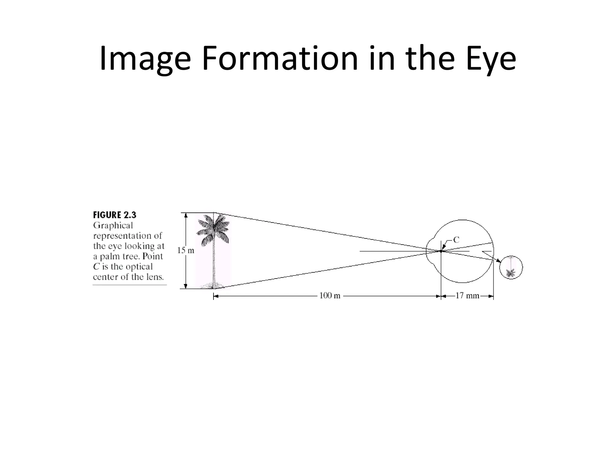
Digital Image Processing
Image formation
• What is an Image?
Picture, photograph
Visual data
Usually two or three dimensional
What is a digital image?
An image which is “discretized,”, i.e., defined on a
discrete grid
Two-dimensional collection of light values (or gray
values)
Basic Terminology
What is a digital image?
• A representation of the original image by a discrete set of data
points
• Each of these data points is called a picture element, commonly
referred to as a “pixel”
Digital Image Generation
Generating a digital image
• Place a regular 2D grid over an image and read the colour value
• at the intersections of the grid => set of data points for that image
What is digital image processing?
Digital image processing is the study of representation
and manipulation of pictorial information by a
computer.
Improve pictorial information for better clarity
(human interpretation)
Automatic machine processing of scene data
(interpretation by a machine/non-human, storage,
transmission)
What is image interpetation?
Assign meaning to an ensemble of recognized
objects
Digital Image
Digital image = a multidimensional
array of numbers (such as intensity image)
or vectors (such as color image)
Each component in the image
called pixel associates with
the pixel value (a single number in
the case of intensity images or a
vector in the case of color images).












39
87
15
32
22
13
25
15
37
26
6
9
28
16
10
10












39
65
65
54
42
47
54
21
67
96
54
32
43
56
70
65












99
87
65
32
92
43
85
85
67
96
90
60
78
56
70
99
Important steps in a typical image processing system
Step 1: Image acquisition
Step 2: Discretization / digitalization; Quantization ; Compression
Step 3: Image enhancement and restoration
Step 4: Image segmentation
Capturing visual data by an image sensor
Convert data into discrete form; compress for efficient
storage/transmission
Improving image quality (low contrast, blur noise)
Partition image into objects or constituent parts
Chapter 1: Introduction
 Image enhancement and restoration both involve
techniques to make a better image
 Image compression involves development of
techniques to make smaller files, while still retaining
high quality images
ELEMENTS OF VISUAL PERCEPTION
Human Visual Perception
The Human Eye
• Diameter: 20 mm
• 3 membranes enclose the eye
– Cornea & sclera
– Choroid
– Retina
The Choroid
• The choroid contains blood vessels for eye nutrition
and is heavily pigmented to reduce extraneous light
entrance and backscatter.
• It is divided into the ciliary body and the iris
diaphragm, which controls the amount of light that
enters the pupil (2 mm ~ 8 mm).
The Lens
• The lens is made up of fibrous cells and is
suspended by fibers that attach it to the ciliary
body.
• It is slightly yellow and absorbs approx. 8% of
the visible light spectrum.
• The lens contains 60-70% water, 6% of fat.
The Retina
• The retina lines the entire posterior portion.
• Discrete light receptors are distributed over
the surface of the retina:
– cones (6-7 million per eye) and
– rods (75-150 million per eye)
Cones
• Cones are located in the fovea and are sensitive to
color.
• Each one is connected to its own nerve end.
• Cone vision is called photopic (or bright-light vision
or day vision).
• There are three types of cones: Red, Green, and Blue
Rods
 Rods are giving a general, overall picture of the field
of view
 They see only brightness (not color) and are not
involved in color vision.
• Several rods are connected to a single nerve and are
sensitive to low levels of illumination (scotopic or
dim-light vision or night vision).
MESOPIC VISION
• Intermediate region of illumination – between
dim and bright light
• Both rods and cones are active
Receptor Distribution
• The distribution of receptors is radially
symmetric about the fovea.
• Cones are most dense in the center of the
fovea while rods increase in density from the
center out to approximately 20% off axis and
then decrease.
Cones & Rods
The Fovea
• The fovea is circular (1.5 mm in diameter) but can be
assumed to be a square sensor array (1.5 mm x 1.5
mm).
• The density of cones: 150,000 elements/mm2 ~
337,000 for the fovea.
• A CCD imaging chip of medium resolution needs 5
mm x 5 mm for this number of elements
BLIND SPOT
• Blind spot is the region of emergence of the
optic nerve from the eye
• Place on the retina where optic nerve
connects, and which consists of no light
sensors
Image Formation in the Eye
• The eye lens (if compared to an optical lens) is
flexible.
• It gets controlled by the fibers of the ciliary
body and to focus on distant objects it gets
flatter (and vice versa).
Image Formation in the Eye
• Distance between the center of the lens and
the retina (focal length):
– varies from 17 mm to 14 mm (refractive power of
lens goes from minimum to maximum).
• Objects farther than 3 m use minimum
refractive lens powers (and vice versa).
Image Formation in the Eye
• Example:
– Calculation of retinal image of an object
17
100
15 x

mm
x 55
.
2

Image Formation in the Eye
• Perception takes place by the relative
excitation of light receptors.
• These receptors transform radiant energy into
electrical impulses that are ultimately
decoded by the brain.

A representation of the original image by a discrete set of data points

  • 1.
  • 2.
    • What isan Image? Picture, photograph Visual data Usually two or three dimensional What is a digital image? An image which is “discretized,”, i.e., defined on a discrete grid Two-dimensional collection of light values (or gray values)
  • 3.
    Basic Terminology What isa digital image? • A representation of the original image by a discrete set of data points • Each of these data points is called a picture element, commonly referred to as a “pixel”
  • 4.
    Digital Image Generation Generatinga digital image • Place a regular 2D grid over an image and read the colour value • at the intersections of the grid => set of data points for that image
  • 5.
    What is digitalimage processing? Digital image processing is the study of representation and manipulation of pictorial information by a computer. Improve pictorial information for better clarity (human interpretation) Automatic machine processing of scene data (interpretation by a machine/non-human, storage, transmission) What is image interpetation? Assign meaning to an ensemble of recognized objects
  • 6.
    Digital Image Digital image= a multidimensional array of numbers (such as intensity image) or vectors (such as color image) Each component in the image called pixel associates with the pixel value (a single number in the case of intensity images or a vector in the case of color images).             39 87 15 32 22 13 25 15 37 26 6 9 28 16 10 10             39 65 65 54 42 47 54 21 67 96 54 32 43 56 70 65             99 87 65 32 92 43 85 85 67 96 90 60 78 56 70 99
  • 7.
    Important steps ina typical image processing system Step 1: Image acquisition Step 2: Discretization / digitalization; Quantization ; Compression Step 3: Image enhancement and restoration Step 4: Image segmentation Capturing visual data by an image sensor Convert data into discrete form; compress for efficient storage/transmission Improving image quality (low contrast, blur noise) Partition image into objects or constituent parts
  • 8.
  • 9.
  • 10.
     Image enhancementand restoration both involve techniques to make a better image  Image compression involves development of techniques to make smaller files, while still retaining high quality images
  • 11.
  • 12.
    ELEMENTS OF VISUALPERCEPTION Human Visual Perception
  • 13.
    The Human Eye •Diameter: 20 mm • 3 membranes enclose the eye – Cornea & sclera – Choroid – Retina
  • 14.
    The Choroid • Thechoroid contains blood vessels for eye nutrition and is heavily pigmented to reduce extraneous light entrance and backscatter. • It is divided into the ciliary body and the iris diaphragm, which controls the amount of light that enters the pupil (2 mm ~ 8 mm).
  • 15.
    The Lens • Thelens is made up of fibrous cells and is suspended by fibers that attach it to the ciliary body. • It is slightly yellow and absorbs approx. 8% of the visible light spectrum. • The lens contains 60-70% water, 6% of fat.
  • 16.
    The Retina • Theretina lines the entire posterior portion. • Discrete light receptors are distributed over the surface of the retina: – cones (6-7 million per eye) and – rods (75-150 million per eye)
  • 17.
    Cones • Cones arelocated in the fovea and are sensitive to color. • Each one is connected to its own nerve end. • Cone vision is called photopic (or bright-light vision or day vision). • There are three types of cones: Red, Green, and Blue
  • 18.
    Rods  Rods aregiving a general, overall picture of the field of view  They see only brightness (not color) and are not involved in color vision. • Several rods are connected to a single nerve and are sensitive to low levels of illumination (scotopic or dim-light vision or night vision).
  • 19.
    MESOPIC VISION • Intermediateregion of illumination – between dim and bright light • Both rods and cones are active
  • 20.
    Receptor Distribution • Thedistribution of receptors is radially symmetric about the fovea. • Cones are most dense in the center of the fovea while rods increase in density from the center out to approximately 20% off axis and then decrease.
  • 21.
  • 22.
    The Fovea • Thefovea is circular (1.5 mm in diameter) but can be assumed to be a square sensor array (1.5 mm x 1.5 mm). • The density of cones: 150,000 elements/mm2 ~ 337,000 for the fovea. • A CCD imaging chip of medium resolution needs 5 mm x 5 mm for this number of elements
  • 23.
    BLIND SPOT • Blindspot is the region of emergence of the optic nerve from the eye • Place on the retina where optic nerve connects, and which consists of no light sensors
  • 24.
    Image Formation inthe Eye • The eye lens (if compared to an optical lens) is flexible. • It gets controlled by the fibers of the ciliary body and to focus on distant objects it gets flatter (and vice versa).
  • 25.
    Image Formation inthe Eye • Distance between the center of the lens and the retina (focal length): – varies from 17 mm to 14 mm (refractive power of lens goes from minimum to maximum). • Objects farther than 3 m use minimum refractive lens powers (and vice versa).
  • 26.
    Image Formation inthe Eye • Example: – Calculation of retinal image of an object 17 100 15 x  mm x 55 . 2 
  • 27.
  • 28.
    Image Formation inthe Eye • Perception takes place by the relative excitation of light receptors. • These receptors transform radiant energy into electrical impulses that are ultimately decoded by the brain.