Download to read offline

![4
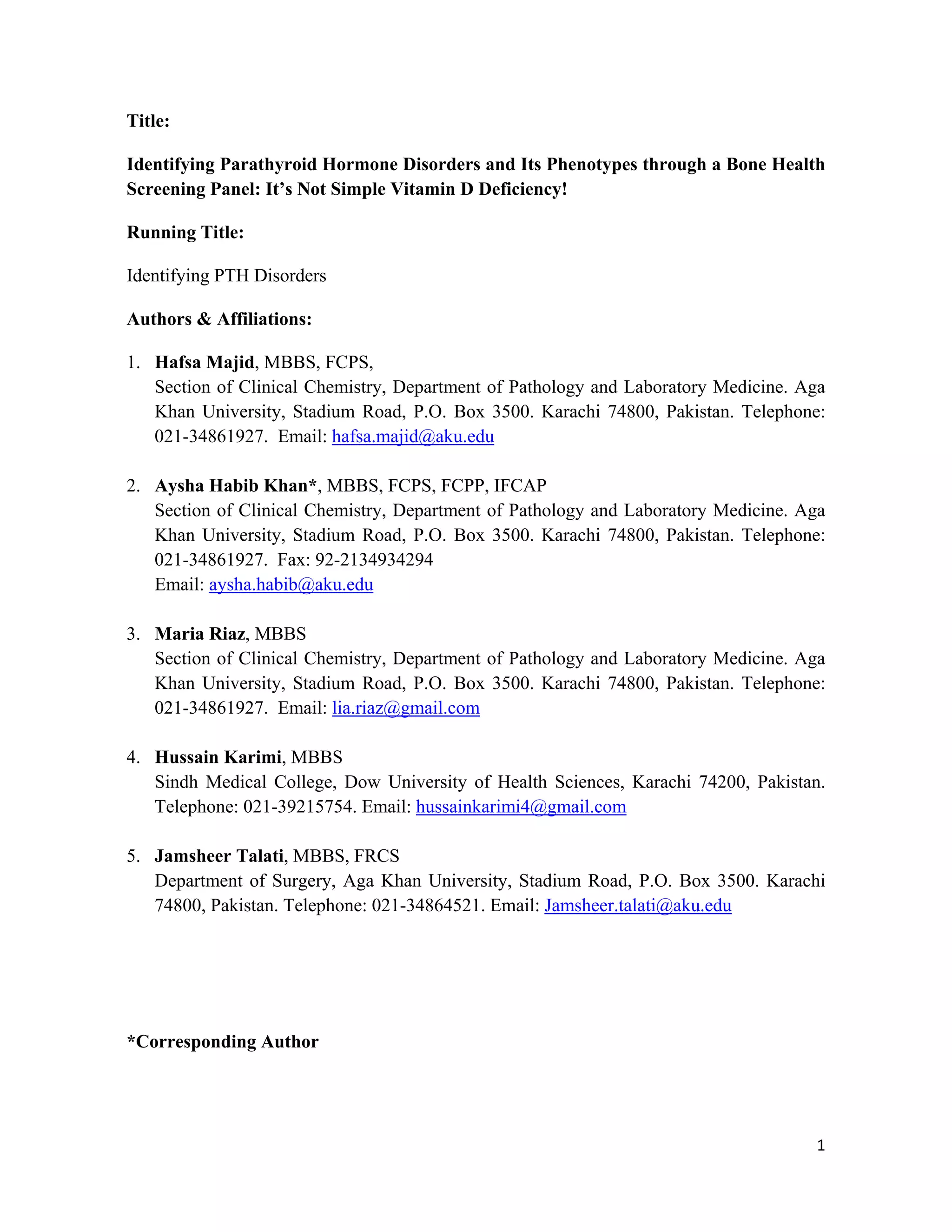
MATERIALS AND METHODS:
Data Collection:
A retrospective analysis was conducted at Section of Clinical Chemistry, Department of
Pathology and Laboratory Medicine, Aga Khan University (AKU) for assessing the
diagnostic utility of BHSP. Laboratory data of patients tested for this panel from 1st
January
2011 till December 31st
2013 was retrieved from integrated laboratory management systems.
Processing and Analysis of Samples in Bone Health Screening Panel:
Venous blood samples were collected after overnight fast (10-12 hours) to standardize
sample collection and to avoid changes due to diurnal variation and diet.
About 10 ml of venous blood was collected into a gel top vacutainer and an EDTA tube.
Calcium, phosphorus, magnesium, alkaline phosphatase, creatinine, albumin were performed
on Advia 1800 Chemistry analyzer (Siemens Healthcare Diagnostics Inc. NY, US) and
plasma iPTH was performed on Immulite 2000 (Siemens Healthcare Diagnostics Inc. NY,
US). Serum 25OHD was determined by electrochemiluminescence immunoassay method on
Liaison auto analyzer (Diasorin the Diagnostic Specialist, Italy). Manufacturer provided
controls were run with each batch of all analytes for internal quality control. External
proficiency was assured by simultaneously analyzing samples from College of American
Pathologist (CAP, USA) three times a year. Serum corrected calcium was used instead of
serum total calcium if low albumin (<4mg/dl) was found. Corrected calcium was measured
using the formula [measured Ca + 0.08 × (4 - albumin)].
Data Analysis:
I. Grouping of Subjects into Hyper and Hypo-functioning States:
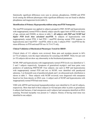
Subjects were grouped into hyper and hypo-functioning states based on the upper and
lower cut-offs of the analytes used in BHSP. The reference ranges used for all
analytes are shown in table 1.
II. Classification of Subjects into Groups of PTH Disorders:
Clinical phenotypes of PTH disorders were grouped based on upper and lower cutoff
of calcium and iPTH after excluding VDD (<20ng/ml) as follows:
1. Primary hyperparathyroidism (PHP) (calcium>10.2mg/dl, iPTH >87pg/ml,
25OHD >20ng/ml).
2. Hypercalcemia with inappropriately normal iPTH (calcium>10.2mg/dl,
25OHD>20ng/ml, iPTH >25pg/ml). Cutoff of >25pg/ml was used for iPTH
based on study of Lungren et al (5).
3. Normocalcemic hyperparathyroidism (NCHP) (calcium 8.6-10.2mg/dl, iPTH
>87pg/ml, 25OHD>20ng/ml).](https://image.slidesharecdn.com/bd2908e7-c285-4c34-9681-846c83298980-160602112910/85/IDENTIFYING-PARATHYROID-HORMONE-DISORDERS-AND-ITS-PHENOTYPES-THROUGH-A-BONE-HEALTH-SCREENING-PANEL-IT-S-NOT-SIMPLE-VITAMIN-D-DEFICIENCY-4-320.jpg)
![5
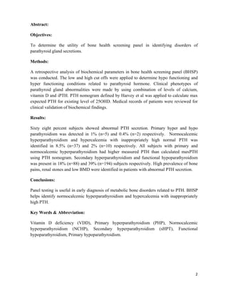
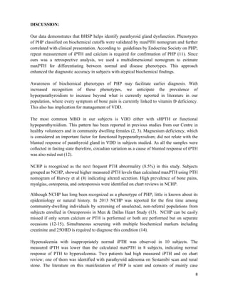
4. Secondary hyperparathyroidism (sHPTH) (25OHD <20ng/ml, calcium
<10.2mg/dl, iPTH>87pg/ml).
5. Functional hypoparathyroidism (25OHD <20ng/ml, calcium <10.2mg/dl,
iPTH 16-87pg/ml).
6. Primary hypoparathyroidism (calcium <8.6mg/dl, iPTH <16pg/ml).
III. Biochemical validation of Hyperparathyroidism by Application of PTH
Nomogram to Calculate maxPTH:
PTH nomogram developed by Harvey et al [120 - (6 × calcium) - (0.5 × 25OHD) +
(0.25 × age)] was used to calculate maxPTH, in subjects identified with NCHP and
hypercalcemia with inappropriately normal PTH (8). This equation calculates subject
specific expected PTH on the basis of his or her total calcium, 25OHD, and age
measured on the same day. The equation predicts a patient-specific upper limit of
normal PTH with 95% confidence interval.
IV. Clinical Validation of Phenotypes of Parathyroid Gland Disorders:
Medical records of subjects enrolled at AKUH for clinical consultation were
reviewed for clinical correlation and validation of biochemical findings by BHSP.
Ethical Consideration:
Study was done in accordance with Helsinki’s ethical code. To maintain confidentiality
coding was given to patients and their original identifications were removed. Exemption was
sought from Institution’s Ethical Review committee (ERC number: 2894-Pat-ERC-14)
Data Analysis:
The statistical analysis was performed using the Statistical Package of Social Sciences
(SPSS) version 19. Frequency was generated for gender and means with standard deviation
(SD) for continuous variables. Analysis of variance was done and mean and SD of all
analytes for five clinical groups were generated.](https://image.slidesharecdn.com/bd2908e7-c285-4c34-9681-846c83298980-160602112910/85/IDENTIFYING-PARATHYROID-HORMONE-DISORDERS-AND-ITS-PHENOTYPES-THROUGH-A-BONE-HEALTH-SCREENING-PANEL-IT-S-NOT-SIMPLE-VITAMIN-D-DEFICIENCY-5-320.jpg)

![12
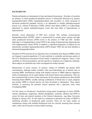
REFERENCES:
1. Fahim F. The magnitude of low bone mineral [corrected] density in middle and old
age women. J Pak Med Assoc. 2005 Nov;55(11):500-2.
2. Dar FJ, Iqbal R, Ghani F, Siddiqui I, Khan AH. Bone health status of premenopausal
healthy adult females in Pakistani females. Arch Osteoporos. 2012, Dec;7(1-2):93-9.
3. Khan AH, Iqbal R, Naureen G, Dar FJ, Ahmed FN. Prevalence of vitamin D
deficiency and its correlates: results of a community-based study conducted in Karachi,
Pakistan. Arch Osteoporos. 2012, Dec;7(1-2):275-82.
4. Lundgren E, Rastad J, Thrufjell E, Akerstrom G, Ljunghall S. Population-based
screening for primary hyperparathyroidism with serum calcium and parathyroid hormone
values in menopausal women. Surgery. 1997 Mar;121(3):287-94.
5. Lundgren E, Hagstrom EG, Lundin J, Winnerback K, Roos J, Ljunghall S, et al.
Primary hyperparathyroidism revisited in menopausal women with serum calcium in the
upper normal range at population-based screening 8 years ago. World J Surg. 2002
Aug;26(8):931-6.
6. Boonen S, Vanderschueren D, Pelemans W, Bouillon R. Primary
hyperparathyroidism: diagnosis and management in the older individual. Eur J Endocrinol.
2004 Sep;151(3):297-304.
7. Lindstedt G, Nystrom E, Lundberg PA, Johansson E, Eggertsen R. Screening of an
elderly population in primary care for primary hyperparathyroidism. Scand J Prim Health
Care. 1992 Sep;10(3):192-7.
8. Harvey A, Hu M, Gupta M, Butler R, Mitchell J, Berber E, et al. A new, vitamin D-
based, multidimensional nomogram for the diagnosis of primary hyperparathyroidism.
Endocr Pract. 2011, Mar-Apr;18(2):124-31.
9. Suh JM, Cronan JJ, Monchik JM. Primary hyperparathyroidism: is there an increased
prevalence of renal stone disease? AJR Am J Roentgenol. 2008 Sep;191(3):908-11.
10. Lumachi F, Motta R, Cecchin D, Ave S, Camozzi V, Basso SM, et al. Calcium
metabolism & hypercalcemia in adults. Curr Med Chem. 2011;18(23):3529-36.
11. Bilezikian JP, Khan AA, Potts Jr JT. Guidelines for the management of asymptomatic
primary hyperparathyroidism: summary statement from the third international workshop. The
Journal of Clinical Endocrinology & Metabolism. 2009;94(2):335-9.](https://image.slidesharecdn.com/bd2908e7-c285-4c34-9681-846c83298980-160602112910/85/IDENTIFYING-PARATHYROID-HORMONE-DISORDERS-AND-ITS-PHENOTYPES-THROUGH-A-BONE-HEALTH-SCREENING-PANEL-IT-S-NOT-SIMPLE-VITAMIN-D-DEFICIENCY-12-320.jpg)
![13
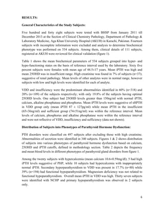
12. Jubiz W, Canterbury JM, Reiss E, Tyler FH. Circadian rhythm in serum parathyroid
hormone concentration in human subjects: correlation with serum calcium, phosphate,
albumin, and growth hormone levels. J Clin Invest. 1972 Aug;51(8):2040-6.
13. Cusano NE, Maalouf NM, Wang PY, Zhang C, Cremers SC, Haney EM, et al.
Normocalcemic hyperparathyroidism and hypoparathyroidism in two community-based
nonreferral populations. J Clin Endocrinol Metab. Jul;98(7):2734-41.
14. Campenni A, Ruggeri RM, Sindoni A, Giovinazzo S, Calbo E, Ieni A, et al.
Parathyroid carcinoma presenting as normocalcemic hyperparathyroidism. J Bone Miner
Metab. May;30(3):367-72.
15. Udelsman R, Lin Z, Donovan P. The superiority of minimally invasive
parathyroidectomy based on 1650 consecutive patients with primary hyperparathyroidism.
Ann Surg. Mar;253(3):585-91.
16. Wallace LB, Parikh RT, Ross LV, Mazzaglia PJ, Foley C, Shin JJ, et al. The
phenotype of primary hyperparathyroidism with normal parathyroid hormone levels: how
low can parathyroid hormone go? Surgery. Dec;150(6):1102-12.
17. Martin KJ, Gonzalez EA. Metabolic bone disease in chronic kidney disease. J Am
Soc Nephrol. 2007 Mar;18(3):875-85.
18. Jensen CE, Tuck SM, Agnew JE, Koneru S, Morris RW, Yardumian A, et al. High
prevalence of low bone mass in thalassaemia major. Br J Haematol. 1998 Dec;103(4):911-5.
19. Haidar R, Musallam KM, Taher AT. Bone disease and skeletal complications in
patients with beta thalassemia major. Bone. 2010, Mar;48(3):425-32.
20. Svara F. Chronic kidney disease-mineral and bone disorder (CKD-MBD): a new term
for a complex approach. J Ren Care. 2009 Mar;35 Suppl 1:3-6.
21. Hamada Y, Fukagawa M. [Chronic kidney disease (CKD) and bone. The mechanisms
of chronic kidney disease--mineral and bone disorder (CKD-MBD)]. Clin Calcium. 2009
Apr;19(4):486-92.
22. Tanko LB, Karsdal MA, Christiansen C, Leeming DJ. Biochemical approach to the
detection and monitoring of metastatic bone disease: What do we know and what questions
need answers? Cancer Metastasis Rev. 2006 Dec;25(4):659-68.](https://image.slidesharecdn.com/bd2908e7-c285-4c34-9681-846c83298980-160602112910/85/IDENTIFYING-PARATHYROID-HORMONE-DISORDERS-AND-ITS-PHENOTYPES-THROUGH-A-BONE-HEALTH-SCREENING-PANEL-IT-S-NOT-SIMPLE-VITAMIN-D-DEFICIENCY-13-320.jpg)

The study investigates the efficacy of a bone health screening panel (BHSP) in identifying parathyroid hormone disorders, revealing that 68% of subjects exhibited abnormal PTH secretion, with various phenotypes identified such as primary hyperparathyroidism and functional hypoparathyroidism. The research emphasizes the utility of simultaneous testing for metabolic bone disorders to enhance diagnostic accuracy, particularly in cases of atypical biochemical findings. The findings advocate for increased awareness and early diagnosis of parathyroid dysfunction, which is often misattributed solely to vitamin D deficiency.


![PERI-PROSTHETIC FRACTURE NAIL-PLATE CONSTRUCT [NPC].pptx](https://cdn.slidesharecdn.com/ss_thumbnails/drarunkumardrmohamedashrafperiprostheticfrasturenail-plateconstructnpc-260209164459-7e9d15a1-thumbnail.jpg?width=640&height=640&fit=bounds)


