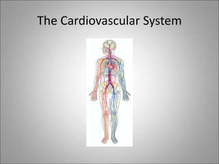
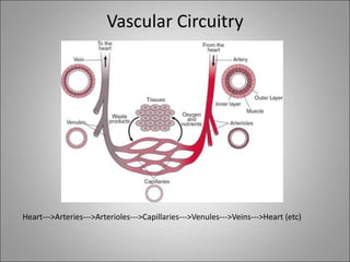
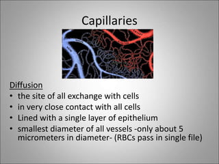
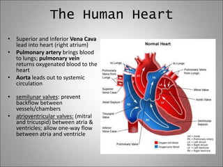
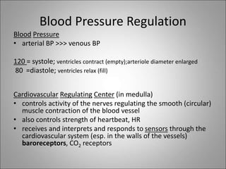
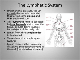
The circulatory system transports blood, nutrients, oxygen, hormones, and waste products throughout the body. Blood contains plasma, red blood cells, white blood cells, and platelets. The heart pumps blood through a closed loop system of arteries, veins, and capillaries. Arteries carry oxygenated blood away from the heart while veins return deoxygenated blood back to the heart. The lymphatic system drains lymph fluid and waste from tissues and returns it to the blood.
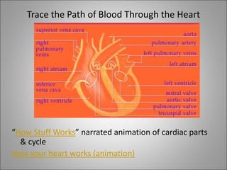
Cardiovascular System + Key Terms + Disease AreaNouman Minhas
Comprehensive presentation on Cardiovascular system.
It covers the Anatomy and Physiology of CV system.
It covers the Related Terms i.e Cardiac Output etc
It covers major diseases related to CV systems .
.............YOU will FIND it USEFUL...................
This presentation is a combination of different slides which I re-purposed. I included a reference of all the slides I used at the end of my presentation.
A powerpoint designed for the South African Life Sciences syllabus for grade 11. Includes information about blood and it's transportation, the human heart, the lymph system etc. Hope it helps :)
1 GNM - Anatomy unit - 4 - CVS by thirumurugan.pptxthiru murugan
By:M. Thiru murugan
Unit – IV:
Heart : Structure, functions including conduction system & cardiac cycle
Blood vessels : Types, Structure and position
Circulation of blood
Blood pressure and pulse
Heart
The circulatory system:
It consisting of blood, blood vessels, and heart.
This supplies oxygen and other nutrients,
Transports hormones
Removes unnecessary waste products.
Heart and its Structure
The heart is a muscular organ about the size of a fist,
located in mediastinum just behind and slightly left of the breastbone (sternum).
The heart pumps blood through the blood vessels (arteries and veins called the cardiovascular system).
Structure of heart:
Layers of the heart (3)
Chambers of the heart (4)
Valves of the heart (4)
Blood vessels of the heart (5)
3 layers of the heart:
Epicardium/pericardium: outer protective layer of the heart. Visceral and parietal (pericardial fluid). Protection for the heart and big vessels and prevent collapse of heart,
Myocardium: muscular middle layer wall of the heart. Responsible for keeping the heart pumping blood around the body.
Endocardium: the inner layer of the heart. Regulate blood flow through the chambers of the heart and pass the electrical impulses
Chambers of the heart:
The atria: These are the 2 upper chambers, which receive blood. RA / LA
The ventricles: These are the 2 lower chambers, which discharge blood. RV/ LV
A wall of tissue called the septum separates the left and right atria called atrial septum and the left and right ventricle called ventricular septum.
Valves in the heart:
There are four valves
Two-atrio ventricular valves: The 2 types: bicuspid (mitral) - LA & LV, and tricuspid valves - RA & RV.
Two-semilunar valves: The aortic valves and the pulmonary valve.
Major blood vessels of the heart
There are 5 major blood vessels
Pulmonary artery
Pulmonary veins
Aorta[artery]
Inferior vena cava [IVC] veins
Superior vena cava [SVC] veins
Functions of heart:
Pumping oxygenated blood to the body parts.
Pumping nutrients and other vital substances
Receiving deoxygenated blood and carrying metabolic waste products from the body
Pumping deoxygenated blood to the lungs for oxygenation.
Maintaining blood pressure.
Conduction system
The electrical conduction system that controls the heart rate.
This system generates electrical impulses and conducts them throughout the muscle of the heart, stimulating the heart to contract and pump blood.
The electrical pulses determine the order in which the chambers contract & the heart rate
Conductive system consist of:
SA Node
AV Node
Bundle of his or His Bundles – bundle of branches
( right and left)
4. Purkinje fibres
Sinoatrial node (SA) : also known as the pace maker of the heart and Located in the upper wall of the right atrium
Made up of both muscle and nervous tissue
Here the electrical impulse begins
Atrioventricular (AV) node:
located between the atria and ventricles of the heart
The electrical impulse is carried fr
Introduction:
RNA interference (RNAi) or Post-Transcriptional Gene Silencing (PTGS) is an important biological process for modulating eukaryotic gene expression.
It is highly conserved process of posttranscriptional gene silencing by which double stranded RNA (dsRNA) causes sequence-specific degradation of mRNA sequences.
dsRNA-induced gene silencing (RNAi) is reported in a wide range of eukaryotes ranging from worms, insects, mammals and plants.
This process mediates resistance to both endogenous parasitic and exogenous pathogenic nucleic acids, and regulates the expression of protein-coding genes.
What are small ncRNAs?
micro RNA (miRNA)
short interfering RNA (siRNA)
Properties of small non-coding RNA:
Involved in silencing mRNA transcripts.
Called “small” because they are usually only about 21-24 nucleotides long.
Synthesized by first cutting up longer precursor sequences (like the 61nt one that Lee discovered).
Silence an mRNA by base pairing with some sequence on the mRNA.
Discovery of siRNA?
The first small RNA:
In 1993 Rosalind Lee (Victor Ambros lab) was studying a non- coding gene in C. elegans, lin-4, that was involved in silencing of another gene, lin-14, at the appropriate time in the
development of the worm C. elegans.
Two small transcripts of lin-4 (22nt and 61nt) were found to be complementary to a sequence in the 3' UTR of lin-14.
Because lin-4 encoded no protein, she deduced that it must be these transcripts that are causing the silencing by RNA-RNA interactions.
Types of RNAi ( non coding RNA)
MiRNA
Length (23-25 nt)
Trans acting
Binds with target MRNA in mismatch
Translation inhibition
Si RNA
Length 21 nt.
Cis acting
Bind with target Mrna in perfect complementary sequence
Piwi-RNA
Length ; 25 to 36 nt.
Expressed in Germ Cells
Regulates trnasposomes activity
MECHANISM OF RNAI:
First the double-stranded RNA teams up with a protein complex named Dicer, which cuts the long RNA into short pieces.
Then another protein complex called RISC (RNA-induced silencing complex) discards one of the two RNA strands.
The RISC-docked, single-stranded RNA then pairs with the homologous mRNA and destroys it.
THE RISC COMPLEX:
RISC is large(>500kD) RNA multi- protein Binding complex which triggers MRNA degradation in response to MRNA
Unwinding of double stranded Si RNA by ATP independent Helicase
Active component of RISC is Ago proteins( ENDONUCLEASE) which cleave target MRNA.
DICER: endonuclease (RNase Family III)
Argonaute: Central Component of the RNA-Induced Silencing Complex (RISC)
One strand of the dsRNA produced by Dicer is retained in the RISC complex in association with Argonaute
ARGONAUTE PROTEIN :
1.PAZ(PIWI/Argonaute/ Zwille)- Recognition of target MRNA
2.PIWI (p-element induced wimpy Testis)- breaks Phosphodiester bond of mRNA.)RNAse H activity.
MiRNA:
The Double-stranded RNAs are naturally produced in eukaryotic cells during development, and they have a key role in regulating gene expression .
Earliest Galaxies in the JADES Origins Field: Luminosity Function and Cosmic ...Sérgio Sacani
We characterize the earliest galaxy population in the JADES Origins Field (JOF), the deepest
imaging field observed with JWST. We make use of the ancillary Hubble optical images (5 filters
spanning 0.4−0.9µm) and novel JWST images with 14 filters spanning 0.8−5µm, including 7 mediumband filters, and reaching total exposure times of up to 46 hours per filter. We combine all our data
at > 2.3µm to construct an ultradeep image, reaching as deep as ≈ 31.4 AB mag in the stack and
30.3-31.0 AB mag (5σ, r = 0.1” circular aperture) in individual filters. We measure photometric
redshifts and use robust selection criteria to identify a sample of eight galaxy candidates at redshifts
z = 11.5 − 15. These objects show compact half-light radii of R1/2 ∼ 50 − 200pc, stellar masses of
M⋆ ∼ 107−108M⊙, and star-formation rates of SFR ∼ 0.1−1 M⊙ yr−1
. Our search finds no candidates
at 15 < z < 20, placing upper limits at these redshifts. We develop a forward modeling approach to
infer the properties of the evolving luminosity function without binning in redshift or luminosity that
marginalizes over the photometric redshift uncertainty of our candidate galaxies and incorporates the
impact of non-detections. We find a z = 12 luminosity function in good agreement with prior results,
and that the luminosity function normalization and UV luminosity density decline by a factor of ∼ 2.5
from z = 12 to z = 14. We discuss the possible implications of our results in the context of theoretical
models for evolution of the dark matter halo mass function.
Slide 1: Title Slide
Extrachromosomal Inheritance
Slide 2: Introduction to Extrachromosomal Inheritance
Definition: Extrachromosomal inheritance refers to the transmission of genetic material that is not found within the nucleus.
Key Components: Involves genes located in mitochondria, chloroplasts, and plasmids.
Slide 3: Mitochondrial Inheritance
Mitochondria: Organelles responsible for energy production.
Mitochondrial DNA (mtDNA): Circular DNA molecule found in mitochondria.
Inheritance Pattern: Maternally inherited, meaning it is passed from mothers to all their offspring.
Diseases: Examples include Leber’s hereditary optic neuropathy (LHON) and mitochondrial myopathy.
Slide 4: Chloroplast Inheritance
Chloroplasts: Organelles responsible for photosynthesis in plants.
Chloroplast DNA (cpDNA): Circular DNA molecule found in chloroplasts.
Inheritance Pattern: Often maternally inherited in most plants, but can vary in some species.
Examples: Variegation in plants, where leaf color patterns are determined by chloroplast DNA.
Slide 5: Plasmid Inheritance
Plasmids: Small, circular DNA molecules found in bacteria and some eukaryotes.
Features: Can carry antibiotic resistance genes and can be transferred between cells through processes like conjugation.
Significance: Important in biotechnology for gene cloning and genetic engineering.
Slide 6: Mechanisms of Extrachromosomal Inheritance
Non-Mendelian Patterns: Do not follow Mendel’s laws of inheritance.
Cytoplasmic Segregation: During cell division, organelles like mitochondria and chloroplasts are randomly distributed to daughter cells.
Heteroplasmy: Presence of more than one type of organellar genome within a cell, leading to variation in expression.
Slide 7: Examples of Extrachromosomal Inheritance
Four O’clock Plant (Mirabilis jalapa): Shows variegated leaves due to different cpDNA in leaf cells.
Petite Mutants in Yeast: Result from mutations in mitochondrial DNA affecting respiration.
Slide 8: Importance of Extrachromosomal Inheritance
Evolution: Provides insight into the evolution of eukaryotic cells.
Medicine: Understanding mitochondrial inheritance helps in diagnosing and treating mitochondrial diseases.
Agriculture: Chloroplast inheritance can be used in plant breeding and genetic modification.
Slide 9: Recent Research and Advances
Gene Editing: Techniques like CRISPR-Cas9 are being used to edit mitochondrial and chloroplast DNA.
Therapies: Development of mitochondrial replacement therapy (MRT) for preventing mitochondrial diseases.
Slide 10: Conclusion
Summary: Extrachromosomal inheritance involves the transmission of genetic material outside the nucleus and plays a crucial role in genetics, medicine, and biotechnology.
Future Directions: Continued research and technological advancements hold promise for new treatments and applications.
Slide 11: Questions and Discussion
Invite Audience: Open the floor for any questions or further discussion on the topic.
Cancer cell metabolism: special Reference to Lactate PathwayAADYARAJPANDEY1
Normal Cell Metabolism:
Cellular respiration describes the series of steps that cells use to break down sugar and other chemicals to get the energy we need to function.
Energy is stored in the bonds of glucose and when glucose is broken down, much of that energy is released.
Cell utilize energy in the form of ATP.
The first step of respiration is called glycolysis. In a series of steps, glycolysis breaks glucose into two smaller molecules - a chemical called pyruvate. A small amount of ATP is formed during this process.
Most healthy cells continue the breakdown in a second process, called the Kreb's cycle. The Kreb's cycle allows cells to “burn” the pyruvates made in glycolysis to get more ATP.
The last step in the breakdown of glucose is called oxidative phosphorylation (Ox-Phos).
It takes place in specialized cell structures called mitochondria. This process produces a large amount of ATP. Importantly, cells need oxygen to complete oxidative phosphorylation.
If a cell completes only glycolysis, only 2 molecules of ATP are made per glucose. However, if the cell completes the entire respiration process (glycolysis - Kreb's - oxidative phosphorylation), about 36 molecules of ATP are created, giving it much more energy to use.
IN CANCER CELL:
Unlike healthy cells that "burn" the entire molecule of sugar to capture a large amount of energy as ATP, cancer cells are wasteful.
Cancer cells only partially break down sugar molecules. They overuse the first step of respiration, glycolysis. They frequently do not complete the second step, oxidative phosphorylation.
This results in only 2 molecules of ATP per each glucose molecule instead of the 36 or so ATPs healthy cells gain. As a result, cancer cells need to use a lot more sugar molecules to get enough energy to survive.
introduction to WARBERG PHENOMENA:
WARBURG EFFECT Usually, cancer cells are highly glycolytic (glucose addiction) and take up more glucose than do normal cells from outside.
Otto Heinrich Warburg (; 8 October 1883 – 1 August 1970) In 1931 was awarded the Nobel Prize in Physiology for his "discovery of the nature and mode of action of the respiratory enzyme.
WARNBURG EFFECT : cancer cells under aerobic (well-oxygenated) conditions to metabolize glucose to lactate (aerobic glycolysis) is known as the Warburg effect. Warburg made the observation that tumor slices consume glucose and secrete lactate at a higher rate than normal tissues.
Unveiling the Energy Potential of Marshmallow Deposits.pdf
5786327.ppt
1. Chapter 47-1, 2
Circulatory System
Blood: the "chemical highway" that
connects the many cells of an organism
•Carries nutrients, oxygen to each cell
•Carries wastes away from cells (CO2, urea)
to kidneys, lungs, skin
•Carries enzymes, hormones, water,
antibodies around body for distribution
2. Components of Blood
•Men, on average, have
about 10-12 pints
•Women, on average, have
about 8-10 pints
•each cc (cubic mL) of blood
contains about 4 million
RBCs and 7,000 WBCs
3. Plasma
Plasma = 60% of blood; straw-colored fluid
• about 90% of the plasma is water
• carries most CO2, nutrients, plasma proteins,
hormones, etc.
plasma proteins:
• albumin (to maintain high osmotic potential)
• fibrinogen (clotting)
• globulins (immunity, antibodies)
4. Red Blood Cells
• Called “erythrocytes”
• Transport oxygen; about 30 trillion in
bloodstream
• contain HEMOGLOBIN (red pigment protein)
which acts like an "oxygen magnet"; iron
• non-nucleated (cannot repair themselves)
• short lifespan (120-130 days)
• Replaced from bone marrow stem cells
5. White Blood Cells
• called “leukocytes”
• 1 or 2 WBC for every 1,000 RBC
• larger than RBC, have nucleus, no hemoglobin,
colorless
• Can migrate outside of vessels (RBC’s cannot) to
directly cleanse tissues (lymph)
• Some act by: phagocytosis of bacteria/viruses/etc.
• Others make antibodies (proteins) that attack
microorganisms
• "Pus"-dead WBC; new WBC form in spleen, bone
marrow
6. Platelets
• colorless, oval/irregular shaped
• smaller than RBCs
• "little bags of chemicals" that function in
clotting and plugging up breaks in vessels
Blood Clotting- a complex series of chemical reactions, involving platelets and plasma
"clotting factors". Often referred to as the "clotting cascade".
Thromboplastin
(produced in liver)
Prothrombin------------------------------>Thrombin
Fibrinogen---------------------------------------------> Fibrin
15 CF’s involved in this conversation (Hemophiliacs usually
genetically lack Factor VIII)
clotting animation
9. Arteries
• thick, muscular walls,
• elastic, to allow for high pressure
• made of layers of
endothelium/muscle/connective tissues
• Narrow down into smaller arterioles
10. Veins
• carry blood back toward the heart
• thinner walls (than arteries)
• less elastic (less pressure to withstand)
• have valves to prevent backflow
• endothelium, muscle, connective tissue layers
• narrow down into smaller diameter venules
12. Capillaries
Diffusion
• the site of all exchange with cells
• in very close contact with all cells
• Lined with a single layer of epithelium
• smallest diameter of all vessels -only about 5
micrometers in diameter- (RBCs pass in single file)
13. Atherosclerosis
•…is a disease affecting arterial
blood vessels.
•Main cause: cholesterol buildup
in arteries (plaques)
•It is commonly referred to as
"hardening" of the arteries.
• It is caused by the formation of
multiple plaques within the
arteries
•This often leads not only to
clogs, but also hypertension
(high blood pressure), which
both may result in heart attacks.
15. Evolution of the Heart
• simplest (in earthworms)
are enlarged, muscular
portions of a blood vessel =
"aortic arches"
•Mollusks= tube shaped heart with open
circulation
•Vertebrates= multi-chambered heart with
closed circulation
•multiple chambers serve to separate
oxygenated/ deoxygenated blood
•Atrium (receiving chamber)
•Ventricle (pumping chamber)
16. The Human Heart
is made of epithelial, nerve, connective tissues and
cardiac muscle (infatiguable)
Four chambers (2 atria,
2 ventricles)
septum- dividing wall
between right and left
side of heart
valves at strategic
locations prevent
backflow
separation into
chambers prevents
mixing oxygenated,
deoxygenated blood
Do right and left seem
backward? That's because
you're looking at an
illustration of somebody
else's heart. To think about
how your own heart works,
imagine wearing this
illustration on your chest.
Did you know that your heart
beats over 100,000 times a
day? Get "heart smart" by
checking out these amazing
heart facts.
17. The Human Heart
• Superior and Inferior Vena Cava
lead into heart (right atrium)
• Pulmonary artery brings blood
to lungs; pulmonary vein
returns oxygenated blood to the
heart
• Aorta leads out to systemic
circulation
• semilunar valves: prevent
backflow between
vessels/chambers
• atrioventricular valves: (mitral
and tricuspid) between atria &
ventricles; allow one-way flow
between atria and ventricle
18. Trace the Path of Blood Through the Heart
“How Stuff Works” narrated animation of cardiac parts
& cycle
How your heart works (animation)
19. Initiation and Regulation of Heartbeat
• Cardiac muscle is able to initiate its own
impulse
• Sinoatrial Node (SA)- "pacemaker" in Right
Atrium; initiates beat through both Atria;
stimulates another area near center of
heart called AV Node
• Atrioventicular Node (AV)...then impulse is
carried to the ventricles by the BH
• Bundle of His (BH) = like the transfer of an
electrical current along a circuit
• Heart Rate (bpm) is modified by the
Autonomic Nervous System (vagus nerve)
– Parasympathetic Nerves- slow down HR
– Sympathetic Nerves- increase HR
20. 1. Oxygen-poor blood (shown in
blue) flows from the body into
the right atrium.
2. Blood flows through the right
atrium into the right ventricle.
3. The right ventricle pumps the
blood to the lungs, where the
blood releases waste gases and
picks up oxygen.
4. The newly oxygen-rich blood
(shown in red) returns to the
heart and enters the left atrium.
5. Blood flows through the left
atrium into the left ventricle.
6. The left ventricle pumps the
oxygen-rich blood to all parts of
the body.
To assess heart rate
(BPM), take your arterial
pulse!
@ rest?
After exercise?
21. How much blood does the heart
pump?
• The heart pumps the equivalent of 5,000 to 6,000
quarts (about 6800 L) of blood each day!
• Total volume of blood pumped by heart per minute =
“cardiac output”
• Cardiac Output = Heart Rate x Stroke volume
• (L/min) = (BPM) x (L/beat)
• [ex: 72 BPM x 0.07 L/beat = 5 L/min.]
22. Blood Pressure Regulation
Blood Pressure
• arterial BP >>> venous BP
120 = systole; ventricles contract (empty);arteriole diameter enlarged
80 =diastole; ventricles relax (fill)
Cardiovascular Regulating Center (in medulla)
• controls activity of the nerves regulating the smooth (circular)
muscle contraction of the blood vessel
• also controls strength of heartbeat, HR
• receives and interprets and responds to sensors through the
cardiovascular system (esp. in the walls of the vessels)
baroreceptors, CO2 receptors
23. The Lymphatic System
• Under arterial pressure, the BP
exceeds the osmotic potential,
thus forcing some plasma and
WBC out into tissues.
• This “lymphatic fluid” is collected
by Lymph vessels which clean the
tissue/ cellular debris (such as
bacteria) out of the body cells.
• Lymph flows thru Lymph Nodes
to be cleaned
• These also make Lymphocytes
(WBC’s)
• Lymph re-enters the circulatory
vessels via the Subclavian Veins in
the neck (back into bloodstream)