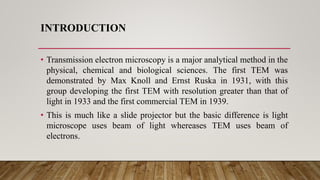
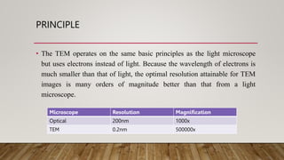
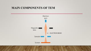
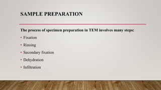
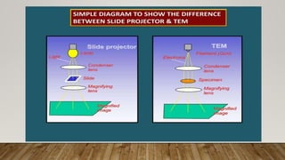
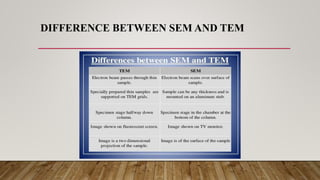
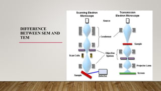
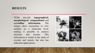
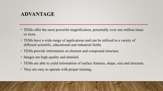
POLYMER CHARACTERIZATION 4102
Transmission
Electron Microscope
Presented by
Faizan Alam 18-NTU-1005
M Qasim 18-NTU-1021
Presented to
Dr. Zakariya Zubair
CONTENTS
• Introduction
• Principal
• Parts
• Sample preparation
• Working
• Application
• Advantage & Disadvantage
INTRODUCTION
• Transmission electron microscopy is a major analytical method in the
physical, chemical and biological sciences. The first TEM was
demonstrated by Max Knoll and Ernst Ruska in 1931, with this
group developing the first TEM with resolution greater than that of
light in 1933 and the first commercial TEM in 1939.
• This is much like a slide projector but the basic difference is light
microscope uses beam of light whereases TEM uses beam of
electrons.
WHAT IS TEM
• TEM is microscopy technique
in which a beam of electrons
is transmitted through an ultra
thin specimen, interacting
with the specimen as it passes
through.
PRINCIPLE
• The TEM operates on the same basic principles as the light microscope
but uses electrons instead of light. Because the wavelength of electrons is
much smaller than that of light, the optimal resolution attainable for TEM
images is many orders of magnitude better than that from a light
microscope.
Microscope Resolution Magnification
Optical 200nm 1000x
TEM 0.2nm 500000x
MAIN COMPONENTS OF TEM
• The Gun – which produces electron.
• The condenser system – which perform the probe
• The sample – sample preparation is important, and time consuming.
• Image formation – use of image plane or back focal plane.
• Intermediate lens – transmitting and magnifying the first enlarged diffraction or
image pattern to projector lens.
• Projection of the image viewing and recording.
MAIN COMPONENTS OF TEM
WORKING
• In a TEM the electron beam is focused on the sample using the condenser
lens system.
• This produces an image which is focused by the objective lens to a point.
• This image is then magnified by a series of projector lenses to vary the size
of the image on a fluorescent screen.
• Changing the current of an electromagnetic lens alters its focal length altering
magnification.
DIAGRAM TO PRESENT TEM WORKING
SAMPLE PREPARATION
The process of specimen preparation in TEM involves many steps:
• Fixation
• Rinsing
• Secondary fixation
• Dehydration
• Infiltration
DIFFERENCE BETWEEN SEM AND TEM
DIFFERENCE
BETWEEN SEM AND
TEM
RESULTS
• TEMs provide topographical,
morphological, compositional and
crystalline information. The
images allow researchers to view
samples on a molecular level,
making it possible to analyze
structure and texture. This
information is useful in the study of
crystals and metals, but also has
industrial applications.
•
GRAPH
APPLICATIONS
• Medical
• Life sciences
• Nanotechnology
• Forensic analysis
• Biological and material research
• Gemology and metallurgy
• Industry and education
ADVANTAGE
• TEMs offer the most powerful magnification, potentially over one million times
or more.
• TEMs have a wide-range of applications and can be utilized in a variety of
different scientific, educational and industrial fields.
• TEMs provide information on element and compound structure.
• Images are high-quality and detailed.
• TEMs are able to yield information of surface features, shape, size and structure.
• They are easy to operate with proper training.
DISADVANTAGE
• TEMs are large and very expensive
• Laborious sample preparation
• Operation and analysis requires special training
• TEMs require special housing and maintenance
• Images are black and white
5,21 (TEM).pptx
5,21 (TEM).pptx

5,21 (TEM).pptx

  • 2.
    POLYMER CHARACTERIZATION 4102 Transmission ElectronMicroscope Presented by Faizan Alam 18-NTU-1005 M Qasim 18-NTU-1021 Presented to Dr. Zakariya Zubair
  • 3.
    CONTENTS • Introduction • Principal •Parts • Sample preparation • Working • Application • Advantage & Disadvantage
  • 4.
    INTRODUCTION • Transmission electronmicroscopy is a major analytical method in the physical, chemical and biological sciences. The first TEM was demonstrated by Max Knoll and Ernst Ruska in 1931, with this group developing the first TEM with resolution greater than that of light in 1933 and the first commercial TEM in 1939. • This is much like a slide projector but the basic difference is light microscope uses beam of light whereases TEM uses beam of electrons.
  • 5.
    WHAT IS TEM •TEM is microscopy technique in which a beam of electrons is transmitted through an ultra thin specimen, interacting with the specimen as it passes through.
  • 6.
    PRINCIPLE • The TEMoperates on the same basic principles as the light microscope but uses electrons instead of light. Because the wavelength of electrons is much smaller than that of light, the optimal resolution attainable for TEM images is many orders of magnitude better than that from a light microscope. Microscope Resolution Magnification Optical 200nm 1000x TEM 0.2nm 500000x
  • 7.
    MAIN COMPONENTS OFTEM • The Gun – which produces electron. • The condenser system – which perform the probe • The sample – sample preparation is important, and time consuming. • Image formation – use of image plane or back focal plane. • Intermediate lens – transmitting and magnifying the first enlarged diffraction or image pattern to projector lens. • Projection of the image viewing and recording.
  • 8.
  • 9.
    WORKING • In aTEM the electron beam is focused on the sample using the condenser lens system. • This produces an image which is focused by the objective lens to a point. • This image is then magnified by a series of projector lenses to vary the size of the image on a fluorescent screen. • Changing the current of an electromagnetic lens alters its focal length altering magnification.
  • 10.
    DIAGRAM TO PRESENTTEM WORKING
  • 11.
    SAMPLE PREPARATION The processof specimen preparation in TEM involves many steps: • Fixation • Rinsing • Secondary fixation • Dehydration • Infiltration
  • 13.
  • 14.
  • 15.
    RESULTS • TEMs providetopographical, morphological, compositional and crystalline information. The images allow researchers to view samples on a molecular level, making it possible to analyze structure and texture. This information is useful in the study of crystals and metals, but also has industrial applications. •
  • 16.
  • 17.
    APPLICATIONS • Medical • Lifesciences • Nanotechnology • Forensic analysis • Biological and material research • Gemology and metallurgy • Industry and education
  • 18.
    ADVANTAGE • TEMs offerthe most powerful magnification, potentially over one million times or more. • TEMs have a wide-range of applications and can be utilized in a variety of different scientific, educational and industrial fields. • TEMs provide information on element and compound structure. • Images are high-quality and detailed. • TEMs are able to yield information of surface features, shape, size and structure. • They are easy to operate with proper training.
  • 19.
    DISADVANTAGE • TEMs arelarge and very expensive • Laborious sample preparation • Operation and analysis requires special training • TEMs require special housing and maintenance • Images are black and white