
Recommended
More Related Content
What's hot
What's hot (20)
Similar to 40484967
Similar to 40484967 (20)
Recently uploaded
Recently uploaded (20)
40484967
- 1. Modeling Early Retinal Development with Human Embryonic and Induced Pluripotent Stem Cells Author(s): Jason S. Meyer, Rebecca L. Shearer, Elizabeth E. Capowski, Lynda S. Wright, Kyle A. Wallace, Erin L. Mcmillan, Su-chun Zhang, David M. Gamm, James Thomsan Reviewed work(s): Source: Proceedings of the National Academy of Sciences of the United States of America, Vol. 106, No. 39 (Sep. 29, 2009), pp. 16698-16703 Published by: National Academy of Sciences Stable URL: http://www.jstor.org/stable/40484967 . Accessed: 02/02/2012 02:51 Your use of the JSTOR archive indicates your acceptance of the Terms & Conditions of Use, available at . http://www.jstor.org/page/info/about/policies/terms.jsp JSTOR is a not-for-profit service that helps scholars, researchers, and students discover, use, and build upon a wide range of content in a trusted digital archive. We use information technology and tools to increase productivity and facilitate new forms of scholarship. For more information about JSTOR, please contact support@jstor.org. National Academy of Sciences is collaborating with JSTOR to digitize, preserve and extend access to Proceedings of the National Academy of Sciences of the United States of America. http://www.jstor.org
- 2. earlyretinal Modeling development withhuman embryonic induced and stem pluripotent cells Jason S. Meyer3,Rebecca L Shearer3, Elizabeth E. Capowski8, Lynda S. Wright8, Kyle A. Wallace3, ErinL McMillan8, Su-ChunZhang3b, and David M. Gamma'cd'1 aStem Cell Research Program,Waisman Center,bDepartmentsof Anatomy and Neurology,department of Ophthalmology and Visual Sciences, and dEye 1500 Highland Avenue, University Wisconsin-Madison,Madison Wl 53705 Research Institute, of Edited by JamesThomson, University Wisconsin,Madison, Wl, and approved July 2009 (received for review May 15, 2009) of 23, Human pluripotent stem cells have the potentialto provide compre- and maturation follows sequenceand timecourse highly a hensivemodel systemsforthe earlieststages of human ontogenesis. reminiscent normal of retinal development. Furthermore, the To serve in this capacity, these cells must undergo a targeted, process retinal of differentiation be selectively could altered via stepwise differentiation process that follows a normaldevelopmen- of manipulation endogenous developmental signaling pathways. tal timeline.Here we demonstratethe abilityof both human embry- We theninvestigated whether same culture the method was onicstemcells(hESCs)and inducedpluripotent stem(iPS) cellsto meet capable ofgenerating identical an cohort developing of retinal these requirements human retinogenesis.Upon differentiation, celltypes for from human induced stem a pluripotent cells, recently hESCsinitially yieldeda highly enrichedpopulation of earlyeye field described sourceof pluripotent cells derived stem from skin cells. Thereafter, subset of cells acquired features of advancing a fibroblasts 11). Cell populations (10, expressing morphologic retinal differentiation a sequence and timecourse that mimicked in in features and/or markers theeyefield, of retinalpigment epithe- vivo human retinaldevelopment.Applicationof this culturemethod lium,neural retinalprogenitors, photoreceptorprecursors,and to a human iPS cell line also generated retina-specific types at cell photoreceptors observed differentiating iPS cell were in human comparable times in vitro. Lastly,altering endogenous signaling cultures time at points predicted results by using hESCs.These duringdifferentiation affectedlineage-specific gene expression in a findings support roleforhuman a pluripotent cellsas in stem mannerconsistentwith established mechanismsof early neural and vitromodel to systems investigate mechanisms involved retinal in retinal cell fate determination.These findings should aid in the anddifferentiationofindividual retinal types. cell specification investigation the molecularevents governingretinalspecification of fromhuman pluripotent stem cells. Results Eye Specification Human Field from Stem The Embryonic Cells. appear- study human of development is limited a lackofmodel anceofeyefield by cellswithin primitive anterior neuroepithelium systems canreproduce precise that the and of sequence timing is thefirst phaseinthestepwise production a retinal of pheno- cellular molecular and events occurduring that human embryo- type from undifferentiated an pluripotent cell(12,13)(Fig. stem genesis, organogénesis, tissue and differentiation. However, the L4). Previous reports have demonstrated hESC-derived that advent human of pluripotent celltechnology stem affords a unique neuroectodermal willadoptanterior cells neuroepithelial char- opportunity to follow full of cell the course lineage-specific pro- acteristics theabsence exogenous in of signaling molecules (14, duction vitro 2). The retina in (1, an provides optimal system to 15).In thecurrent study (Fig.'B), hESCswere differentiated as investigate potential to itswell-defined conserved free-floating this due and hESC aggregates and prompted adhereto to developmental program theavailabilitymarkers distin- laminin-coated and of to culture dishes permit to neuralrosette forma- guish eachmajor of stage early retinogenesis. In addition, human tion. 16 of After days differentiation, rosette-containing colonies embryonic cells stem (hESCs)displaypropensitya toproduce cells were to as removed grow neurospheres. During this mechanically with retinal characteristics Onecriterion assessing (3,4). for hESC- hESCsrapidly expression thepluripotency lost of process, genes based model is to developmental systemsthecapacity recapitulate Oct4andNanog acquired and of factors in the embryo a associated in expression transcription the normal maturation sequencepresent with field eye specification Six3, (Rx, Sixò, Lhx2, Til), controlled, stepwise fashion such (1,2). Preferably, systems should anterior neural induction of to test effects developmental the neural specification (Otx2)andgeneral the alsoprovide opportunity for cell (Pax6,Soxl,Sox2)(Fig.1C). In RT-PCRexperiments, was Pax6 stimuli enrich early populations, and thereby reducing present a doublet cell unidentified lineages. as band, reflecting expression both the of the contamination undesired from and/or To date,hESC studies have focused the derivation on Pax6(-5a) and Pax6(+5a) isoforms. appropriate of and The staging lineage this of early population further cell was supported by subsets retinal populations, emphasis thepro- theabsenceof the of cell with on duction either of retinal ormore mature cells photoreceptor precursor-specific transcrip- progenitors 6) (5, tion factor Crx and the spinalcord-associated transcription such retinal as pigment epithelium (RPE) (3, 4) or photorecep- factor HoxB4,as wellas markers other of germ layers suchas tors Many these (7). of studies various used exogenous factors to within brachyury and (mesoderm) alpha-fetoprotein (endoderm). Im- the increase percentage early of cell retinal types present showedthatnearly cells within all these the mixed of hESCs. population differentiating However, despite munocytochemistry these recent advances, ability hESCsto produce highly the of a enriched population cells at the earliest of stage of retinal Author contributions:J.S.M.,E.E.C., and D.M.G. designed research; J.S.M.,R.L.S.,K.A.W., specification can that progress through each of thekeydevel- and E.L.M. performed research; S.-C.Z. contributed new reagents/analytictools; J.S.M., opmental stagesof the retinahas yet to be demonstrated. R.L.S., E.E.C., L.S.W., K.A.W., S.-C.Z., and D.M.G. analyzed data; and J.S.M.,L.S.W., and Moreover, timing onsetof selectedstagesin retinal D.M.G. wrote paper. the of the development varied has widely among published human pluri- The authors declare no conflictof interest. potent stemcell differentiation protocols, noneof which ap- This article is a PNAS DirectSubmission. proximated timeline normal the of human retinogenesis (5-9). See Commentaryon page 16543. We addressed these issues first examining major by each step 1To whom correspondence should be addressed. E-mail:dgamm@wisc.edu. in thedevelopment definitive of cell retinal populations from This article contains supporting informationonline at www.pnas.org/cgi/content/full/ hESCs.In doing wedemonstrated cellfate so, that specification 0905245106/DCSupplemental. 16698-16703 | PNAS | September29, 2009 | vol.106 | no. 39 www.pnas.org/cgi/doi/10.1073/pnas.0905245106
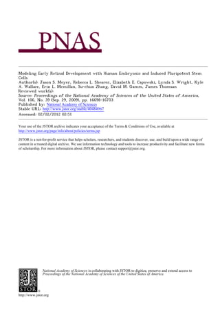
- 3. < ■z. !E o u h ¡i s Fig. 2. Highly derivation eye fieldphenotypes efficient of from hESCs. RT-PCR (A) analysisshowingthe onsetof Pax6and Rxgene expression concomitant of and loss Oct4. (B and 0 qPCR analysis Oct4 gene expression and Pax6 and Rxgene of (8) expression Values were expressedas foldchange relative undifferentiated (Q. to hESCs.(D) Immunocytochemical analysis cellsat day 10 showinguniform of coex- pressionof Pax6 and Rx (merged image includes ToPro-3nuclearstain).(£) FACS analysisconfirming rapidlossofOct4expression the onsetofbothPax6and the and Rxprotein expression.Negative controls FACS for analyses indicated thewhite are by histograms. (Fand G) qPCR (f) and Western (G) the analysis demonstrating endog- enous expression the BMP and Wnt antagpnists of Nogginand Dkk-1.(H) qPCR Fig. 1. Commitment toward a retinal lineage occurs as a stepwise process, showingthe nearcompletelossof Pax6and Rxgene expression cellstreatedwith in beginningwiththe establishment the eye fieldwithinthe anteriorneuroep- of BMP4and Wnt3A.(Scale bar,40 /am.) ithelium. Each majorstage inretinogenesiscanbe distinguished partbythe (A) in expressionof varioustranscription factors.(B) Schematicof the differentiation protocol used to generate cells of a retinallineage. (Q RT-PCR analysisof the The onsetof Pax6 and Rx expression detected day6, when was by changes in gene expressiontoward an eye fieldfate throughthe first days of 16 approximately of all cellsexpressed (D-F) Immunocytochemistry typicalhESC aggregates 10 days differentiation. of 25% thesefactors. Expression of Pax6 and Rx surpassed demonstrating expressionof the anteriorneuraltran- afterdifferentiation, the 90% ofcellsbyday 10 ofdifferentiation scriptionfactor Otx2 (D), the eye field transcription factor Lhx2 (£), and the to and increased greater than95% byday 16. Conversely, protein definitiveneuraltranscriptionfactorSox1 (F). (Scale bar, 200 pirn.)expression Oct4 decreasedto an undetectable of levelbyday10 of differentiation. generation a highpercentage cellswith The of of eyefield characteristicstheabsenceofexogenous in Wntand BMP coloniesexpressed Lhx2(Fig. ID), Otx2(Fig. IE), and Soxl (Fig. antagonists prompted further investigation the endogenous into IF) by day 10 of differentiation. expression Dkk-1and Nogginin differentiating cultures. of hESC Eyefield cellsareoften characterized thecoexpression Pax6 by of Both geneswere up-regulated during fieldspecification eye (Fig. Western de- the and Rx (7, 16). Therefore, gene and protein expression ofthese 2F) as determined qPCR. Furthermore, by analysis tected proteinexpressionof Dkk-1 and Noggin at day 10 of twotranscription factors examined further was in detail.RT-PCR differentiation Addition Wnt3A BMP4 tocultures of and and quantitative PCR (qPCR) analysesrevealed the onset of overthefirst (Fig.2G).differentiation 10 daysof abolished boththeexpres- expressionof both Pax6 and Rx withinthe firstfew days of sion of Pax6 and Rx (Fig. 2H) and the appearanceof neuroepi- differentiation 2A-C), which was closelycorrelated withloss thelialcolonies was also (Fig. (Fig. SI). Endogenous FGF signaling ofOct4expression. theprotein On nearly cellscoexpressed involved the acquisition earlyeye fieldfeatures, level, all in of since the bothPax6 and Rx within daysof differentiationdetermined addition SU5402, a potentand specific 10 as of inhibitor theFGFR1 of by immunocytochemistry ID). Cell populations (Fig. were then receptor, to a complete led loss ofbothPax6 and Rx expression at analyzed FACS overthefirst daysofdifferentiation IE). by 16 (Fig. day 10 of differentiation S2). (Fig. Meyeret al. PNAS | September29. 2009 | voi. 106 | no. 39 | 16699
- 4. of Acquisition Optic and Vesicle Optic CupCellPhenotypes. next The phase in retinal specification vivooccurswiththeformation in of theopticvesicles from pairedeye fields. thisstage,all cells the At that giveriseto either neuralretina theRPE express will the or the transcription factor Mitf of + (17). The subset Mitf cellsdestined to becomeneuralretina subsequently down-regulate inresponse Mitf to theonsetof ChxlO(also calledVsx2) expression 19). When (18, eye fieldrosettes were liftedand grownas neurospheres, near uniform expression Mitfprotein of was observedwithin14.7 ± 2.1% of all spheresby day 30 of differentiation 14). qPCR (Fig. analysisfurther demonstrated that gene expression Mitfin- of creasedfrom 16 to day37 ofdifferentiation 3B). Next, day (Fig. the relationship betweenMitfand ChxlO protein expression was ex- amined overtime. ChxlOexpression only was rarely observed day at 30 (Fig. 3C). Coexpression Mitf of and ChxlOwas prevalent day by 40 (Fig. 3D), followed mutually by exclusive expression ChxlO of and Mitfbyday50 as Mitfexpression diminished within ChxlO+ neurospheres (Fig. 3E). qPCR analysis that confirmed ChxlOgene expression delayedrelative Mitf(Fig. 3F). Similar Mitf, was to to ChxlO protein was eventually detectedin nearlyall cells of the subsetof neurospheres whichitwas expressed in (Fig. 3G). Quan- tification ChxlO proteinexpression of demonstrated that18.0 ± 3.3% of all neurospheres containedChxl0+ cells by day 40 of differentiation (Fig. S14), and withinthese ChxlO-expressing spheres, 90.7% ± 5.2% of cells expressed ChxlOby day 50 (Fig. S3B). ByFACS, 26% oftheentire culture cell population expressed ChxlOat day40 (Fig. 3H). The remaining ChxlO-negative neuro- spheres derived from early field population the eye cell maintained a neuralfateas indicated expression Soxl and ßlll-tubulin by of (Fig. S4 A-F, and J). Non-retinal neurospheres also expressed forebrain markers, including Otx2(Fig. S4 G-I, and/), butdid not express endoderm (alpha-fetoprotein), mesoderm(brachyury), hindbrain (HoxB4), or midbrain (En-1) markers (Fig. S4/). Amongthoseindividual cellsthatexpressed ChxlO, greater than 99% maintained expression Pax6,which a requirement early of is of retinal progenitor cells (20) (Fig. 31). Furthermore, manyof the + ChxlO clusters were arrangedin rosetteswith cells oriented radially awayfroma core thatwas positive the tight for junction protein ZO-1 (Fig. 37), a feature associatedwithprogenitor pop- ulations (14). Whileclusters contained that Chxl0+ cellsincluded a smallnumber ßlll tubulin+neurons of (Fig.3K),thesecellsrarely coexpressed ChxlO.Thus,ChxlOexpression associatedwas witha neural cell type that had not yet acquired a matureneuronal phenotype. Fig. 3. Acquisition of optic vesicle and optic cup cell phenotypes. {A) Mitf Giventhepotential roleof FGF signaling thespecification in of protein expression in neurospheres after30 days of differentiation. qPCR (B) theneuralretina(21), we nextexamined effect thespecific the of analysisof Mitfgene expressionover the first days of differentiation. 80 (C-£) The Immunocytochemical analyses of the time course of Mitfand ChxlO protein FGF inhibitor SU5402, on Mitfand ChxlOgene expression. additionof SU5402 to adherenthESC cultures the optic expression in neurospheres at 30 (Q, 40 (D), or 50 (£) days of differentiation. during (F) qPCR analysis of ChxlO gene expression over the first days of differen- 80 vesicleand opticcup stagesofdifferentiation 16-40) resulted (days tiation. (G) Uniform Chx10 expressionthroughouta subset of neurospheresby in an 11.8-fold increasein Mitfgene expression day 40, as at day 40. (H) FACSanalysisdemonstratingthe percentage of all cells expressing measuredby qPCR (Fig. 3L). By contrast, ChxlO expression was ChxlO at day 40. (I) Immunocytochemicalanalysis showed all Chx10+ cells reduced15.9-fold a result thistreatment. as of coexpressed Pax6 at day 40. (J) Rosettes of Chx10+ cells expressed the tight junction proteinZO-1 withintheircore. (K) Rare Chx10+ cells coexpressed j3lll DifferentiationRetinal Types of Cell from hESC-Derived Retinal Pro- tubulin at day 40. (/.) qPCR demonstrating increased Mitf expression and genitors.The RPE is the first differentiatedretinalcell typeto correspondingdecreased ChxlO expression in adherent culturestreated with + the FGFinhibitor SU5402. qPCR values were expressed as fold change relative appear during retinogenesis,arisingfroma populationof Mitf (Scale bars, 500 to culturesat day 16 (B and F) or day 10 (L) of differentiation. andPax6+ cellspresent theouterlayer theearly in of opticcup (21). /mm panels A and G; 50 /¿m panels C-E,J,and K; and 75 /xm panel /;blue in in in WhenPax6+/Rx+ eyefieldrosettes weremaintained adherent as stain in A and G is Hoechst nuclear dye.) cultures,distinct of patches polygonal, pigmented wereinitially cells observed approximately 30 ofhESC differentiation 44). at day (Fig. These cellsmaintained expression thetranscription of factor Mitf, while also expressing RPE-associatedtight the junctionprotein Prolongedmaintenance the hESC-derivedretinalprogen- of ZO-1 (Fig.AB).Atday40 ofdifferentiation, analysis FACS revealed itorsas neurospheresallowed for further maturation these of that25% of all adherent cellsexpressed Mitf,and 77% of all cells cells towarda photoreceptor phenotype.Among the first dif- expressed Pax6 (Fig. AC). RT-PCR analysis demonstrated main- ferentiatedneuralretinalphenotypes observedduring develop- tainedexpression Pax6 in thiscell population of overtime, well as mentare cone photoreceptors 23), whoseprecursors (22, express as theacquisition moremature of RPE-associated markers suchas the primitivecone and rod photoreceptor-specifictranscription RPE65 and bestrophin (Fig. AD). factorCrx (24). By day 80 of differentiation, ± 3.1% of all 19.4 16700 | www.pnas.org/cgi/doi/10.1073/pnas.0905245106 Meyeret al.
- 5. >- oc < h- z LU cell lineingreater the detail.Upon differentiation, appearances of O 'J the iPS cell colonies, iPS cell aggregates, neural rosettesand m ut t/i neurospheres indistinguishable those hESCs (Fig.S7). were from of Duringdifferentiation, immunocytochemistry revealedearlyeye field cellscoexpressing Pax6and Rxbyday10 (Fig.6A). Thesecells also expressed fullcomplement eye fieldand neuroepithelial a of transcription factors (Fig. S8). Discretepopulations Mitf cells of 4- wereobserved uponfurther differentiationeyefield of coloniesas neurospheres (Fig. 6B). Like theirhESC counterparts, manyof these iPS cell neurospheres appeared to lose Mitfexpression in favorof ChxlO expression (Fig. 6C), yielding neurospheres that werehighly enriched Chxl0+ cells(Fig. 6D). Amongthetotal for population, 12.9 ± 4.3% ofall neurospheres expressed ChxlOat 40 days of differentiation, withinwhich90.1 ± 1.2% of all cells expressedChxlO. Over time,photoreceptor markers appeared, such as the rod-and cone-specific transcription factor Crx,which was present 14.4 ± 5.1% ofall neurospheres day80 (Fig. 6E). in by Similar the expression earliermarkers retinal to of of differentia- tion,Crx+ cells were commonwithin individual positive neuro- spheres, constituting ± 9.3% of cells.At day80,44.6 ± 8.1% 65.5 of cellswithin Crx+ clusters expressed recoverin (Fig. 6F) and/or opsin (Fig. 6 G and H). As with hESCs, recoverin and opsin expression was not found in Crx-negative cells. PCR analysis Fig. 4. Generation of retinal pigment epithelium. (A) Photomicrographof confirmed sequence and timing gene expression these the of of adherent culturesshowing pigmented, hexagonal RPE-like cells. (6) Immuno- markers, alongwith early ofOct4expression the loss (Fig.61).RPE staining revealing expression of MitfwithinRPE-like cells, as well as the tight cellswerepresent within cellcultures well, iPS as with pigmentation junction protein ZO-1. (Q FACS analysis demonstratingthe percentage of all first apparent approximately 35 ofdifferentiation typical ¿S at day and adherent cells expressing Mitf and Pax6 at day 40 of differentiation.(D) RT-PCR studies showing expressionof genes associated withan RPEfate. (Scale monolayers arising day 50 (Fig. 67). Like hESC-derived by RPE, thesecellspossessedmorphological characteristics mature of RPE bars, 100 /im.) and expressed Mitfand ZO-1 (Fig. 6K). Discussion neurospherescontained Crx+ photoreceptor precursors(Fig. The results 63.0 ± 7.6% ofall cellsexpressed presented heredemonstrate thathumanpluripotent 5^4).Within theseneuropheres, Crx. Furthermore, 46.4 ± 7.9% of Crx+ cells expressedmore stemcells can adopt signature features associatedwithall major mature photoreceptormarkers,such as recoverin (Fig. 5B) stages of earlyeye and retinaldevelopment, whilefollowing an and/or cone photoreceptor-specific the expected timeline for human retinal development(22, 23). proteinopsin (Fig. 5C). Recoverin and opsin expression was not observed in Crx- Althoughpreviousreports have shownthathESCs can acquire retinal characteristics various times duringdifferentiation at negativecells. used to identify retinalcell types To analyze the time course and sequential acquisition of (3-7), manymarkers primitive are also expressedin otherdevelopingneuralcells. This makes neuroretinal-and photoreceptor-associated gene expression,it difficult unequivocallyassign immature to cell typesto the RT-PCR analysis was performed(Fig. 5D). Throughoutthe retinallineagewithout knowledgeof theirdynamic behaviorin differentiation process fromday 16 through day 80, Pax6 geneculture.Thus, it is important monitor to each stage of cellular expressionwas detected. Rx gene expressionwas also present maturation ensurethatcritical to checkpoints are developmental earlyin differentiation, followedby the consecutiveexpression met in order and withina predictabletimeframe. of ChxlO,Crx,and opsin.Overall,thetiming expression the of of In the first weeksof humandevelopment, portion the few a of gene and proteinmarkers used in thisstudy coincidedwiththat primitive anterior neuroepithelium risetotheeyefield gives (13, 16, of normalhumanretinaldevelopment(22, 23) (Fig. S5). of 26, 27), a cell populationcharacterized the expression by During normal retinogenesis, Pax6(+5a) isoformis ex- the numerous transcription factors including Pax6,Rx, Six3,Six6,Til, pressedin increasing abundancerelative total Pax6 (25). RT- to and Lhx2.We have demonstrated the production Pax6+/ that of PCR results fromthe presentstudysimilarly suggested thatthe Rx+ cellsis highly efficient, 95% ofall cellscoexpressing with these Pax6(-l-5a) isoform becamemoreprevalent during hESC differen- essential factors. efficiencylikely inpartto This is due transcription tiation(Figs.2A and 5D). To verify observation, this qPCR of thea relativelack of influencefromendogenousBMP and Wnt Pax6(+5a) isoform relative totalPax6expression performed to was signaling, since both pathways knownto antagonizeneural are (Fig. S6). This analysis confirmed onsetof Pax6(+5a) expres- the specification (28, 29). In support thistheory, of increasing expres- sionbetweendays4 and 16 of differentiation demonstrated and a sionofBMP andWntantagonists and (Noggin Dkk-1, respectively) relative in increase theexpression thisisoform of betweendays60 was observedin hESC cultures shortly after onsetof differen- the and 70, whichcorresponded the appearanceof photoreceptor- to tiation.Early exposureof differentiating hESCs to recombinant likecells in culture. BMP4 andWnt3aeliminated expression Pax6and Rx,as well the of as the subsequent formation neuralrosettes. of Differentiation of Retinal Cell Types from Human iPS Cells. To AlthoughPax6 and Rx have been used to identify retinal determine potential stepwise the for derivation retinal types progenitor of cell cells in differentiating cultures(7, 16), during ESC from human cells, appliedthehESC differentiation iPS we protocol development are thesefactors intially coexpressed a broadregion in to fourdifferent humaniPS cell lines.Consistent witha previous of the anterior neuralplate thatincludes eye fieldand future the report (11), considerable variation found theability these forebrain was in of (16). Thereafter, Pax6+/Rx+ cellsbecome restricted to linesto producePax6+ neuroectodermal at day 10 of differ- morespecific cells areasofthedeveloping CNS (16), predominantly the entiation, efficiencies with rangingfrom to 56% ofthetotalcell retina(26, 30). In the presentstudy, 5% the majority the early of population. Based on theseresults, choseto study IMR90-4 we the Pax6+/Rx+ populationdid not subsequently adopt cellularphe- Meyeret al. PNAS | September29, 2009 | vol.106 | no. 39 | 16701
- 6. Fig. 5. Generationof earlyphotoreceptorphenotypes.(A) Immunocytochem- ¡cal detection of cells expressing the photoreceptor-specif transcription ic factor Crx at 80 days of differentiation. and O Expressionof the photoreceptor (B protein recoverin(B) and the cone photoreceptor-specific protein opsin (O among Crx-expressing cells at day 80. (D) RT-PCRdemonstratedthe stepwise acquisitionofa cone photoreceptor fatefrom eye fieldpopulation.(Scale bars, an 50 /xm.) Blue stain in A is Hoechst nucleardye. notypes theoptic of vesicle opticcupdespite or retaining anterior an neuralidentity. Therefore, enriched the Pax6+/Rx+ cell popula- in tionderived thisstudy mostcloselyresembled primitive a stage ofhuman field eye development, which the preceded appearance of committed retinal progenitors. Afterthe optic vesiclesevaginatefromthe paired eye fields, expression Mitfoccursthroughout fatedto become retina of cells (12, 17). However,the decisionto differentiate towardeithera neuralretina RPE fateis revealedduring late opticvesicle or the and optic cup stages,in part via differential expression the of transcription factorChxlO (18, 19). Neural retinalprogenitors destinedforthe innerlayerof the opticcup expressChxlO and down-regulate in response FGFs secreted theoverlying Fig.6. Stepwise retinalspecificationfromhuman ¡PScells. {A) Variousstages Mitf to by surfaceectoderm. Thus, ChxlO is the earliestspecific marker of of retinaldifferentiationwere observed, beginningwith Pax6+/Rx+ eye field neuralretinal progenitor cells (19). Conversely, cells destined for cells by day 10. (B-D) Mitf+ and Chx10+ cells, indicativeof the optic vesicle/ theouterlayer theopticcup remain of + Mitf and ChxlO-negative optic cup stages, are evident by day 40. (£) By day 80, clusterswere present and subsequently differentiate into RPE. Our resultsprovide containing Chx10+ retinal progenitors and Crx+ photoreceptor precursor cells expressed the photoreceptor protein evidencethathESCs proceed through analogousstagesof early cells. {F-H) Many Crx-expressing recoverin(F) and the cone-specificproteinopsin (G and H). (/)RT-PCR analysis retinaldifferentiation,indicated the spatiotemporal as by expres- sionofMitf ChxlOinneurospheres. and inhibition demonstrating the stepwise expression of retina- and photoreceptor- of associated Furthermore, genes in differentiating ¡PS cells over time. (J and K) RPE cells endogenous FGF signaling during opticvesicleand opticcup derived fromiPS cells acquired a typicalhexagonal morphologyand pigmen- the stagesof hESC differentiation in resulted a profound increasein tation (J)and expressed Mitfand ZO-1 {K). (Scale bars, 50 /am.)Blue stain in B Mitf geneexpression a corresponding and decreasein ChxlOgene and D is Hoechst nuclear dye; blue stain in F is To-Pro-3nuclear dye. expression. This suggeststhat mechanisms governing cell fate choicein thedeveloping retina mayalso function differentiating in hESC cultures. differentiation. observationis consistent This withnormalret- After adopting a retinalfate, individual neurospheres yielded a inal development,where early cell types often give rise to high percentageof photoreceptor precursors a time frame multipledistinct in progeny the same lineage. However,there of predicted by normal humanretinogenesis. withAs earlierstages of now exist opportunitiesto introduceexogenous factorsfor retinaldifferentiation, was achievedwithout additionof definedtimeperiodsto augment this the production retinal types of cell specificexogenous agents.Previously, retinoic and taurine acid had at specificdevelopmental stages. Such precisionis likelyto be been used to inducedifferentiationphotoreceptor-like (7). of cells important, since a single factorcan have diverse effectson By eliminating such agents,the presentsystem suitedforthe cellularfatechoice depending thestageofdevelopment is on (31). investigation of endogenousfactors and mechanisms thataffect For example,we observed thatearlyinhibition endogenous of differentiation maturation specific and of retinal types. cell in FGF signaling differentiating hESCs resulted a loss of eye in Taking intoaccounttheentire hESC populationpresent the field specification, at whereas later inhibition differentially regu- startof the differentiation process,we observed a decrease in latedgenesimportant theinduction RPE and neuralretina for of targetedcell production witheach subsequentstage of retinal progenitors.Manipulation of the culture environment with 16702 | www.pnas.org/cgi/doi/10.1073/pnas.0905245106 Meyer al. et
- 7. | signaling factors may also alter the time course of retinalcell In summary, haveshownthathESCs meetthecriteria 2) we (1, differentiation fromhESCs. This is suggestedby the striking to serve as a comprehensive vitromodel system human in for in differences the timing photoreceptor of markerexpression retinogenesis. Usingan identical culture method, humaniPS cells observedin previousstudies,in whichthe onset of Crx expres- showa similar potential, although variation occurbetween can lines. sion rangedfromone to thirteen weeks (6, 7). On a broaderlevel,thisstudy a supports roleforpluripotent stem Giventheability hESCs to mimic of normalhumanretinogen- cells to testconceptsin humandevelopmental biologythatwere sourceofhumanpluripotent previously extrapolated fromanimalmodels.In turn, thisability esis,we investigated whether another stem similar A previous could narrow gap between understanding humandevel- the our of cells,iPS cells, displayed a potential. report opmentand thatof othermammalian species. byYu et al. (11) showedthathumaniPS cell linesdiffered their in earlyexpression Pax6, a finding of confirmed here. Since Pax6 Methods expression necessary retinal is for development, is notsurprising Maintenance of hESCs and Human iPS Cells. Pluripotent stem cells were it thatone of the highest Pax6-expressing lines fromthat study, maintained as previouslydescribed for hESCs (15). Detailed protocols are IMR90-4, wasefficient producing at retinal populations. cell Other available in the 5/Text iPS celllinesdisplayed reduced competency to produceneuraland retinal types, phenomenon observed Hirami al. (8). cell a also by et Differentiation hESCs and Human iPS Cells. The initial differentiationof of Therefore, present techniques forderiving cells from iPS somatic hESCs and human iPS cells toward an eye field fate was performed with modificationsto previouslydescribed protocols (15, 36). Thereafter,a chem- cells do not always yielduniform lineagecompetencies between ically-defined retinaldifferentiation medium was used to promote the step- lines. wise production of retina-specific types fromfree-floatingneurospheres cell A detailedknowledge the stagesand timecourseof retinal (36). Detailed protocols are available in the 5/Text of differentiation hESCs and iPS cells not only providesan from opportunity studyfundamental to questionsof human retinal RT-PCR.RT-PCRand qPCR experiments were performed as previouslyde- development, mayalso aid efforts use pluripotent but to stemcell scribed (36). More detailed methods, including primer sequences, can be found in the SI Text as well as Table S1. for derivatives pharmaceutical testing and retinalrepopulation A studies. previous report MacLaren et al. (32) demonstrated Immunocytochemistry. aggregates were plated onto coversiips,fixedwith by Cell that a cellsfrom specific stageof mouseretinal development were 4% paraformaldehyde,and then immunostained as described (15). Detailed capable of functionally integrating degenerateadult mouse procedures are provided in the SI Textas well as Table S2. into J 5 retinas. Morerecently, Lamba et al. (33) notedsimilar resultsusing a mixture retinal of cell typesderivedfromhESCs. The hESC FACS. Staining and sorting of cells were performed as previouslydescribed Detailed procedures are provided in the 5/Text differentiation protocoldescribedin the presentstudyprovides (15). access to humanretinal cells at all majorstagesof retinal devel- opment. The nearabsenceof contamination from non-neural cell WesternAnalysis.Western blots were 5/Textas well previouslydescribed (36). Detailed procedures are provided in performedas as Table S2. types and the potentialto enrichfor discreteretinalcell types further to the possibleclinicalutility thesedifferentiatingACKNOWLEDGMENTS. We thank B. Hu, T. Lavaute, and M. Pankratz for add of cultures. potential iPS cellsto generate The for multiple retinalcell technical assistance,and the staffat WiCell forpreparation of MEF. Thiswork types willaid inthedevelopment invitroof models human of retinal was supported by National Institutes of Health Grants K08EY015138 (to D.M.G.) and R01NS045926 (to S.C.Z.), the Foundation Fighting Blindness degenerative diseasesand stimulate investigation customized (D.M.G.), the Walsh Consortium(D.M.G.), the LincyFoundation (D.M.G.), and into cell for stem therapies patients afflicted thesedisorders 35). by (34, a Retina Research Foundation Gamewell Professorship D.M.G.) (to 1. KellerG (2005) Embryonic stemcell differentiation: Emergenceof a new era in biology 19. Rowan S, Chen CM, Young TL, FisherDE, Cepko CL (2004) Transdifferentiation the of and medicine. Genes Dev 19:1129-1155. retina into pigmented cells in ocular retardation mice defines a new functionof the 2. Pera MF,TrounsonAO (2004) Human embryonic stemcells: Prospectsfordevelopment. homeodomain gene Chx10. Development 131:5139-5152. Development 131:5515-5525. 20. Belecky-AdamsT, et al. (1997) Pax-6, Prox 1, and ChxiO homeobox gene expression 3. KlimanskayaI,et al. (2004) Derivationand comparative assessment ot retinalpigment correlates with phenotypic fate of retinal precursorcells. Invest Ophthalmol Vis Sci epitheliumfromhuman embryonic stemcells usingtranscriptomics. CloningStem Cells 38:1293-1303. 6:217-245. 21. Muller F, RohrerH, Vogel-Hopker A (2007) Bone morphogenetic proteinsspecifythe 4. VuglerA, et al. (2008) Elucidatingthe phenomenon of HESC-denved RPE:anatomy ot retinal pigment epithelium in the chickembryo. Development 134:3483-3493. cell genesis, expansion and retinaltransplantation.Exp Neurol 214:347-361. 22. BarishakY (2001) in Embryology the Eye and itsAdnexa (Karger,New York) 2nd Ed. of 5. Banin E, et al. (2006) Retinal incorporationand differentiation neural precursors of 23. FinlayBL(2008) The developing and evolvingretina:Usingtimeto organize form.Brain derived fromhuman embryonicstem cells. Stem Cells 24:246-257. Res 1192:5-16. 6. Lamba DA, KarlMO, Ware CB, Reh TA (2006) Efficient generation of retinalprogenitor 24. Chen S, et al. (1997) Crx,a novel Otx-likepaired-homeodomain protein,binds to and cells fromhuman embryonicstem cells. Proc Nati Acad Sci USA 103:12769-12774. transactivatesphotoreceptor cell-specific genes. Neuron 19:1017-1030. 7. Osakada F,et al. (2008) Toward the generation of rod and cone photoreceptorsfrom 25. PinsonJ,Mason JO,SimpsonTI, PriceDJ(2005) Regulation of the Pax6: Pax6(5a) mRNA mouse, monkeyand human embryonicstem cells. Nat Biotechnol 26:215-224. ratio in the developing mammalian brain. BMC Dev Biol 5:13. 8. HiramiY, et al. (2009) Generation of retinal cells from mouse and human induced 26. BaileyTJ, al. (2004) Regulation of vertebrateeye development by Rxgenes. IntJDev et pluripotentstem cells. Neurosci Lett. Biol 48:761-770. 9. Klassen H, ReubinoffB (2008) Stem cells in a new light.Nat Biotechnol 26:187-188. 27. Zuber ME, GestriG, ViczianAS, BarsacchiG, Harris WA (2003) Specification the verte- of 10. Takahashi K, et al. (2007) Induction of pluripotent stem cells from adult human brate eye by a networkof eye fieldtranscription factors.Development 130:5155-5167. fibroblastsby defined factors.Cell 131:861-872. 28. GlinkaA, et al. (1998) Dickkopf- is a memberof a new family secreted proteinsand 1 of 11. Yu J, al. (2007) Induced pluripotentstemcell linesderivedfromhuman somatic cells. et functionsin head induction. Nature 391:357-362. Science 318:1917-1920. 29. Lamb TM, et al. (1993) Neural induction by the secreted polypeptide noggin. Science 12. Chow RL,Lang RA(2001 ) Earlyeye development in vertebrates.Annu Rev Cell Dev Biol 262:713-718. 17:255-296. 30. Furukawa T, Kozak CA, Cepko CL (1997) Rax, a novel paired-type homeobox gene, 13. Li H, TierneyC, Wen L,Wu JY,Rao Y (1997) A single morphogeneticfield gives riseto shows expression in the anteriorneural fold and developing retina.Proc Nati Acad Sci two retina primordia under the influence of the prechordal plate. Development USA 94:3088-3093. 124:603-615. 31. Esteve P, Bovolenta P (2006) Secreted inducers in vertebrate eye development: More 14. Elkabetz Y, et al. (2008) Human ES cell-derivedneural rosettes reveal a functionally functionsfor old morphogens. CurrOpin Neurobiol 16:13-19. distinctearly neural stem cell stage. Genes Dev 22:152-165. 32. MacLaren RE,et al. (2006) Retinal repair by transplantationof photoreceptor precur- 15. Pankratz MT, et al. (2007) Directed neural differentiation human embryonicstem of sors. Nature 444:203-207. cells via an obligated primitive anteriorstage. Stem Cells 25:151 1-1520. 33. Lamba DA, Gust J, Reh TA (2009) Transplantation of human embryonicstem cell- 16. Mathers PH, JamrichM (2000) Regulation of eye formation by the Rx and Pax6 derived photoreceptors restoressome visual functionin Crx-deficient mice. Cell Stem homeobox genes. Cell Mol LifeSci 57:186-194. Cell 4:73-79. 17. BhartiK, LiuW, Csermely BertuzziS, ArnheiterH (2008) Alternativepromoteruse in T, 34. Ebert AD, et al. (2009) Induced pluripotentstem cells froma spinal muscularatrophy eye development: the complex role and regulation of the transcription factor MITF. patient. Nature 457:277-280. Development 135:1169-1178. 35. Park IH,et al. (2008) Disease-specificinduced pluripotentstem cells. Cell 134:877-886. 18. HorsfordDJ,et al. (2005) Chx10 repressionof Mitfis required forthe maintenance of 36. Gamm DM, et al. (2008) A novel serum-freemethod for cultunng human prenatal mammalian neuroretinalidentity.Development 132:177-187. retinal pigment epithelial cells. Invest Ophthalmol Vis Sci 49:788-799. Meyeret al. PNAS | September29, 2009 | vol.106 | no. 39 | 16703