Downloaded 92 times
![654 CHAPTER 35 Pregnancy-Related Hypertension
factors for preeclampsia are similar in nulliparous and parous Obesity is a risk factor for preeclampsia.28,51 In the National Insti-
women.22 tute of Child Health and Human Development (NICHD) study of
Although preeclampsia was thought to be more common among aspirin to prevent preeclampsia in low-risk pregnancies,31 the inci-
women of lower socioeconomic status, this impression may be a con- dence of preeclampsia increased with maternal body mass index. Even
sequence of the associations of preeclampsia with age, race, and parity. in women of normal weight, there is a linear relationship between
Studies of pregnant women in Scotland23 from Aberdeen,24 Finland,25 pre-pregnancy body mass index and the frequency of preeclampsia.52
and Israel26 found that preeclampsia was not related to socioeconomic The mechanism may be related to increased insulin resistance, because
status. Eclampsia, in contrast, is clearly more common in women of preeclampsia is more common in another setting of increased insulin
lower socioeconomic status,23,25,26 related to the lack of availability of resistance: gestational diabetes.53 With a threefold increased risk for
quality obstetric care for indigent women. Remarkably, preeclampsia obese women and with 35% to 50% of women of reproductive age in
and eclampsia were once thought to occur more frequently in women the United States being obese, obesity has become a major attributable
of higher socioeconomic status.18 risk factor for preeclampsia, which is associated with more than one
There is a relationship between the extremes of childbearing age third of cases of preeclampsia.
and the incidence of eclampsia and preeclampsia. Because most first Certain conditions of pregnancy increase the risk of preeclampsia.
pregnancies occur in young women, most cases of preeclampsia and The incidence is increased among parous and nulliparous women with
eclampsia occur in this age group, but the association with young multiple gestations, although to a larger degree in the latter.36,54 In a
maternal age is lost when parity is considered. In the studies cited,23,25,26 study of 34,374 pregnancies with singleton, twin, triplet, or quadruplet
a higher incidence of preeclampsia was found in older women inde- pregnancies, the incidence of preeclampsia increased with each addi-
pendent of parity. tional fetus. The incidences were 6.7%, 12.7%, 20.0%, and 19.6%,
The relationship of preeclampsia and eclampsia to race is compli- respectively.55 The disease process may be initiated earlier and may be
cated by the higher prevalence of chronic hypertension in African more severe in these cases.54
Americans and the difficulty in differentiating preeclampsia from Preeclampsia affects 70% of women with large, rapidly growing
unrecognized preexisting chronic hypertension. Some studies indicate hydatidiform moles and occurs earlier than usual during gestation.56
a relationship.26,27 In a small case-control study of carefully defined In cases of preeclampsia occurring before 24 weeks’ gestation, hyda-
preeclampsia, black race was a significant risk factor only in nullipa- tidiform mole should be suspected and sought.
rous women (odds ratio [OR] = 12.3; 95% confidence interval [CI], An interesting variant of preeclampsia is the mirror syndrome, in
1.6 to 100.8).28 Other studies support a more modest increased risk in which the mother’s peripheral edema mirrors the fetal hydrops. It
African-American women.29,30 Studies that include the more severe occurs with fetal hydrops, although not with erythroblastosis uncom-
forms of preeclampsia more often suggest an increased incidence plicated by hydrops. The incidence approaches 50% of pregnancies
among African-American women.28 complicated by hydrops. The mirror syndrome is not confined to
In contrast, the incidence of rigorously defined preeclampsia did hydrops resulting from isoimmunization. In one series, mirror syn-
not differ by race after other risk factors were controlled in two large, drome occurred in 9 of 11 pregnancies with hydropic infants of non-
prospective trials of medical prophylaxis that enrolled 294731 and immune origin.57 This condition can manifest early in pregnancy with
431432 nulliparous women. Maternal nonwhite race appears to be severe signs and symptoms of preeclampsia, and it has resolved with
related more to the severity than the incidence of disease. treatment of the underlying process.58-60 Proteinuria is massive, and
A diverse array of medical disorders that often coexist with preg- blood pressure elevation and edema are marked. Eclampsia occurs
nancy, including diabetes, chronic hypertension, chronic renal disor- rarely (see Chapter 26).
ders, and rheumatologic conditions, have been associated with
preeclampsia. The existence and severity of diabetes have been
associated with an increased risk for preeclampsia, and diabetic Short-Term Prognosis for Preeclampsia
microvascular disease further increases this risk. This relationship PERINATAL MORTALITY
has been found in Sweden33 and in the United States.34 Both The perinatal mortality rate is increased in infants of preeclamptic
studies33,34 demonstrated that the risk of preeclampsia was approxi- women.61-63 In a study that examined 10,614,679 singleton pregnancies
mately 20% and 21% in 491 and 462 pregnancies, respectively. This in the United States from 1995 to 1997 after 24 weeks’ gestation, the
estimate is far more modest than the 50% incidence reported in his- relative risk for fetal death was 1.4 for infants born to women with any
torical cohorts.18 The preeclampsia risk increased according to the of the gestational hypertensive disorders and 2.7 for those born to
severity of disease, with an 11% to 12% risk among women with class women with chronic hypertensive disorders compared with low-risk
B diabetes and 21% to 23% with class C and D diabetes. Microvascular controls. Causes of perinatal death are placental insufficiency and
disease increased this risk to 36% to 54% in diabetics with class F or abruptio placentae,64 which cause intrauterine death before or during
R disease.33,34 labor, and prematurity. Predictably, the mortality rate is higher for
Chronic renal insufficiency and hypertension are well-recognized infants of women with more severe forms of the disorder. At any level
risk factors. Of women with hypertension antedating pregnancy, 25% of disease severity, the perinatal mortality rate is greatest for women
develop preeclampsia.35,36 Renal insufficiency with33,37 and without dia- with preeclampsia superimposed on preexisting vascular disease.
betes38-40 also is an important risk factor.38,40 The stillbirth rate attributable to preeclampsia has declined dra-
Connective tissue disorders such as systemic lupus erythe- matically in the past 35 years. However, infants born of preeclamptic
matosus41,42 and antiphospholipid antibody syndrome43-45 have been pregnancies continue to have an approximately twofold increased risk
reported as risk factors for preeclampsia. With lupus, the risk is par- for neonatal death.65 Although neonatal survival rates have improved
ticularly elevated with hypertension or nephropathy.46,47 However, dramatically, delivery before 34 weeks’ gestation continues to be associ-
data concerning an association between isolated antiphospholipid ated with an increased risk of long-range neurologic disability (see
antibodies and preeclampsia have been conflicting, with some Chapter 58).
studies demonstrating no relationship48,49 and others confirming the Growth restriction is more common in infants born to preeclamp-
association.44,50 tic women (see Chapter 34) and more pronounced with increasing](https://image.slidesharecdn.com/4-u1-0-b978-1-4160-4224-2-50038-7-docpdf-120121090843-phpapp01/85/4-u1-0-b978-1-4160-4224-2-50038-7-docpdf-4-320.jpg)
![656 CHAPTER 35 Pregnancy-Related Hypertension
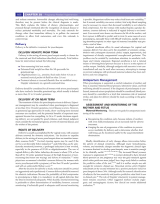
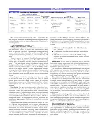
100 TABLE 35-3 SIGNS AND SYMPTOMS OF
PREECLAMPSIA OR ECLAMPSIA
90
Cerebral Blurred vision
Headache Amaurosis
80
Percentages surviving
Dizziness Gastrointestinal
Tinnitus Nausea
70 Drowsiness Vomiting
Change in respiratory rate Epigastric pain
60 Tachycardia Hematemesis
Fever Renal
50 Visual Oliguria
Diplopia Anuria
40 Scotomata Hematuria
Hemoglobinuria
30
dyslipidemia,92 altered angiogenic factors,93 and increased antibodies
10 20 30 40 45
to the angiotensin-2 receptor.94 These data may explain the common
Years
risk factors for preeclampsia and cardiovascular disease, but alternative
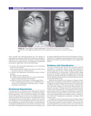
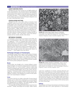
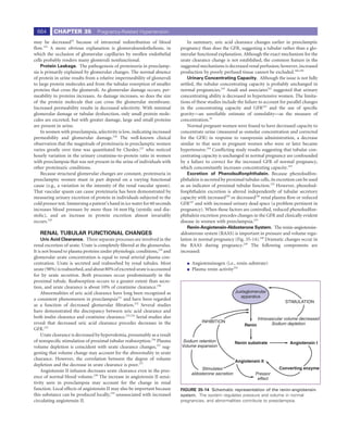
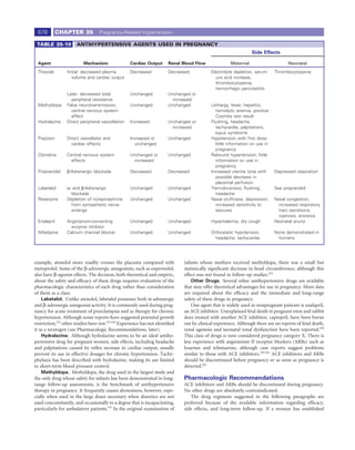
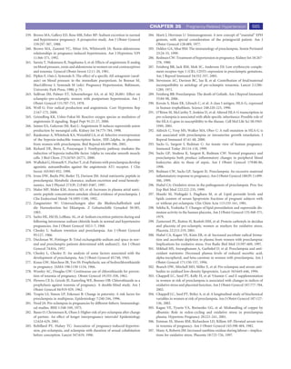
FIGURE 35-3 Eclampsia survivorship. Survival times are plotted
explanations, such as that preeclampsia causes vascular injury that
for women with eclampsia in the first pregnancy (solid line) and those increases cardiovascular risk or that normal pregnancies have a protec-
with eclampsia in a later pregnancy (dashed line). Survival of women tive effect, cannot be excluded.
with first-pregnancy eclampsia was not different from survival of a
control group. (From Chesley LC, Annitto JE, Cosgrove RA: The
remote prognosis of eclamptic women: Sixth periodic report. Am J Clinical Presentation
Obstet Gynecol 124:446, 1976, Courtesy of the American College of Preeclampsia can manifest with a wide spectrum of disease, ranging
Obstetricians and Gynecologists.) from life-threatening neurologic, renal, hepatic, and coagulation
abnormalities to mild findings of preeclampsia with minimal end-
organ involvement. The fetus may be severely compromised by the
Scottish investigators reported a fourfold increased risk of subsequent maternal condition and by extreme preterm delivery or only minimally
hypertension in nulliparous women with preeclampsia2,76,77 (OR = affected. These variations have puzzled clinicians and researchers for
3.98; CI, 2.82 to 5.61). Funai and colleagues78 described excess long- many years. An understanding of the pathophysiology of the disorder
term mortality in women with prior preeclampsia that was largely provides insight into the diverse clinical presentations.
attributed to a threefold increase in deaths due to cardiovascular
disease. Other reports support a link between preeclampsia and mater- Symptoms
nal ischemic heart disease,79,80 which is sometimes evident 20 years Most women with early preeclampsia are asymptomatic. The absence
after the preeclamptic pregnancy and coincident with the onset of of symptoms is the rationale for frequent obstetric visits in late preg-
menopause.78,80 A family history of cardiovascular disease increases the nancy. In most cases, signs such as increased blood pressure and pro-
association between preeclampsia and cardiovascular outcomes.81 teinuria antedate overt symptoms.
Obesity is a known risk factor for preeclampsia and cardiovascular The various symptoms associated with preeclampsia, especially
disease. Although controlling for obesity attenuates the increased risk preeclampsia of increasing severity, are listed in Table 35-3. Because
of death for postmenopausal women, this risk is not fully explained by preeclampsia is a disease of generalized poor perfusion, the diversity
obesity alone.82 of symptoms related to many organ systems is not surprising. Symp-
The relationships among obesity, insulin resistance, and preeclamp- toms suggesting hepatic, neurologic, and visual involvement are par-
sia are part of an interesting relationship of preeclampsia to the meta- ticularly worrisome. They include epigastric pain, “stomach upset,” and
bolic or insulin resistance syndrome.83 This syndrome predisposes to pain penetrating to the back. Headache and mental confusion indicate
cardiovascular disease in later life and consists of obesity, hypertension, poor cerebral perfusion and may be precursors of convulsions. Visual
dyslipidemia (i.e., increased low-density lipoprotein [LDL] cholesterol, symptoms ranging from scotomata to blindness indicate retinal arte-
decreased high-density lipoprotein [HDL] cholesterol, and increased rial spasm and edema. Symptoms suggesting congestive heart failure
triglycerides), and increased uric acid, all of which are found in women or abruptio placentae also represent significant complications of pre-
with preeclampsia.83 Other conditions predisposing to later-life cardio- eclampsia. Other symptoms, such as tightness of hands and feet and
vascular disease—including elevated levels of homocysteine,84 evidence paresthesias resulting from medial or ulnar nerve compression, may
of androgen excess (including polycystic ovarian syndrome),85 elevated alarm the patient but have little prognostic significance.
testosterone levels,86 male fat distribution (i.e., increased waist-to-hip
ratio),87 and lipoprotein lipase mutations88—are also linked to an Signs
increased risk for preeclampsia. Signs of preeclampsia usually antedate symptoms. The most common
Women who appear normal years after a preeclamptic pregnancy sequence is increased blood pressure followed by proteinuria.18
may nevertheless demonstrate subtle metabolic and cardiovascular
abnormalities. Compared with women with uncomplicated pregnan- BLOOD PRESSURE CHANGE
cies, formerly preeclamptic women have evidence of endothelial dys- An increase in blood pressure is required for the diagnosis of
function,89,90 higher blood pressures,89 increased insulin resistance,91 preeclampsia. Blood pressure variation in normal pregnancy can](https://image.slidesharecdn.com/4-u1-0-b978-1-4160-4224-2-50038-7-docpdf-120121090843-phpapp01/85/4-u1-0-b978-1-4160-4224-2-50038-7-docpdf-6-320.jpg)
![CHAPTER 35 Pregnancy-Related Hypertension 659
EN
BM
R
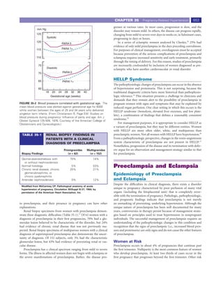
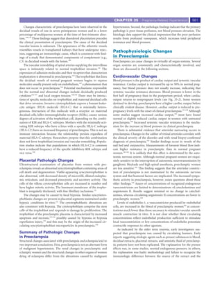
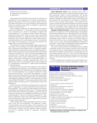
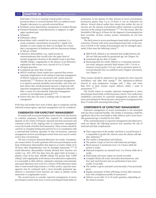
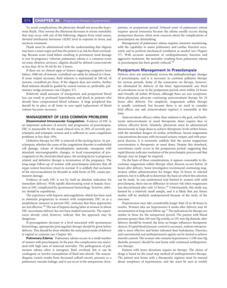
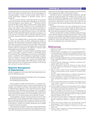
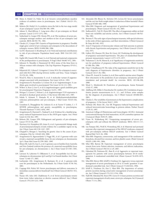
FIGURE 35-6 Glomerular changes in preeclampsia are identified A
by light microscopy. The enlarged glomerulus completely fills
Bowman’s capsule. Diffuse edema of the glomerular wall is indicated Ep Cy
by the vacuolated appearance. The visible capillary loops are
extremely narrow, and there are virtually no red blood cells in the
capillary tuft.
En
Glomerular, tubular, and arteriolar changes have been described. The
glomerular lesion is considered by some to be pathognomonic of pre-
eclampsia and eclampsia, but identical changes have been seen in pla- BS R
cental abruption without evident preeclampsia.128 This change is not
En
seen in any other form of hypertension.
Ep
GLOMERULAR CHANGES
Changes seen by light microscopy in glomeruli that are character-
istic of preeclampsia include103 decreased glomerular size, with protru-
sion of the glomerular tuft into the proximal tubule. The diameter of P
the glomerular capillary lumen is decreased and contains few blood
cells. The endothelial-mesangial cells have increased cytoplasmic
B
volume and can contain lipoid droplets (Fig. 35-6).
Electron microscopic examination of glomeruli provides more evi- FIGURE 35-7 Electron photomicrographs of renal glomeruli.
dence that the primary pathologic change occurs in endothelial cells, A, Normal anatomy. B, Biopsy specimen from a preeclamptic woman.
which are greatly increased in size and can occlude the capillary lumen; Endothelial cells (En) are markedly enlarged, obstruct the capillary
their cytoplasm contains electron-dense material.129 The basement lumen, and contain electron-dense inclusions. The basement
membrane bordering the epithelial cell may be slightly thickened, and membrane (BM) is slightly thickened with inclusions, but the
it also contains electron-dense material. The epithelial cell podocytes epithelial foot processes (EP) are normal. BM, basement membrane;
are not altered (Fig. 35-7). These changes are collectively called glo- BS, Bowman’s space; Cy, cytoplasmic inclusions; EN, capillary
endothelial cells that line the glomeruli; Ep, renal epithelial cells; L,
merular capillary endotheliosis.
capillary lumen containing red blood cells; P, podocytes; R, red blood
Characteristic glomerular changes occur in 70% of primiparas
cell. (From McCartney CP: Pathological anatomy of acute
but in only 14% of multiparas with a diagnosis of preeclampsia.17 The hypertension of pregnancy. Circulation 30[Suppl II]:37, 1964. By
more likely the diagnosis of preeclampsia, the more common the glo- permission of the American Heart Association, Inc.)
merular lesion. As the clinical condition worsens, the magnitude of the
glomerular lesion increases. The glomerular lesions are reversible after
delivery and are not present in subsequent biopsy specimens obtained
5 to 10 weeks later.103 NONGLOMERULAR CHANGES
The glomerular changes correlate more consistently with protein- Pathologic changes in renal tubules include dilatation of proximal
uria than with hypertension, suggesting that proteins identified immu- tubules with thinning of the epithelium,123 tubular necrosis,103 enlarge-
nohistochemically may be trapped in the glomerulus. These staining ment of the juxtaglomerular apparatus,131 and hyaline deposition in
patterns are not found in other renal disorders with proteinuria. The renal tubules.123 Fat deposition in women with prolonged heavy pro-
glomerular changes of preeclampsia can be mimicked in animal studies teinuria has been reported.123 Necrosis of the loop of Henle, a change
by reducing the renal concentration of vascular endothelial growth that correlates with the degree of hyperuricemia, has also been
factor (VEGF), which usually exists in high concentration in this tissue described.131
by increasing the synthesis of the VEGF antagonist soluble Fms-like Thickening of renal arterioles may be seen in preeclampsia, espe-
tyrosine kinase 1 (sFlt1).130 cially in women with preexisting hypertension. Unlike the glomerular](https://image.slidesharecdn.com/4-u1-0-b978-1-4160-4224-2-50038-7-docpdf-120121090843-phpapp01/85/4-u1-0-b978-1-4160-4224-2-50038-7-docpdf-9-320.jpg)
![660 CHAPTER 35 Pregnancy-Related Hypertension
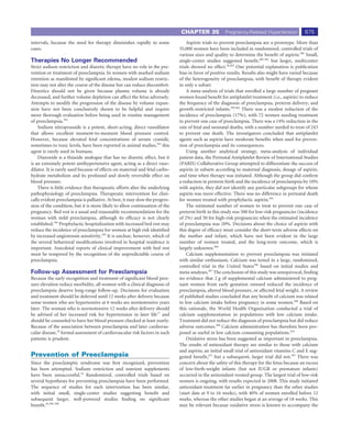
Spinal
Endometrium
Basal
Radial
Arcuate
Myometrium
A
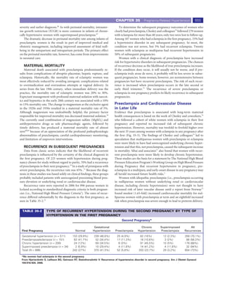
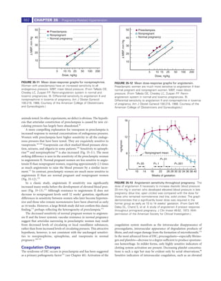
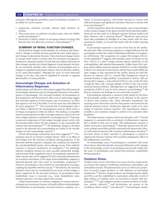
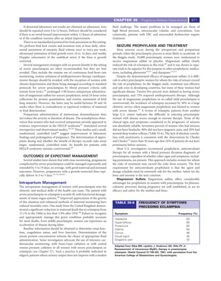
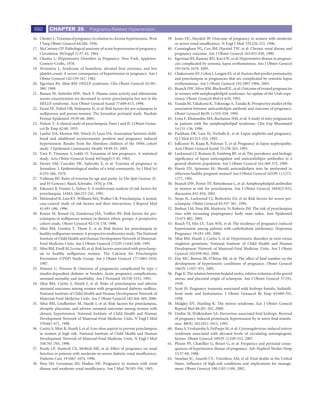
FIGURE 35-8 Schematic representation of uterine arteries. The
characteristic changes occur in the decidual vessels supplying the
placental site in a normal pregnancy. (From Okkels H, Engle ET:
Studies of the finer structure of the uterine vessels of the Macacus
monkey. Acta Pathol Microbiol Scand 15:150, 1938.)
lesion, it does not regress after delivery,103 suggesting that the arteriolar
change results from coincident disease, not preeclampsia.
B
Vascular Changes in the Placental Site FIGURE 35-9 Spiral arterial changes in normal pregnancy. A, In
The characteristic changes in the decidual vessels supplying the pla- the section of spiral arterioles at the junction of the endometrium and
cental site in normal pregnancy are depicted in Figure 35-8. In normal myometrium in a nonpregnant woman, notice the inner elastic lamina
pregnancy, the spiral arteries (Fig. 35-9) increase greatly in diameter.132 and smooth muscle. B, In a section of a spiral arteriole at the same
Morphologically, the endothelium is replaced by trophoblast, and the scale and from the same location during pregnancy, notice the
internal elastic lamina and smooth muscle of the media are replaced markedly increased diameter and absence of inner elastic lamina and
by trophoblast and an amorphous matrix-containing fibrin (see smooth muscle. (From Sheppard BL, Bonnar J: Uteroplacental
Fig. 35-9).133 These changes occur originally in the decidual portion of arteries and hypertensive pregnancy. In Bonnar J, MacGillivray I,
Symonds G [eds]: Pregnancy Hypertension. Baltimore, University Park
the spiral arteries but extend into the myometrium as pregnancy
Press, 1980, p 205.)
advances and can even involve the distal portion of the uterine radial
artery. The basal arteries are not affected. These morphologic changes
are considered to be a vascular reaction to the trophoblast, occurring
directly or humorally, that results in increased perfusion of the placen-
tal site.
In placental-site vessels of women with preeclampsia, the normal
physiologic changes do not occur, or they are limited to the decidual
portion of the vessels. Myometrial segments of spiral arteries retain the
nonpregnant component of intima and smooth muscle, and the diam-
eter of these arteries is about 40% that of vessels in normal preg-
nancy.134 Spiral arterioles in decidua and myometrium and basal and F
radial arterioles may become necrotic, with components of the normal
vessel wall replaced by amorphous material and foam cells, a change
called acute atherosis (Fig. 35-10).135 This lesion is best seen in the basal
arteries because they do not undergo the normal changes of pregnancy.
It is also present in decidual and myometrial spiral arteries and can
progress to vessel obliteration. The obliterated vessels correspond to
areas of placental infarction.
Failed vascular remodeling and atherotic changes may be seen
with fetal growth restriction in women without clinical evidence of
FIGURE 35-10 Atherosis. Numerous lipid-laden cells (L) and fibrin
preeclampsia. Atherotic changes occur in decidual vessels of some dia- deposition (F) are present in the media of this occluded decidual
betic women,136 and failed vascular remodeling is present in about vessel. (From Sheppard BL, Bonnar J: Uteroplacental arteries and
one third of women who experience preterm labor.137 It appears hypertensive pregnancy. In Bonnar J, MacGillivray I, Symonds G
that abnormal invasion may be necessary but is not sufficient to cause [eds]: Pregnancy Hypertension. Baltimore, University Park Press,
preeclampsia. 1980, p 205.)](https://image.slidesharecdn.com/4-u1-0-b978-1-4160-4224-2-50038-7-docpdf-120121090843-phpapp01/85/4-u1-0-b978-1-4160-4224-2-50038-7-docpdf-10-320.jpg)
![CHAPTER 35 Pregnancy-Related Hypertension 669
Clinical management is dictated by the overt clinical signs of pre- fetus with growth restriction. Fetal jeopardy, rather than lung maturity,
eclampsia. Proteinuria—the most valid clinical indicator of preeclamp- is the fetal criterion to determine delivery when preeclampsia occurs
sia—is often a late change, sometimes even preceded by seizures, and remote from term.
it is therefore not useful for early recognition of disease. Although
rapid weight gain and edema of the hands and face suggest fluid and Expectant Management of Severe
sodium retention characteristic of preeclampsia, they are not univer- Preeclampsia Remote from Term
sally present in or uniquely characteristic of preeclampsia. These signs Prolonged expectant antepartum management of women with severe
are at most a reason for close observation of blood pressure and preeclampsia is not practiced in most centers. With improvements in
urinary protein levels. Early recognition of preeclampsia is necessarily neonatal care, many clinicians regard delivery of women with severe
based primarily on diagnostic blood pressure increases in the late preeclampsia beyond 32 to 34 weeks’ gestation to be in the best inter-
second and early third trimesters compared with pressures in early ests of the mother and fetus. When gestational age is critical (<32
pregnancy. Blood pressure changes without proteinuria undoubtedly weeks), the physician may consider control of maternal blood pressure
occur in some normal women and in some with underlying renal or along with meticulous observation of maternal and fetal conditions.
vascular disease. Because the goal of early diagnosis is to identify This approach requires personnel and facilities for very close assess-
patients requiring more careful observation, overdiagnosis is prefera- ment of both patients.
ble to underdiagnosis. The initial evaluation and management of a woman suspected to
After blood pressure changes diagnostic of preeclampsia occur, evi- have severe preeclampsia between 24 and 32 to 34 weeks’ gestation
dence of multiorgan involvement should be sought through laboratory includes the following components:
assessment. A 24-hour or timed urine specimen should be collected,343
regardless of findings on urine dipstick evaluation.9 Because of the The pregnant woman is admitted to the hospital.
hectic protein excretion characteristic of the disorder,344 24-hour urine A course of antenatal corticosteroids is administered (see
collections may reveal excretion of more than 300 mg of protein, even Chapter 23). Barring rapid deterioration of the maternal or
with only trace proteinuria identified on the dipstick evaluation.9 fetal status, reasonable efforts should be made to delay delivery
Platelet count and liver enzyme tests should also be obtained.2 To rule for 48 hours to complete a full course of antenatal
out fulminant progression, repeated examination of pressure and corticosteroids. Neonates from preeclamptic pregnancies may
urinary protein is suggested within 24 hours. The frequency of subse- have a reduced incidence of respiratory distress syndrome, but
quent observations is determined by these initial observations and the this does not justify withholding antenatal corticosteroid
ensuing clinical progression. If the condition appears stable, once- or therapy.345,346
twice-weekly observations may be appropriate. Any evidence of pro- Seizure prophylaxis is undertaken with magnesium sulfate.
gression merits more frequent observations, perhaps in the hospital. Blood pressure is monitored at least every 1 to 2 hours.
The appearance of proteinuria is an especially important sign of pro- Fluid intake and urine output are strictly monitored.
gression and requires frequent observation. A 24-hour urine collection is used to determine protein
If deterioration in laboratory findings, symptoms, or clinical signs excretion and creatinine clearance.
occurs, the decision to continue the pregnancy is determined day by Laboratory studies include a complete blood cell count with a
day. Subjective evidence of central nervous system involvement (i.e., platelet count and smear and determinations of electrolytes,
headache, disorientation, and visual symptoms) and hepatic distention creatinine, ALT, AST, lactic acid dehydrogenase (LDH), uric
(i.e., abdominal pain and right upper quadrant or epigastric tender- acid, and albumin. A coagulopathy profile (i.e., prothrombin
ness) indicates worsening preeclampsia. Important clinical signs are time [PT], partial thromboplastin time [PTT], and fibrinogen)
blood pressure, urinary output, and fluid retention as evidenced by should be obtained if the ALT and AST values are more than
daily weight increase. twice normal or if the platelet count is less than 100,000 cells/μL.
Laboratory studies are performed at intervals of no less often than Assessment of fetal well-being includes a nonstress test,
every 48 hours. Tests should include a platelet count and fibrin split amniotic fluid volume determination, and estimation of fetal
products, urinary protein excretion and serum creatinine levels, and size. If growth restriction is recognized, umbilical artery
serum levels of transaminases. Doppler velocimetry is suggested.
Fetal Observation. Assessment of fetal well-being is required to
determine whether continuing the pregnancy is safe (see Chapter 21). After the complete assessment of the fetus and mother, the safety
With the diagnosis of gestational hypertension, fetal assessment for size and potential utility of expectant management should be reassessed
by sonography and for function by nonstress testing is indicated. After daily. Several factors mandate delivery regardless of gestational age.
the diagnosis of preeclampsia is made, it is mandatory to monitor the Under these circumstances, the initial dose of antenatal steroids should
fetal condition. Ultrasound evaluation of fetal weight and amniotic be administered, but pregnancy should not be unnecessarily prolonged
fluid volume and a nonstress test of the fetal heart rate should be per- to give the second dose.
formed. Alternatively, a complete biophysical profile may be performed.
Doppler velocimetry is not recommended unless fetal growth restric- CONTRAINDICATIONS TO
tion is identified. EXPECTANT MANAGEMENT
As long as the maternal condition is mild and stable, weekly moni- Immediate delivery should be considered if any of the following
toring of the fetus appears to be adequate. Unfortunately, no test of conditions are present:
fetal well-being is satisfactory when the mother’s condition is unstable,
and testing should be repeated whenever the maternal status changes. Maternal hemodynamic instability (e.g., shock)
Management of fetal growth restriction, a common complication of Non-reassuring fetal test results (e.g., persistently abnormal
preeclampsia, is discussed in Chapter 34. Amniotic fluid testing for fetal heart rate testing, estimated fetal weight less than the 5th
fetal lung maturity (see Chapter 23) may aid the decision to deliver the percentile for gestational age, oligohydramnios with amniotic](https://image.slidesharecdn.com/4-u1-0-b978-1-4160-4224-2-50038-7-docpdf-120121090843-phpapp01/85/4-u1-0-b978-1-4160-4224-2-50038-7-docpdf-19-320.jpg)

![CHAPTER 35 Pregnancy-Related Hypertension 681
63. Conde-Agudelo A, Belizan JM, Diaz-Rossello JL: Epidemiology of 89. Agatisa PK, Ness RB, Roberts JM, et al: Impairment of endothelial
fetal death in Latin America. Acta Obstet Gynecol Scand 79:371-378, function in women with a history of preeclampsia: An indicator of
2000. cardiovascular risk. Am J Physiol Heart Circ Physiol 286:H1389-H1393,
64. Naeye RL, Friedman EA: Causes of perinatal death associated with gesta- 2004.
tional hypertension and proteinuria. Am J Obstet Gynecol 133:8-10, 90. Chambers JC, Ueland PM, Obeid OA, et al: Improved vascular endothelial
1979. function after oral B vitamins: An effect mediated through reduced
65. Basso O, Rasmussen S, Weinberg CR, et al: Trends in fetal and infant sur- concentrations of free plasma homocysteine. Circulation 102:2479-2483,
vival following preeclampsia. JAMA 296:1357-1362, 2006. 2000.
66. Odegard RA, Vatten LJ, Nilsen ST, et al: Preeclampsia and fetal growth. 91. Laivuori H, Tikkanen MJ, Ylikorkala O: Hyperinsulinemia 17 years after
Obstet Gynecol 96:950-955, 2000. preeclamptic first pregnancy. J Clin Endocrinol Metab 81:2908-2911,
67. Lopez-Llera M, Hernandez Horta JL: Perinatal mortality in eclampsia. J 1996.
Reprod Med 8:281-287, 1972. 92. Hubel CA, Snaedal S, Ness RB, et al: Dyslipoproteinaemia in postmeno-
68. Sibai BM, McCubbin JH, Anderson GD, et al: Eclampsia. I. Observations pausal women with a history of eclampsia. BJOG 107:776-784, 2000.
from 67 recent cases. Obstet Gynecol 58:609-613, 1981. 93. Wolf M, Hubel CA, Lam C, et al: Preeclampsia and future cardiovascular
69. Pritchard JA, Pritchard SA: Standardized treatment of 154 consecutive disease: Potential role of altered angiogenesis and insulin resistance. J Clin
cases of eclampsia. Am J Obstet Gynecol 123:543-552, 1975. Endocrinol Metab 89:6239-6243, 2004.
70. Berman S: Observations in the toxemic clinic, Boston Lying-In Hospital, 94. Hubel CA, Wallukat G, Wolf M, et al: Agonistic angiotensin II type 1
1923-1930. Obstet Gynecol 203:361, 1930. receptor autoantibodies in postpartum women with a history of pre-
71. Sibai BM, el-Nazer A, Gonzalez-Ruiz A: Severe preeclampsia-eclampsia in eclampsia. Hypertension 49:612-617, 2007.
young primigravid women: Subsequent pregnancy outcome and remote 95. Buchbinder A, Sibai BM, Caritis S, et al: Adverse perinatal outcomes are
prognosis. Am J Obstet Gynecol 155:1011-1016, 1986. significantly higher in severe gestational hypertension than in mild pre-
72. Hjartardottir S, Leifsson BG, Geirsson RT, Steinthorsdottir V: Recurrence eclampsia. Am J Obstet Gynecol 186:66-71, 2002.
of hypertensive disorder in second pregnancy. Am J Obstet Gynecol 96. Dieckman W: The Toxemias of Pregnancy, 2nd ed. St Louis, CV Mosby,
194:916-920, 2006. 1952.
73. Sibai BM, Mercer B, Sarinoglu C: Severe preeclampsia in the second tri- 97. Douglas KA, Redman CW: Eclampsia in the United Kingdom. BMJ
mester: Recurrence risk and long-term prognosis. Am J Obstet Gynecol 309:1395-400, 1994.
165(Pt 1):1408-1412, 1991. 98. Sibai BM: Eclampsia. VI. Maternal-perinatal outcome in 254 consecutive
74. Chesley SC, Annitto JE, Cosgrove RA: The remote prognosis of eclamptic cases. Am J Obstet Gynecol 163:1049-1054; discussion 1054-1055, 1990.
women. Sixth periodic report. Am J Obstet Gynecol 124:446-459, 1976. 99. Conde-Agudelo A, Kafury-Goeta AC: Epidemiology of eclampsia in
75. Irgens HU, Reisaeter L, Irgens LM, Lie RT: Long term mortality of mothers Colombia. Int J Gynaecol Obstet 61:1-8, 1998.
and fathers after pre-eclampsia: Population based cohort study. BMJ 100. Macgillivray I: Some observations on the incidence of pre-eclampsia. J
323:1213-1217, 2001. Obstet Gynaecol Br Emp 65:536-539, 1958.
76. Chesley LC, Annitto JE, Cosgrove RA: The remote prognosis of eclamptic 101. Tervila L, Goecke C, Timonen S: Estimation of gestosis of pregnancy
women. Sixth periodic report. Am J Obstet Gynecol 124:448, 1976. (EPH-gestosis). Acta Obstet Gynecol Scand 52:235-443, 1973.
77. Wilson BJ, Watson MS, Prescott GJ, et al: Hypertensive diseases of preg- 102. Ferrazzani S, Caruso A, De Carolis S, et al: Proteinuria and outcome of
nancy and risk of hypertension and stroke in later life: Results from cohort 444 pregnancies complicated by hypertension. Am J Obstet Gynecol
study. BMJ 326:845, 2003. 162:366-371, 1990.
78. Funai EF, Friedlander Y, Paltiel O, et al: Long-term mortality after pre- 103. Pollak VE, Nettles JB: The kidney in toxemia of pregnancy: A clinical and
eclampsia. Epidemiology 16:206-215, 2005. pathologic study based on renal biopsies. Medicine (Baltimore) 39:469-
79. Smith GC, Pell JP, Walsh D: Pregnancy complications and maternal risk 526, 1960.
of ischaemic heart disease: A retrospective cohort study of 129,290 births. 104. Jaffe G, Schatz H: Ocular manifestations of preeclampsia. Am J Ophthal-
Lancet 357:2002-2006, 2001. mol 103(Pt 1):309-315, 1987.
80. Arnadottir GA, Geirsson RT, Arngrimsson R, et al: Cardiovascular death 105. Roberts JM, Bodnar LM, Lain KY, et al: Uric acid is as important as pro-
in women who had hypertension in pregnancy: A case-control study. teinuria in identifying fetal risk in women with gestational hypertension
BJOG 112:286-292, 2005. [see comment]. Hypertension 46:1263-1269, 2005.
81. Ness RB, Markovic N, Bass D, et al: Family history of hypertension, heart 106. Many A, Hubel CA, Roberts JM: Hyperuricemia and xanthine oxidase in
disease, and stroke among women who develop hypertension in preg- preeclampsia, revisited. Am J Obstet Gynecol 174(Pt 1):288-291, 1996.
nancy. Obstet Gynecol 102:1366-7131, 2003. 107. Johnson RJ, Kang DH, Feig D, et al: Is there a pathogenetic role for uric
82. Samuels-Kalow ME, Funai EF, Buhimschi C, et al: Prepregnancy body acid in hypertension and cardiovascular and renal disease? Hypertension
mass index, hypertensive disorders of pregnancy, and long-term maternal 41:1183-1190, 2003.
mortality. Am J Obstet Gynecol 197:490 e1-e6, 2007. 108. Martin JN Jr, Blake PG, Perry KG Jr, et al: The natural history of HELLP
83. Barden AE, Beilin LJ, Ritchie J, et al: Does a predisposition to the metabolic syndrome: Patterns of disease progression and regression. Am J Obstet
syndrome sensitize women to develop pre-eclampsia? J Hypertens Gynecol 164(Pt 1):1500-1509; discussion 1509-1513, 1991.
17:1307-1315, 1999. 109. Burrows RF, Kelton JG: Thrombocytopenia at delivery: A prospective
84. Powers RW, Evans RW, Majors AK, et al: Plasma homocysteine concentra- survey of 6715 deliveries. Am J Obstet Gynecol 162:731-734, 1990.
tion is increased in preeclampsia and is associated with evidence of endo- 110. Redman CW, Bonnar J, Beilin L: Early platelet consumption in pre-
thelial activation. Am J Obstet Gynecol 179(Pt 1):1605-1611, 1998. eclampsia. BMJ 1:467-469, 1978.
85. Fridstrom M, Nisell H, Sjoblom P, Hillensjo T: Are women with polycystic 111. Weiner CP, Brandt J: Plasma antithrombin III activity: An aid in the
ovary syndrome at an increased risk of pregnancy-induced hypertension diagnosis of preeclampsia-eclampsia. Am J Obstet Gynecol 142:275-281,
and/or preeclampsia? Hypertens Pregnancy 18:73-80, 1999. 1982.
86. Acromite MT, Mantzoros CS, Leach RE, et al: Androgens in preeclampsia. 112. Redman CW, Denson KW, Beilin LJ, et al: Factor-VIII consumption in
Am J Obstet Gynecol 180(Pt 1):60-63, 1999. pre-eclampsia. Lancet 2:1249-1252, 1977.
87. Sattar N, Clark P, Holmes A, et al: Antenatal waist circumference and 113. Lok CA, Nieuwland R, Sturk A, et al: Microparticle-associated P-selectin
hypertension risk. Obstet Gynecol 97:268-271, 2001. reflects platelet activation in preeclampsia. Platelets 18:68-72, 2007.
88. Hubel CA, Roberts JM, Ferrell RE: Association of pre-eclampsia with 114. Hutt R, Ogunniyi SO, Sullivan MH, Elder MG: Increased platelet volume
common coding sequence variations in the lipoprotein lipase gene. Clin and aggregation precede the onset of preeclampsia. Obstet Gynecol
Genet 56:289-296, 1999. 83:146-149, 1994.](https://image.slidesharecdn.com/4-u1-0-b978-1-4160-4224-2-50038-7-docpdf-120121090843-phpapp01/85/4-u1-0-b978-1-4160-4224-2-50038-7-docpdf-31-320.jpg)
![682 CHAPTER 35 Pregnancy-Related Hypertension
115. Hayashi M, Inoue T, Hoshimoto K, et al: Characterization of five marker labor and preterm ruptured membranes. Am J Obstet Gynecol 168:585-
levels of the hemostatic system and endothelial status in normotensive 591, 1993.
pregnancy and pre-eclampsia. Eur J Haematol 69:297-302, 2002. 138. Lichtig C, Deutsch M, Brandes J: Immunofluorescent studies of the endo-
116. Knopp R: Lipid Metabolism in Pregnancy. New York, Springer-Verlag, metrial arteries in the first trimester of pregnancy. Am J Clin Pathol
1991. 83:633-636, 1985.
117. Hubel C, Roberts J: Lipid metabolism and oxidative stress. In Lindheimer 139. Nadji P, Sommers SC: Lesions of toxemia in first trimester pregnancies.
M, Roberts J, Cunningham F (eds): Chesley’s Hypertensive Disorders in Am J Clin Pathol 59:344-349, 1973.
Pregnancy. Stamford, CT, Appleton & Lange, 1999, p 453. 140. Kitzmiller JL, Benirschke K: Immunofluorescent study of placental bed
118. Hubel CA, McLaughlin MK, Evans RW, et al: Fasting serum triglycerides, vessels in pre-eclampsia of pregnancy. Am J Obstet Gynecol 115:248-251,
free fatty acids, and malondialdehyde are increased in preeclampsia, are 1973.
positively correlated, and decrease within 48 hours post partum. Am J 141. Cross JC, Werb Z, Fisher SJ: Implantation and the placenta: Key pieces of
Obstet Gynecol 174:975-982, 1996. the development puzzle. Science 266:1508-1518, 1994.
119. Lorentzen B, Endresen M, Clausen T, et al: Fasting serum free fatty acids 142. Zhou Y, Damsky CH, Chiu K, et al: Cytotrophoblast expression of integrin
and triglycerides are increased before 20 weeks of gestation in women who extracellular matrix receptors is altered in preeclampsia. In Soars MJ,
later develop preeclampsia. Hypertens Pregnancy 13:103, 1994. Handwerger S, Talamantes F (eds): Trophoblast Cells: Pathways for
120. Rosing U, Samsioe G, Olund A, et al: Serum levels of apolipoprotein A-I, Maternal-Embryonic Communication. New York, Springer-Verlag, 1993,
A-II and HDL-cholesterol in second half of normal pregnancy and in p 109.
pregnancy complicated by pre-eclampsia. Horm Metab Res 21:376-382, 143. Zhou Y, Fisher SJ, Janatpour M, et al: Human cytotrophoblasts adopt a
1989. vascular phenotype as they differentiate. A strategy for successful endo-
121. Hubel CA, Shakir Y, Gallaher MJ, et al: Low-density lipoprotein particle vascular invasion? J Clin Invest 99:2139-2151.
size decreases during normal pregnancy in association with triglyceride 144. Zhou Y, Damsky CH, Fisher SJ: Preeclampsia is associated with failure of
increases. J Soc Gynecol Investig 5:244-250, 1998. human cytotrophoblasts to mimic a vascular adhesion phenotype. One
122. Sattar N, Bendomir A, Berry C, et al: Lipoprotein subfraction concentra- cause of defective endovascular invasion in this syndrome? J Clin Invest
tions in preeclampsia: Pathogenic parallels to atherosclerosis. Obstet 99:2152-2164, 1997.
Gynecol 89:403-408, 1997. 145. Caniggia I, Grisaru-Gravnosky S, Kuliszewsky M, et al: Inhibition of TGF-
123. Sheehan H, Lynch J: Pathology of Toxemia in Pregnancy. London, beta 3 restores the invasive capability of extravillous trophoblasts in pre-
Churchill Livingstone, 1973. eclamptic pregnancies. J Clin Invest 103:1641-1650, 1999.
124. Naheedy MH, Biller J, Schiffer M, et al: Toxemia of pregnancy: Cerebral 146. Librach CL, Feigenbaum SL, Bass KE, et al: Interleukin-1 beta regulates
CT findings. J Comput Assist Tomogr 9:497-501, 1985. human cytotrophoblast metalloproteinase activity and invasion in vitro. J
125. Belfort MA, Varner MW, Dizon-Townson DS, et al: Cerebral perfusion Biol Chem 269:17125-17131, 1994.
pressure, and not cerebral blood flow, may be the critical determinant 147. Bass KE, Morrish D, Roth I, et al: Human cytotrophoblast invasion is up-
of intracranial injury in preeclampsia: A new hypothesis. Am J Obstet regulated by epidermal growth factor: Evidence that paracrine factors
Gynecol 187:626-634, 2002. modify this process. Dev Biol 164:550-561, 1994.
126. Schmorl G: Zur pathologischen Anatomie Untersuchung uber Puerperal- 148. Caniggia I, Winter J, Lye SJ, Post M: Oxygen and placental development
Eklampsie. Verhandl Dtsch Gesellsch Gyneakol 1901:203, 1901. during the first trimester: Implications for the pathophysiology of pre-
127. Sheehan H: Pathologic lesions in the hypertensive toxaemias of pregnancy. eclampsia. Placenta 21(Suppl A):S25-S30, 2000.
In Hammond J, Browne F, Wolstenholm G (eds): Toxaemias of Pregnancy, 149. Genbacev O, Joslin R, Damsky CH, et al: Hypoxia alters early gestation
Human and Veterinary. Philadelphia, Blakiston, 1950, p 16. human cytotrophoblast differentiation/invasion in vitro and models the
128. Thomson D, Paterson WG, Smart GE, et al: The renal lesions of toxaemia placental defects that occur in preeclampsia. J Clin Invest 97:540-550,
and abruptio placentae studied by light and electron microscopy. J Obstet 1996.
Gynaecol Br Commonw 79:311-320, 1972. 150. Hiby SE, Walker JJ, O’Shaughnessy KM, et al: Combinations of maternal
129. Spargo B, McCartney CP, Winemiller R: Glomerular capillary endothelio- KIR and fetal HLA-C genes influence the risk of preeclampsia and repro-
sis in toxemia of pregnancy. Arch Pathol 68:593-599, 1959. ductive success. J Exp Med 200:957-965, 2004.
130. Maynard SE, Min JY, Merchan J, et al: Excess placental soluble fms-like 151. Jones CJ, Fox H: An ultrastructural and ultrahistochemical study of the
tyrosine kinase 1 (sFlt1) may contribute to endothelial dysfunction, human placenta in maternal pre-eclampsia. Placenta 1:61-76, 1980.
hypertension, and proteinuria in preeclampsia [see comment]. J Clin 152. Fox H, Kharkongor NF: The effect of hypoxia on human trophoblast in
Invest 111:649-658, 2003. organ culture. J Pathol 101:v, 1970.
131. Altchek A, Albright NL, Sommers SC: The renal pathology of toxemia of 153. Allaire AD, Ballenger KA, Wells SR, et al: Placental apoptosis in preeclamp-
pregnancy. Obstet Gynecol 31:595-607, 1968. sia. Obstet Gynecol 96:271-276, 2000.
132. Ramsey E, Harris H: Comparison of uteroplacental vasculature and cir- 154. Huppertz B, Kingdom J, Caniggia I, et al: Hypoxia favours necrotic versus
culation in the rhesus monkey and man. Carnegie Contrib Embryol 38:59- apoptotic shedding of placental syncytiotrophoblast into the maternal
70, 1966. circulation. Placenta 24:181-190, 2003.
133. Brosens IA, Robertson WB, Dixon HG: The role of the spiral arteries 155. Hung TH, Skepper JN, Charnock-Jones DS, Burton GJ: Hypoxia-
in the pathogenesis of preeclampsia. Obstet Gynecol Annu 1:177-191, reoxygenation: A potent inducer of apoptotic changes in the human pla-
1972. centa and possible etiological factor in preeclampsia. Circ Res 90:1274-1281,
134. Khong TY, De Wolf F, Robertson WB, Brosens I: Inadequate maternal 2002.
vascular response to placentation in pregnancies complicated by pre- 156. Goswami D, Tannetta DS, Magee LA, et al: Excess syncytiotrophoblast
eclampsia and by small-for-gestational age infants. BJOG 93:1049-1059, microparticle shedding is a feature of early-onset pre-eclampsia, but not
1986. normotensive intrauterine growth restriction. Placenta 27:56-61, 2006.
135. Zeek PM, Assali NS: Vascular changes in the decidua associated with 157. Bosio PM, McKenna PJ, Conroy R, O’Herlihy C: Maternal central hemo-
eclamptogenic toxemia of pregnancy. Am J Clin Pathol 20:1099-1109, dynamics in hypertensive disorders of pregnancy. Obstet Gynecol 94:978-
1950. 984, 1999.
136. Kitzmiller JL, Watt N, Driscoll SG: Decidual arteriopathy in hypertension 158. Easterling TR, Benedetti TJ, Schmucker BC, Millard SP: Maternal hemo-
and diabetes in pregnancy: Immunofluorescent studies. Am J Obstet dynamics in normal and preeclamptic pregnancies: A longitudinal study.
Gynecol 141:773-779, 1981. Obstet Gynecol 76:1061-1069, 1990.
137. Arias F, Rodriquez L, Rayne SC, Kraus FT: Maternal placental vasculopa- 159. Mabie WC, Ratts TE, Sibai BM: The central hemodynamics of severe pre-
thy and infection: Two distinct subgroups among patients with preterm eclampsia. Am J Obstet Gynecol 161(Pt 1):1443-1448, 1989.](https://image.slidesharecdn.com/4-u1-0-b978-1-4160-4224-2-50038-7-docpdf-120121090843-phpapp01/85/4-u1-0-b978-1-4160-4224-2-50038-7-docpdf-32-320.jpg)

![CHAPTER 35 Pregnancy-Related Hypertension 687
355. Chammas MF, Nguyen TM, Li MA, et al: Expectant management of severe 378. Pritchard JA: The use of the magnesium ion in the management of
preterm preeclampsia: Is intrauterine growth restriction an indication for eclamptogenic toxemias. Surg Gynecol Obstet 100:131-140, 1955.
immediate delivery? Am J Obstet Gynecol 183:853-858, 2000. 379. Pritchard JA, Stone SR: Clinical and laboratory observations on eclampsia.
356. Chari RS, Friedman SA, O’Brien JM, Sibai BM: Daily antenatal testing in Am J Obstet Gynecol 99:754-765, 1967.
women with severe preeclampsia. Am J Obstet Gynecol 173:1207-1210, 380. Appleton MP, Kuehl TJ, Raebel MA, et al: Magnesium sulfate versus phe-
1995. nytoin for seizure prophylaxis in pregnancy-induced hypertension. Am J
357. Magann EF, Martin RW, Isaacs JD, et al: Corticosteroids for the enhance- Obstet Gynecol 165(Pt 1):907-913, 1991.
ment of fetal lung maturity: Impact on the gravida with preeclampsia and 381. Crowther C: Magnesium sulphate versus diazepam in the management of
the HELLP syndrome. Aust N Z J Obstet Gynaecol 33:127-131, 1993. eclampsia: A randomized controlled trial. BJOG 97:110-117, 1990.
358. Magann EF, Martin JN Jr. Complicated postpartum preeclampsia-eclamp- 382. Lucas MJ, Leveno KJ, Cunningham FG: Magnesium sulfate versus phe-
sia. Obstet Gynecol Clin North Am 22:337-356, 1995. nytoin for the prevention of eclampsia-reply. N Engl J Med 333:1639,
359. Martin JN Jr, Perry KG Jr, Blake PG, et al: Better maternal outcomes are 1995.
achieved with dexamethasone therapy for postpartum HELLP (hemolysis, 383. Prasad K, Al-Roomi K, Krishnan PR, Sequeira R: Anticonvulsant therapy
elevated liver enzymes, and thrombocytopenia) syndrome. Am J Obstet for status epilepticus. Cochrane Database Syst Rev (4):CD003723,
Gynecol 177:1011-1017, 1997. 2005.
360. O’Brien JM, Milligan DA, Barton JR: Impact of high-dose corticosteroid 384. Lindenstrom E, Boysen G, Nyboe J: Influence of systolic and diastolic
therapy for patients with HELLP (hemolysis, elevated liver enzymes, and blood pressure on stroke risk: A prospective observational study. Am J
low platelet count) syndrome. Am J Obstet Gynecol 183:921-924, 2000. Epidemiol 142:1279-1290, 1995.
361. O’Brien JM, Shumate SA, Satchwell SL, et al: Maternal benefit of cortico- 385. Martin JN Jr, Thigpen BD, Moore RC, et al: Stroke and severe preeclampsia
steroid therapy in patients with HELLP (hemolysis, elevated liver enzymes, and eclampsia: A paradigm shift focusing on systolic blood pressure [see
and low platelet count) syndrome: Impact on the rate of regional anes- comment]. Obstet Gynecol 105:246-254, 2005.
thesia. Am J Obstet Gynecol 186:475-479, 2002. 386. Greer I, Walker J, Bjornsson S, et al: Second line therapy with nifedipine
362. Tompkins MJ, Thiagarajah S: HELLP (hemolysis, elevated liver enzymes, in severe pregnancy induced hypertension. Clin Exp Hypertens B 8:277,
and low platelet count) syndrome: The benefit of corticosteroids. Am J 1989.
Obstet Gynecol 181:304-309, 1999. 387. Mabie WC, Gonzalez AR, Sibai BM, Amon E: A comparative trial of
363. Yalcin OT, Sener T, Hassa H, et al: Effects of postpartum corticosteroids labetalol and hydralazine in the acute management of severe hypertension
in patients with HELLP syndrome. Int J Gynaecol Obstet 61:141-148, complicating pregnancy. Obstet Gynecol 70(Pt 1):328-333, 1987.
1998. 388. Benedetti TJ, Quilligan EJ: Cerebral edema in severe pregnancy-induced
364. Magann EF, Bass D, Chauhan SP, et al: Antepartum corticosteroids: Disease hypertension. Am J Obstet Gynecol 137:860-862, 1980.
stabilization in patients with the syndrome of hemolysis, elevated liver 389. Howie PW, Prentice CR, Forbes CD: Failure of heparin therapy to affect
enzymes, and low platelets (HELLP). Am J Obstet Gynecol 171:1148-1153, the clinical course of severe pre-eclampsia. BJOG 82:711-717, 1975.
1994. 390. Cotton DB, Benedetti TJ: Use of the Swan-Ganz catheter in obstetrics and
365. Barrilleaux PS, Martin JN Jr, Klauser CK, et al: Postpartum intravenous gynecology. Obstet Gynecol 56:641-645, 1980.
dexamethasone for severely preeclamptic patients without hemolysis, 391. Ehrenberg HM, Mercer BM: Abbreviated postpartum magnesium sulfate
elevated liver enzymes, low platelets (HELLP) syndrome: A randomized therapy for women with mild preeclampsia: A randomized controlled
trial. Obstet Gynecol 105:843-848, 2005. trial. Obstet Gynecol 108:833-838, 2006.
366. Hall DR, Odendaal HJ, Kirsten GF, Smith J, Grove D: Expectant manage- 392. Goodlin RC, Cotton DB, Haesslein HC: Severe edema-proteinuria-
ment of early onset, severe pre-eclampsia: Perinatal outcome. BJOG hypertension gestosis. Am J Obstet Gynecol 132:595-598, 1978.
107:1258-1264, 2000. 393. Naulty J, Cefalo RC, Lewis PE: Fetal toxicity of nitroprusside in the preg-
367. Haddad B, Deis S, Goffinet F, et al: Maternal and perinatal outcomes nant ewe. Am J Obstet Gynecol 139:708-711, 1981.
during expectant management of 239 severe preeclamptic women between 394. Mathews DD: A randomized controlled trial of bed rest and sedation or
24 and 33 weeks’ gestation. Am J Obstet Gynecol 190:1590-1595; discus- normal activity and non-sedation in the management of non-albuminuric
sion 1595-1597, 2004. hypertension in late pregnancy. BJOG 84:108-114, 1977.
368. Leitch CR, Cameron AD, Walker JJ: The changing pattern of eclampsia 395. Hauth JC, Cunningham FG, Whalley PJ: Management of pregnancy-
over a 60-year period. BJOG 104:917-922, 1997. induced hypertension in the nullipara. Obstet Gynecol 48:253-259,
369. Altman D, Carroli G, Duley L, et al: Do women with pre-eclampsia, and 1976.
their babies, benefit from magnesium sulphate? The Magpie trial: A ran- 396. Levine RJ, Hauth JC, Curet LB, et al: Trial of calcium to prevent pre-
domised placebo-controlled trial. Lancet 359:1877-1890, 2002. eclampsia. N Engl J Med 337:69-76, 1997.
370. Lucas MJ, Leveno KJ, Cunningham FG: A comparison of magnesium 397. Poston L, Briley AL, Seed PT, et al, for the Vitamins in Pre-eclampsia Trial:
sulfate with phenytoin for the prevention of eclampsia. N Engl J Med Vitamin C and vitamin E in pregnant women at risk for pre-eclampsia
333:201-205, 1995. (VIP trial): Randomised placebo-controlled trial [see comment]. Lancet
371. Duley L, Henderson-Smart D: Magnesium sulphate versus phenytoin for 367:1145-1154, 2006.
eclampsia. Cochrane Database Syst Rev (4):CD000128, 2003. 398. Rumbold AR, Crowther CA, Haslam RR, et al: Vitamins C and E and the
372. Which anticonvulsant for women with eclampsia? Evidence from the Col- risks of preeclampsia and perinatal complications [see comment]. N Engl
laborative Eclampsia Trial. Lancet 345:1455-1463, 1995. J Med 354:1796-1806, 2006.
373. Duley L, Henderson-Smart D: Magnesium sulphate versus diazepam for 399. Duley L, Henderson-Smart DJ, Knight M, King JF: Antiplatelet agents for
eclampsia. Cochrane Database Syst Rev (4):CD000127, 2003. preventing pre-eclampsia and its complications. Cochrane Database Syst
374. Alexander JM, McIntire DD, Leveno KJ, Cunningham FG: Selective mag- Rev (1):CD004659, 2004.
nesium sulfate prophylaxis for the prevention of eclampsia in women with 400. Beaufils M, Uzan S, Donsimoni R, Colau JC: Prevention of pre-eclampsia
gestational hypertension. Obstet Gynecol 108:826-832, 2006. by early antiplatelet therapy. Lancet 1:840-842, 1985.
375. Chesley LC: Parenteral magnesium sulfate and the distribution, plasma 401. Hauth JC, Goldenberg RL, Parker CR Jr, et al: Low-dose aspirin therapy
levels, and excretion of magnesium. Am J Obstet Gynecol 133:1-7, 1979. to prevent preeclampsia. Am J Obstet Gynecol 168:1083-1091; discussion
376. Massey S: Pharmacology of magnesium. Annu Rev Pharmacol Toxicol 1091-1093, 1993.
17:67, 1977. 402. Schiff E, Peleg E, Goldenberg M, et al: The use of aspirin to prevent preg-
377. Chesley LC, Tepper I: Plasma levels of magnesium attained in magnesium nancy-induced hypertension and lower the ratio of thromboxane A2 to
sulfate therapy for preeclampsia and eclampsia. Surg Clin North Am prostacyclin in relatively high risk pregnancies. N Engl J Med 321:351-356,
37:353-367, 1957. 1989.](https://image.slidesharecdn.com/4-u1-0-b978-1-4160-4224-2-50038-7-docpdf-120121090843-phpapp01/85/4-u1-0-b978-1-4160-4224-2-50038-7-docpdf-37-320.jpg)
![688 CHAPTER 35 Pregnancy-Related Hypertension
403. Collaborative Low-dose Aspirin Study in Pregnancy (CLASP) Collabora- 424. Ounsted M, Cockburn J, Moar VA, Redman CW: Maternal hypertension
tive Group: A randomised trial of low-dose aspirin for the prevention and with superimposed pre-eclampsia: Effects on child development at 71/2
treatment of pre-eclampsia among 9364 pregnant women. CLASP (Col- years. BJOG 90:644-649, 1983.
laborative Low-dose Aspirin Study in Pregnancy) Collaborative Group. 425. Oakes G, Walker A, Ehrenkranz R, et al: Effect of propranolol infusion on
Lancet 343:619-629, 1994. the umbilical and uterine circulations of pregnant sheep. Am J Obstet
404. Duley L, Henderson-Smart DJ, Meher S, King JF: Antiplatelet agents for Gynecol 126:1038, 1976.
preventing pre-eclampsia and its complications. Cochrane Database Syst 426. Rane A, Tomson G: Prenatal and neonatal drug metabolism in man. Eur
Rev (2):CD004659, 2007. J Clin Pharmacol 18:9-15, 1980.
405. Askie LM, Duley L, Henderson-Smart DJ, et al: Antiplatelet agents for 427. Christianson R, Page E: Diuretic drugs and pregnancy. Obstet Gynecol
prevention of pre-eclampsia: A meta-analysis of individual patient data. 48:647, 1976.
Lancet 369:1791-1798, 2007. 428. Soffronoff EC, Kaufmann BM, Connaughton JF: Intravascular volume
406. Roberts JM, Catov JM: Aspirin for pre-eclampsia: Compelling data on determinations and fetal outcome in hypertensive diseases of pregnancy.
benefit and risk. Lancet 369:1765-1766, 2007. Am J Obstet Gynecol 127:4-9, 1977.
407. Bucher HC, Guyatt GH, Cook RJ, et al: Effect of calcium supplementation 429. Sibai BM, Grossman RA, Grossman HG: Effects of diuretics on plasma
on pregnancy-induced hypertension and preeclampsia: A meta-analysis volume in pregnancies with long-term hypertension. Am J Obstet Gynecol
of randomized controlled trials. JAMA 275:1113-1117, 1996. 150:831-835, 1984.
408. Villar J, Belizan JM: Same nutrient, different hypotheses: Disparities in 430. Sibai B, Abdella T, Anderson G, et al: Plasma volume Findings in pregnant
trials of calcium supplementation during pregnancy. Am J Clin Nutr women with mild hypertension: Therapeutic considerations. Am J Obstet
71:1375S-1379S, 2000. Gynecol 15:539, 1983.
409. Villar J, Abdel-Aleem H, Merialdi M, et al: World Health Organization 431. Feitelson PJ, Lindheimer MD: Management of hypertensive gravidas. J
randomized trial of calcium supplementation among low calcium intake Reprod Med 8:111-116, 1972.
pregnant women. Am J Obstet Gynecol 194:639-649, 2006. 432. Jandhyala B, Clarke D, Buckley J: Effects of prolonged administration of
410. Hofmeyr GJ, Atallah AN, Duley L: Calcium supplementation during certain antihypertensive agents. J Pharm Sci 63:1497, 1974.
pregnancy for preventing hypertensive disorders and related problems. 433. Sibai BM, Gonzalez AR, Mabie WC, Moretti M: A comparison of labetalol
Cochrane Database Syst Rev (1):CD001059, 2002. plus hospitalization versus hospitalization alone in the management
411. Chappell LC, Seed PT, Briley AL, et al: Effect of antioxidants on the occur- of preeclampsia remote from term. Obstet Gynecol 70(Pt 1):323-327,
rence of pre-eclampsia in women at increased risk: A randomised trial. 1987.
Lancet 354:810-816, 1999. 434. Easterling TR, Brateng D, Schmucker B, et al: Prevention of preeclampsia:
412. Burton GJ, Jauniaux E: Placental oxidative stress: From miscarriage to A randomized trial of atenolol in hyperdynamic patients before onset of
preeclampsia. J Soc Gynecol Investig 11:342-352, 2004. hypertension. Obstet Gynecol 93(Pt 1):725-733, 1999.
413. Sibai BM: Chronic hypertension in pregnancy. Obstet Gynecol 100:369- 435. Plouin PF, Breart G, Maillard F, et al: Comparison of antihypertensive
377, 2002. efficacy and perinatal safety of labetalol and methyldopa in the treatment
414. Neutra R, Neff R: Fetal death in eclampsia. II. The effect of non- of hypertension in pregnancy: A randomized controlled trial. BJOG
therapeutic factors. BJOG 82:390-396, 1975. 95:868-876, 1988.
415. Lin CC, Lindheimer MD, River P, Moawad AH: Fetal outcome in hyper- 436. Pickles CJ, Symonds EM, Broughton Pipkin F: The fetal outcome in a
tensive disorders of pregnancy. Am J Obstet Gynecol 142:255-260, 1982. randomized double-blind controlled trial of labetalol versus placebo in
416. Robertson WB, Brosens I, Dixon HG: The pathological response of the pregnancy-induced hypertension. BJOG 96:38-43, 1989.
vessels of the placental bed to hypertensive pregnancy. J Pathol Bacteriol 437. Redman CW, Beilin LJ, Bonnar J: Treatment of hypertension in pregnancy
93:581-592, 1967. with methyldopa: Blood pressure control and side effects. BJOG 84:419-
417. Sibai BM, Abdella TN, Anderson GD: Pregnancy outcome in 211 patients 426, 1977.
with mild chronic hypertension. Obstet Gynecol 61:571-576, 1983. 438. Rosa FW, Bosco LA, Graham CF, et al: Neonatal anuria with maternal
418. Rey E, Couturier A: The prognosis of pregnancy in women with chronic angiotensin-converting enzyme inhibition. Obstet Gynecol 74(Pt 1):371-
hypertension. Am J Obstet Gynecol 171:410-416, 1994. 374, 1989.
419. Abalos E, Duley L, Steyn DW, Henderson-Smart DJ: Antihypertensive 439. Pietrement C, Malot L, Santerne B, et al: Neonatal acute renal failure sec-
drug therapy for mild to moderate hypertension during pregnancy ondary to maternal exposure to telmisartan, angiotensin II receptor
[update of Cochrane Database Syst Rev (2):CD002252, 2001]. Cochrane antagonist. J Perinatol 23:254-255, 2003.
Database Syst Rev (1):CD002252, 2007. 440. Serreau R, Luton D, Macher MA, et al: Developmental toxicity of the
420. Naden RP, Redman CW: Antihypertensive drugs in pregnancy. Clin Peri- angiotensin II type 1 receptor antagonists during human pregnancy: A
natol 12:521-538, 1985. report of 10 cases. BJOG 112:710-712, 2005.
421. Roberts JM, Perloff DL: Hypertension and the obstetrician-gynecologist. 441. Saji H, Yamanaka M, Hagiwara A, Ijiri R: Losartan and fetal toxic effects.
Am J Obstet Gynecol 127:316-325, 1977. Lancet 357:363, 2001.
422. Kincaid-Smith P, Bullen M, Mills J: Prolonged use of methyldopa in severe 442. Burrows RF, Burrows EA: Assessing the teratogenic potential of angioten-
hypertension in pregnancy. BMJ 1:274-276, 1966. sin-converting enzyme inhibitors in pregnancy. Aust N Z J Obstet Gynae-
423. Duley L, Meher S, Abalos E: Management of pre-eclampsia. BMJ 332:463- col 38:306-311, 1998.
468, 2006.](https://image.slidesharecdn.com/4-u1-0-b978-1-4160-4224-2-50038-7-docpdf-120121090843-phpapp01/85/4-u1-0-b978-1-4160-4224-2-50038-7-docpdf-38-320.jpg)

1) The document discusses classifications of hypertensive disorders during pregnancy, including chronic hypertension, preeclampsia, gestational hypertension, and preeclampsia superimposed on chronic hypertension. 2) Preeclampsia is diagnosed based on new onset of high blood pressure and proteinuria after 20 weeks of gestation. Severe preeclampsia involves additional complications such as organ dysfunction. 3) Gestational hypertension is diagnosed when high blood pressure is found for the first time during pregnancy but without proteinuria, and is a provisional diagnosis until postpartum.


