Learning Objectives
• Definethe lymphoreticular system
• Identify key components and their functions
• Explain the role of phagocytic cells
• Discuss clinical relevance
3.
What is theLymphoreticular System?
• Also known as the reticuloendothelial system
• A network of tissues and cells that help the
body fight infections and clean up cellular
debris
• Includes immune and phagocytic components
4.
Main Functions
• Immunesurveillance
• Phagocytosis (engulfing pathogens and debris)
• Antigen presentation
• Recycling old blood cells
• Filtering lymph and blood
5.
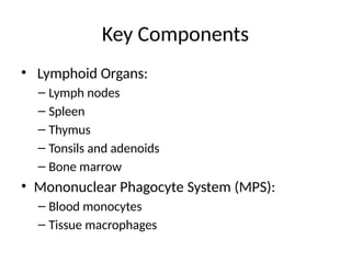
Key Components
• LymphoidOrgans:
– Lymph nodes
– Spleen
– Thymus
– Tonsils and adenoids
– Bone marrow
• Mononuclear Phagocyte System (MPS):
– Blood monocytes
– Tissue macrophages
6.
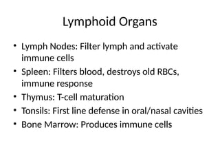
Lymphoid Organs
• LymphNodes: Filter lymph and activate
immune cells
• Spleen: Filters blood, destroys old RBCs,
immune response
• Thymus: T-cell maturation
• Tonsils: First line defense in oral/nasal cavities
• Bone Marrow: Produces immune cells
7.
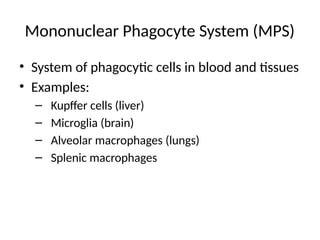
Mononuclear Phagocyte System(MPS)
• System of phagocytic cells in blood and tissues
• Examples:
– Kupffer cells (liver)
– Microglia (brain)
– Alveolar macrophages (lungs)
– Splenic macrophages
8.
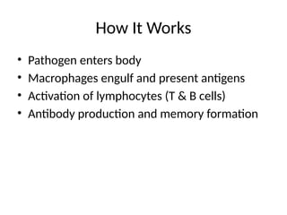
How It Works
•Pathogen enters body
• Macrophages engulf and present antigens
• Activation of lymphocytes (T & B cells)
• Antibody production and memory formation
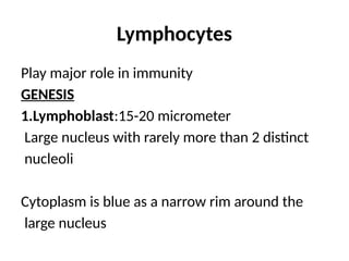
Lymphocytes
Play major rolein immunity
GENESIS
1.Lymphoblast:15-20 micrometer
Large nucleus with rarely more than 2 distinct
nucleoli
Cytoplasm is blue as a narrow rim around the
large nucleus
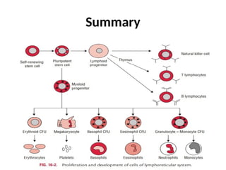
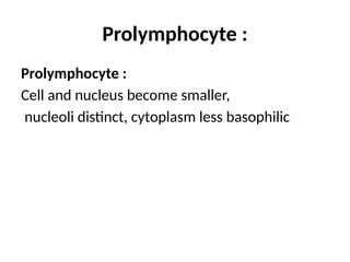
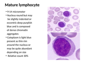
Mature lymphocyte
• 9-14micrometer
• Nucleus round but may
be slightly indented or
eccentric deep purplish
blue and is composed
of dense chromatin
aggregates
• Cytoplasm is light blue
present as thin rim
around the nucleus or
may be quite abundant
depending on size
• Relative count 30%
14.
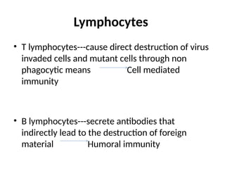
Lymphocytes
• T lymphocytes---causedirect destruction of virus
invaded cells and mutant cells through non
phagocytic means Cell mediated
immunity
• B lymphocytes---secrete antibodies that
indirectly lead to the destruction of foreign
material Humoral immunity
15.
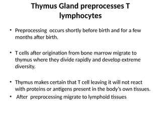
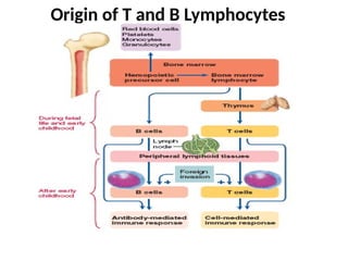
Thymus Gland preprocessesT
lymphocytes
• Preprocessing occurs shortly before birth and for a few
months after birth.
• T cells after origination from bone marrow migrate to
thymus where they divide rapidly and develop extreme
diversity.
• Thymus makes certain that T cell leaving it will not react
with proteins or antigens present in the body’s own tissues.
• After preprocessing migrate to lymphoid tissues
16.
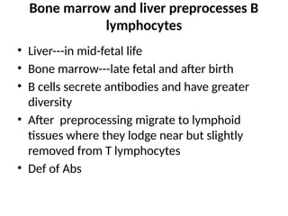
Bone marrow andliver preprocesses B
lymphocytes
• Liver---in mid-fetal life
• Bone marrow---late fetal and after birth
• B cells secrete antibodies and have greater
diversity
• After preprocessing migrate to lymphoid
tissues where they lodge near but slightly
removed from T lymphocytes
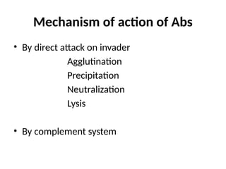
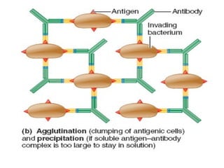
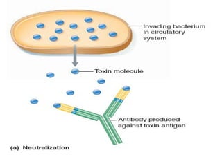
• Def of Abs
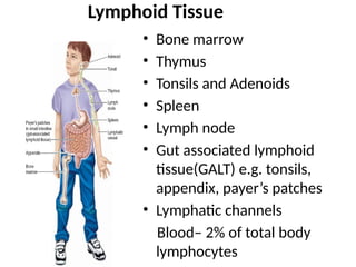
Lymphoid Tissue
• Bonemarrow
• Thymus
• Tonsils and Adenoids
• Spleen
• Lymph node
• Gut associated lymphoid
tissue(GALT) e.g. tonsils,
appendix, payer’s patches
• Lymphatic channels
Blood– 2% of total body
lymphocytes
19.
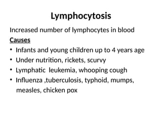
Lymphocytosis
Increased number oflymphocytes in blood
Causes
• Infants and young children up to 4 years age
• Under nutrition, rickets, scurvy
• Lymphatic leukemia, whooping cough
• Influenza ,tuberculosis, typhoid, mumps,
measles, chicken pox
Immunity
The capability toresist almost all type of
organisms or toxins that tend to damage tissues
or organs.
Immune responses may be either
• Innate or non-specific results from general processes
• Acquired or adaptive or specific does not develop
until after the body is first exposed by bacterium,
virus or toxin and often requires weeks or months to
develop
23.
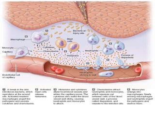
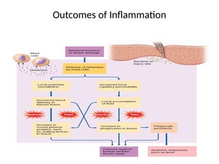
Innate Immunity
• Phagocytosis
•Inflammation
• Acid secretions of stomach and digestive
enzymes
• Skin
• Chemical compounds attached to foreign
organisms and toxins e .g. lysozymes, basic
polypeptides, complement system, natural killer
cells, interferons
24.
Acquired Immunity
Is causedby immune system that form antibodies
and/ or activated lymphocytes that attack and
destroy the specific invading organism or toxin
• Passive immunity--- produced by already made
antibodies or activated T cells from horse or
human serum
• Active immunity--- a person itself produces an
immune reaction in response to the entry of
antigens into the body
25.
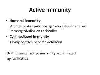
Active Immunity
• HumoralImmunity
B lymphocytes produce gamma globulins called
immnoglobulins or antibodies
• Cell mediated Immunity
T lymphocytes become activated
Both forms of active immunity are initiated
by ANTIGENS
26.
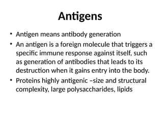
Antigens
• Antigen meansantibody generation
• An antigen is a foreign molecule that triggers a
specific immune response against itself, such
as generation of antibodies that leads to its
destruction when it gains entry into the body.
• Proteins highly antigenic –size and structural
complexity, large polysaccharides, lipids
27.
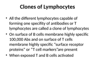
Clones of Lymphocytes
•All the different lymphocytes capable of
forming one specifity of antibodies or T
lymphocytes are called a clone of lymphocytes
• On surface of B cells membrane highly specific
100,000 Abs and on surface of T cells
membrane highly specific “surface receptor
proteins” or “T cell markers”are present
• When exposed T and B cells activated
28.
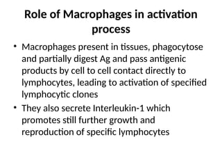
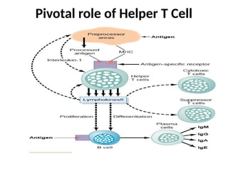
Role of Macrophagesin activation
process
• Macrophages present in tissues, phagocytose
and partially digest Ag and pass antigenic
products by cell to cell contact directly to
lymphocytes, leading to activation of specified
lymphocytic clones
• They also secrete Interleukin-1 which
promotes still further growth and
reproduction of specific lymphocytes
29.
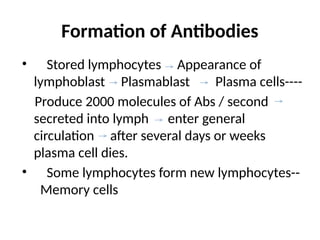
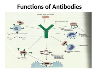
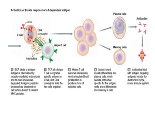
Formation of Antibodies
•Stored lymphocytes Appearance of
lymphoblast Plasmablast Plasma cells----
Produce 2000 molecules of Abs / second
secreted into lymph enter general
circulation after several days or weeks
plasma cell dies.
• Some lymphocytes form new lymphocytes--
Memory cells
Immunization
• By injectingdead organisms– typhoid fever,
whooping cough, diphtheria
• By treating toxins– tetanus, botulism
• By injecting live attenuated organisms–
poliomyelitis, yellow fever, measles, small pox
and many other viral infections
32.
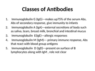
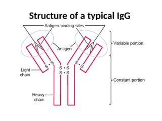
Classes of Antibodies
1.Immunoglobulin G (IgG)---makes up75% of the serum Abs,
Abs of secondary response, give immunity to infants
2. Immunoglobulin A (IgA)---external secretions of body such
as saliva, tears, breast milk, bronchial and intestinal mucus
3. Immunoglobulin E(IgE)---allergic responses
4. Immunoglobulin M (IgM)--- primary immune response, Abs
that react with blood group antigens
5. Immunoglobulin D (IgD)---present on surface of B
lymphocytes along with IgM , role not clear
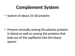
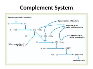
Complement System
• Systemof about 25-30 proteins
• Present normally among the plasma proteins
in blood as well as among the proteins that
leak out of the capillaries into the tissue
spaces
Important Effects of
ComplementSystem
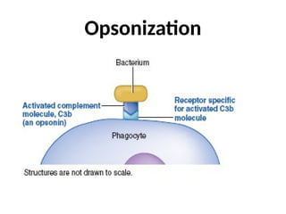
• Opsonization and phagocytosis
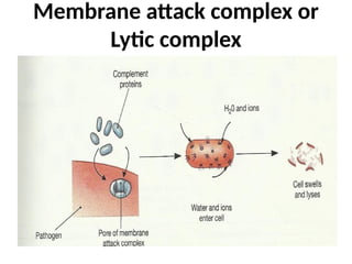
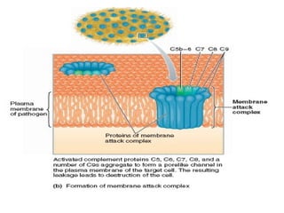
• Lysis
• Neutralization
• Agglutination of viruses
• Chemotaxis
• Activation of mast cells and basophils
• Inflammatory effects
Role of Macrophagesin activation process
• Macrophages present in tissues, phagocytose
and partially digest Ag and pass antigenic
products by cell to cell contact directly to
lymphocytes, leading to activation of specified
lymphocytic clones
• They also secrete Interleukin-1 which
promotes further growth and reproduction of
specific lymphocytes
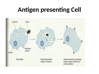
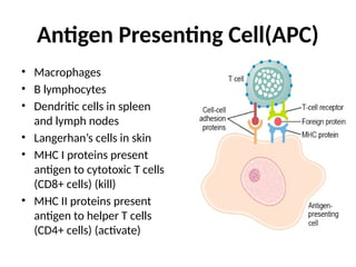
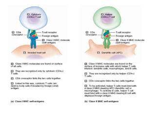
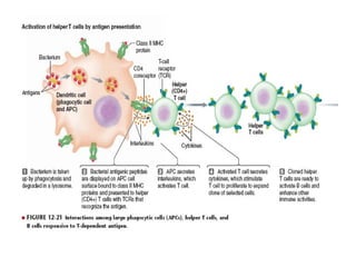
Antigen Presenting Cell(APC)
•Macrophages
• B lymphocytes
• Dendritic cells in spleen
and lymph nodes
• Langerhan’s cells in skin
• MHC I proteins present
antigen to cytotoxic T cells
(CD8+ cells) (kill)
• MHC II proteins present
antigen to helper T cells
(CD4+ cells) (activate)
50.
Types of TCells
• Helper T cells
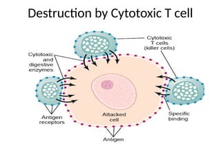
• Cytotoxic T cells– attack and destroy invading
agent or antigen
• Suppressor T Cells– Inhibit or terminate
activities of killer cells, plasma cells or T helper
cells when their activities are no more needed
51.
Lymphokines secreted byHelper T
cells
• Helper T cells serve as the major regulator of virtually all
immune functions, secrete lymphokines e.g. Inerleukin-
2,3,4,5,6
• Granulocyte-monocyte CSF
• Gamma interferon
Regulatory Functions
1. Stimulation of growth and proliferation of Cytotoxic and
suppressor T cells: Interleukin--2
2. Stimulation of B cell growth and differentiation to form
plasma cells and antibodies:IL-4,5,6
3. Activation of macrophage system
4. Feedback stimulatory effect on helper T cells themselves: IL 2
52.
Role of Tcells in activation of B cells
• Usually both the cells are activated
simultaneously
• Helper T cells secrete lymphokines that
activate specific B lymphocytes. Without its
aid quantity of Abs formed is slight
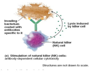
Autoimmune Diseases
Immune Toleranceand role of suppressor T cells
• Rheumatic Fever
• Glomerulonephritis
• Myasthenia gravis
• Rheumatoid arthritis
• Lupus erythematosis
60.
Allergy and Hypersensitivity
•Is an inflammatory immune response to a non-
pathogenic antigen--- allergen
– Delayed hypersensitivity reaction –mediated by activated T
cells e.g ivy toxin
– Immediate hypersensitivity reaction—mediated by Abs
Excess IgE(Reagin Abs) antibodies allergy
• Anaphylaxis
• Urticaria
• Hay fever
• Asthma
61.
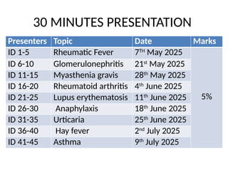
30 MINUTES PRESENTATION
PresentersTopic Date Marks
ID 1-5 Rheumatic Fever 7TH
May 2025
5%
ID 6-10 Glomerulonephritis 21st
May 2025
ID 11-15 Myasthenia gravis 28th
May 2025
ID 16-20 Rheumatoid arthritis 4th
June 2025
ID 21-25 Lupus erythematosis 11th
June 2025
ID 26-30 Anaphylaxis 18th
June 2025
ID 31-35 Urticaria 25th
June 2025
ID 36-40 Hay fever 2nd
July 2025
ID 41-45 Asthma 9th
July 2025
62.
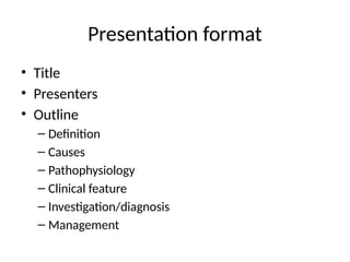
Presentation format
• Title
•Presenters
• Outline
– Definition
– Causes
– Pathophysiology
– Clinical feature
– Investigation/diagnosis
– Management
Editor's Notes
#46 MHC stands for Major Histocompatibility Complex, which refers to a set of proteins found on the surfaces of cells that are essential for the immune system to recognize foreign substances.