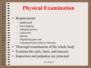
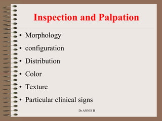
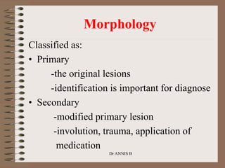
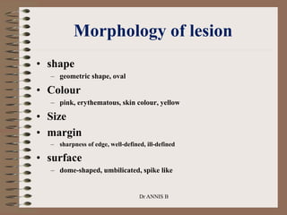
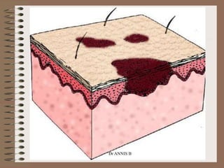
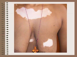
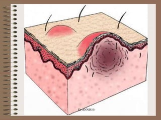
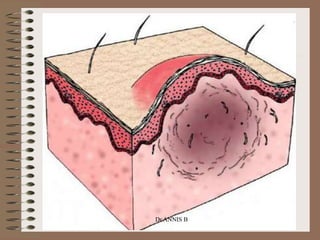
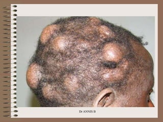
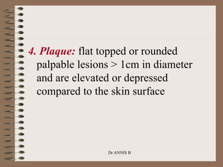
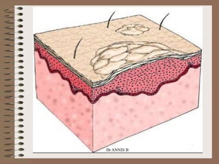
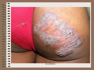
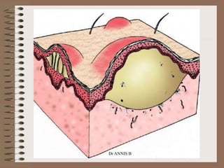
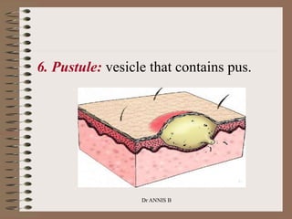
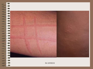
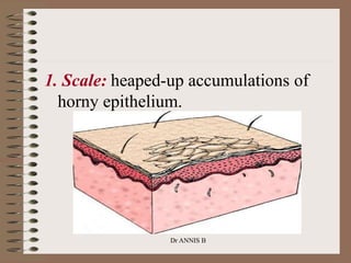
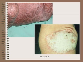
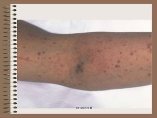
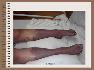
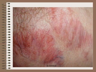
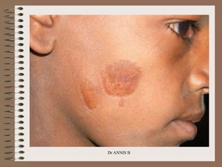
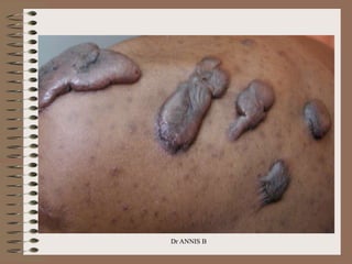
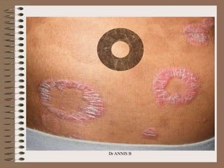
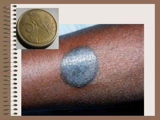
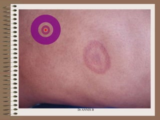
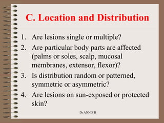
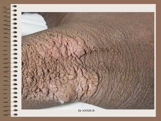
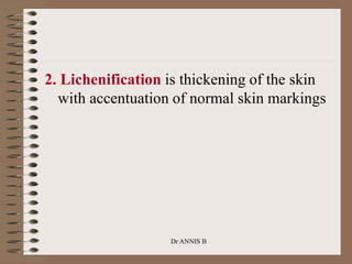
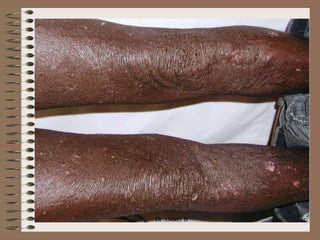
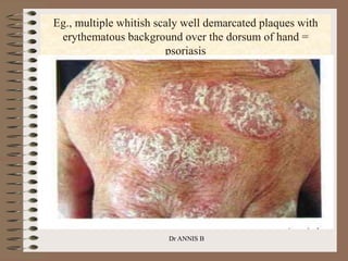
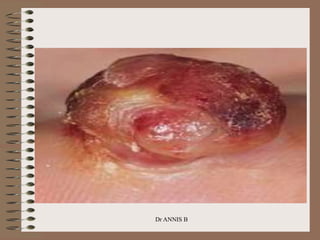
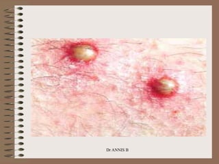
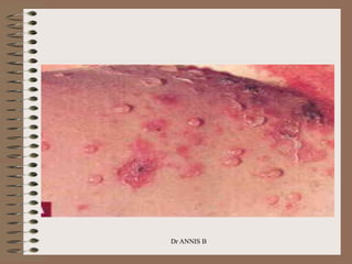
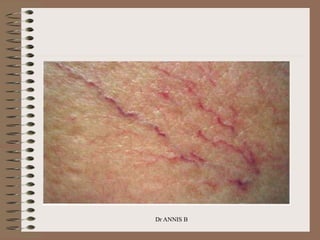
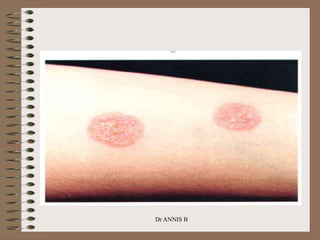
The
Integumentary
System
Diagnosis
Dr ANNIS B
Dr ANNIS B
Components of Dermatological
Evaluation
• History (Subjective)
• Physical Examination (Objective)
• Diagnosis (Assessment)
• Plan
Dr ANNIS B
Dr ANNIS B
• 90% of skin diseases can be properly
diagnosed with meticulous history and
proper physical examination
• The principal steps in physical examination
are inspection and palpation
• Complete cutaneous examination should be
performed
Inspection of the entire skin
Palpation of rashes or localized lesion
-texture, consistency, thickness,
tenderness and temperature
Dr ANNIS B
History Components
• A dermatologic history is similar to
other fields of medicine and includes:
– Chief Complaint:
– History of Present Illness:
• Onset and evolution
• Symptom (itch, pain),
• details of spread
• original morphology
• precipitating and relieving factors such as
climate, sunlight etc.
Dr ANNIS B
Hx….
• Current Treatments
– topical including herbs & systemic
medication
• patient initiated or physician prescribed
– Patient's own perception on the cause of the problem.
• Past Medical History (PMH)
– Allergies
– Medications
Dr ANNIS B
….
• Family History
– Psoriasis
– atopic dermatitis, allergic rhinitis, asthma
– skin cancer- particularly melanoma
• Social History:
– Occupation
– living state
• Review of Systems
Dr ANNIS B
Physical Examination
• Requirements
– undressed
– Good lighting
– Adequate privacy
– Light torch
– Spatula
– Magnifying glass and
– Transparent glass slide for diascopy
• Thorough examination of the whole body
• Examine the nails, hairs, and mucosa
• Inspection and palpation are principal
Dr ANNIS B
Inspection and Palpation
• Morphology
• configuration
• Distribution
• Color
• Texture
• Particular clinical signs
Dr ANNIS B
Classified as:
• Primary
-the original lesions
-identification is important for diagnose
• Secondary
-modified primary lesion
-involution, trauma, application of
medication
Dr ANNIS B
Morphology
Distribution of the lesion:
– Symmetrical Vs asymmetrical
– exposed area Vs sun exposed area
– scalp region
– Hand
– extensor aspect Vs flexor aspect
Dr ANNIS B
Arrangement and
configuration of the lesion
• Grouped
– insect bites, herpes simplex, common warts
• Annular
– granuloma annulare, mycosis fungoides, erythema annulare
centrifugum
• Linear pattern
– Koebner phenomenon, Psoriasis, lichen planus, plane wart,
morphoea, phytophotodermatitis
Dr ANNIS B
Morphology of lesion
• shape
– geometric shape, oval
• Colour
– pink, erythematous, skin colour, yellow
• Size
• margin
– sharpness of edge, well-defined, ill-defined
• surface
– dome-shaped, umbilicated, spike like
Dr ANNIS B
Primary lesions
1. Macule: flat, nonpalpable lesions <
1cm in diameter
 Macules represent a change in color and are
not raised or depressed compared to the skin
surface.
A patch is a large macule.
Dr ANNIS B
Dr ANNIS B
Dr ANNIS B
2. Papule: elevated lesions < 1cm in
diameter that can be felt or palpated.
• solid elevations with no visible fluid
• may be acuminate, rounded, conical,
flat topped, or umbilicated, and may
appear white (as in milium), red (as in
eczema), yellowish (as in xanthoma),
or black (as in melanoma).
Dr ANNIS B
Dr ANNIS B
Dr ANNIS B
3. Nodule: firm papule of >1cm or
lesions that extend into the dermis or
subcutaneous tissue.
Dr ANNIS B
Dr ANNIS B
Dr ANNIS B
4. Plaque: flat topped or rounded
palpable lesions > 1cm in diameter
and are elevated or depressed
compared to the skin surface
Dr ANNIS B
Dr ANNIS B
Dr ANNIS B
5. Vesicle: small, clear, fluid-filled
lesion of < 1cm in diameter. If they
are greater than 1cm in diameter, they
are called bulla or blister.
Dr ANNIS B
Dr ANNIS B
Dr ANNIS B
6. Pustule: vesicle that contains pus.
Dr ANNIS B
Dr ANNIS B
7. Urticaria: a.k.a wheal or hive is
characterized by elevated lesions
caused by localized edema.
Dr ANNIS B
Dr ANNIS B
Secondary morphology
Dr ANNIS B
1. Scale: heaped-up accumulations of
horny epithelium.
Dr ANNIS B
Dr ANNIS B
2. Crust (scab): consists of dried serum,
blood, or pus
Dr ANNIS B
3. Erosion: open area of skin that
results from loss of part or all of the
epidermis
Dr ANNIS B
4. Ulcer: results from loss of the
epidermis and at least part of the
dermis.
Dr ANNIS B
Dr ANNIS B
5. Excoriation
• Punctate or linear abrasion due to
scratching
• Usually involves only the epidermis
• Provides accesses to pyogenic organism
6. Petechia: nonblanchable punctate
focus of hemorrhage.
Dr ANNIS B
Dr ANNIS B
7. Purpura: a larger area of
hemorrhage that may be palpable
Dr ANNIS B
Dr ANNIS B
8. Atrophy: thinning of the skin, which
may appear dry and wrinkled,
resembling cigarette paper
Dr ANNIS B
Dr ANNIS B
9. Scar: area of fibrosis that replaces
normal skin after injury. Some scars
become hypertrophic or thickened and
raised.
Keloid is hypertrophic scar that
extend beyond the original wound
margin.
Dr ANNIS B
Dr ANNIS B
Dr ANNIS B
10. Telangiectasia: a focus of small,
permanently dilated blood vessels
Dr ANNIS B
Dr ANNIS B
B. Configuration
1. Linear lesions take on the shape of a
straight line
Dr ANNIS B
Dr ANNIS B
2. Annular lesions are rings with central
clearing.
Dr ANNIS B
Dr ANNIS B
3. Nummular lesions are circular or coin-
shaped
Dr ANNIS B
Dr ANNIS B
4. Target (iris) lesions appear as rings with
central duskiness
Dr ANNIS B
Dr ANNIS B
5. Serpiginous lesions have linear, branched,
and curving element
Dr ANNIS B
Dr ANNIS B
6. Reticulated lesions have a lacy or
networked pattern
Dr ANNIS B
Dr ANNIS B
7. Herpetiform describes grouped papules or
vesicles
Dr ANNIS B
Dr ANNIS B
8. Zosteriform describes lesions clustered in
a dermatomal distribution
Dr ANNIS B
Dr ANNIS B
C. Location and Distribution
1. Are lesions single or multiple?
2. Are particular body parts are affected
(palms or soles, scalp, mucosal
membranes, extensor, flexor)?
3. Is distribution random or patterned,
symmetric or asymmetric?
4. Are lesions on sun-exposed or protected
skin?
Dr ANNIS B
D. Color
• Pigment (hypo, hyper, de-)
• Violet = Violaceous
• White = Alba
• Red = Erythema
Dr ANNIS B
E. Texture
1. Verrucous lesions have an irregular,
pebbly, or rough surface
Dr ANNIS B
Dr ANNIS B
2. Lichenification is thickening of the skin
with accentuation of normal skin markings
Dr ANNIS B
Dr ANNIS B
3. Induration is deep thickening of the skin
that can result from edema, inflammation,
or infiltration by cancer.
Dr ANNIS B
Dr ANNIS B
4. Umbilicated lesions have a central
indentation
Dr ANNIS B
Dr ANNIS B
F. Particular Clinical Signs
1. Dermatographism is the appearance of an
urticarial wheal after focal pressure.
2. Darier's sign refers to rapid swelling of a lesion
when stroked.
3. Nikolsky's sign is epidermal shearing that occurs
with gentle lateral pressure on seemingly
uninvolved skin.
4. Auspitz sign is the appearance of pinpoint
bleeding after scale is removed from plaques.
5. Koebner phenomenon describes the
development of lesions within areas of trauma.
Dr ANNIS B
DERMATOLOY INVESTIGATION
• SKIN BIOPSY-
• WOOD’S LAMP EXAMINATION
ultraviolet light of 365 nm wavelength is obtained by
passing the beam through a wood’s filter composed of
nickel oxide containing glass
examination should be done in a dark room
microsporum canis-bright green
pityriasis versicolor-yellow
erythrasma-croral red
vitiligo-more white
Dr ANNIS B
Cont’d
• Patch test-tests type 4 hypersensitivity
reaction and it is confirmatory test for
allergic contact dermatitis
• Mycology examination
-Superficial fungi can be identified by
examination of the skin scraping, nail or hair.
The scales, nail or hair should be collected
onto a slide and a drop of 10 to 20 percent
KOH to dissolve the keratin
Dr ANNIS B
Ixs……
• Mite examination-to identify burrow
• CBC, U/A, S/E, SEROLOGY
• Radiology examination
• Clinical Photography
Dr ANNIS B
• Tangential shining of examination torch
to the skin lesions will enhance and
detect elevated skin lesions with ease
• Diascopy consists of pressing a
transparent slide or plastic spatula over
a skin lesion. It is useful to detect the
glassy yellow-brown appearance of
papules in sarcoidosis, tuberculosis and
other granuloma
Special Techniques
Dr ANNIS B
Quiz
Dr ANNIS B
Eg., multiple whitish scaly well demarcated plaques with
erythematous background over the dorsum of hand =
psoriasis
Dr ANNIS B
Dr ANNIS B
Dr ANNIS B
Dr ANNIS B
Dr ANNIS B
Dr ANNIS B
Dr ANNIS B
Dr ANNIS B
Dr ANNIS B
Dr ANNIS B
Dr ANNIS B
Dr ANNIS B
Dr ANNIS B

3morphology.ppt

  • 1.
  • 2.
  • 3.
    Components of Dermatological Evaluation •History (Subjective) • Physical Examination (Objective) • Diagnosis (Assessment) • Plan Dr ANNIS B
  • 4.
    Dr ANNIS B •90% of skin diseases can be properly diagnosed with meticulous history and proper physical examination • The principal steps in physical examination are inspection and palpation • Complete cutaneous examination should be performed
  • 5.
    Inspection of theentire skin Palpation of rashes or localized lesion -texture, consistency, thickness, tenderness and temperature Dr ANNIS B
  • 6.
    History Components • Adermatologic history is similar to other fields of medicine and includes: – Chief Complaint: – History of Present Illness: • Onset and evolution • Symptom (itch, pain), • details of spread • original morphology • precipitating and relieving factors such as climate, sunlight etc. Dr ANNIS B
  • 7.
    Hx…. • Current Treatments –topical including herbs & systemic medication • patient initiated or physician prescribed – Patient's own perception on the cause of the problem. • Past Medical History (PMH) – Allergies – Medications Dr ANNIS B
  • 8.
    …. • Family History –Psoriasis – atopic dermatitis, allergic rhinitis, asthma – skin cancer- particularly melanoma • Social History: – Occupation – living state • Review of Systems Dr ANNIS B
  • 9.
    Physical Examination • Requirements –undressed – Good lighting – Adequate privacy – Light torch – Spatula – Magnifying glass and – Transparent glass slide for diascopy • Thorough examination of the whole body • Examine the nails, hairs, and mucosa • Inspection and palpation are principal Dr ANNIS B
  • 10.
    Inspection and Palpation •Morphology • configuration • Distribution • Color • Texture • Particular clinical signs Dr ANNIS B
  • 11.
    Classified as: • Primary -theoriginal lesions -identification is important for diagnose • Secondary -modified primary lesion -involution, trauma, application of medication Dr ANNIS B Morphology
  • 12.
    Distribution of thelesion: – Symmetrical Vs asymmetrical – exposed area Vs sun exposed area – scalp region – Hand – extensor aspect Vs flexor aspect Dr ANNIS B
  • 13.
    Arrangement and configuration ofthe lesion • Grouped – insect bites, herpes simplex, common warts • Annular – granuloma annulare, mycosis fungoides, erythema annulare centrifugum • Linear pattern – Koebner phenomenon, Psoriasis, lichen planus, plane wart, morphoea, phytophotodermatitis Dr ANNIS B
  • 14.
    Morphology of lesion •shape – geometric shape, oval • Colour – pink, erythematous, skin colour, yellow • Size • margin – sharpness of edge, well-defined, ill-defined • surface – dome-shaped, umbilicated, spike like Dr ANNIS B
  • 15.
    Primary lesions 1. Macule:flat, nonpalpable lesions < 1cm in diameter  Macules represent a change in color and are not raised or depressed compared to the skin surface. A patch is a large macule. Dr ANNIS B
  • 16.
  • 17.
  • 18.
    2. Papule: elevatedlesions < 1cm in diameter that can be felt or palpated. • solid elevations with no visible fluid • may be acuminate, rounded, conical, flat topped, or umbilicated, and may appear white (as in milium), red (as in eczema), yellowish (as in xanthoma), or black (as in melanoma). Dr ANNIS B
  • 19.
  • 20.
  • 21.
    3. Nodule: firmpapule of >1cm or lesions that extend into the dermis or subcutaneous tissue. Dr ANNIS B
  • 22.
  • 23.
  • 24.
    4. Plaque: flattopped or rounded palpable lesions > 1cm in diameter and are elevated or depressed compared to the skin surface Dr ANNIS B
  • 25.
  • 26.
  • 27.
    5. Vesicle: small,clear, fluid-filled lesion of < 1cm in diameter. If they are greater than 1cm in diameter, they are called bulla or blister. Dr ANNIS B
  • 28.
  • 29.
  • 30.
    6. Pustule: vesiclethat contains pus. Dr ANNIS B
  • 31.
  • 32.
    7. Urticaria: a.k.awheal or hive is characterized by elevated lesions caused by localized edema. Dr ANNIS B
  • 33.
  • 34.
  • 35.
    1. Scale: heaped-upaccumulations of horny epithelium. Dr ANNIS B
  • 36.
  • 37.
    2. Crust (scab):consists of dried serum, blood, or pus Dr ANNIS B
  • 38.
    3. Erosion: openarea of skin that results from loss of part or all of the epidermis Dr ANNIS B
  • 39.
    4. Ulcer: resultsfrom loss of the epidermis and at least part of the dermis. Dr ANNIS B
  • 40.
    Dr ANNIS B 5.Excoriation • Punctate or linear abrasion due to scratching • Usually involves only the epidermis • Provides accesses to pyogenic organism
  • 41.
    6. Petechia: nonblanchablepunctate focus of hemorrhage. Dr ANNIS B
  • 42.
  • 43.
    7. Purpura: alarger area of hemorrhage that may be palpable Dr ANNIS B
  • 44.
  • 45.
    8. Atrophy: thinningof the skin, which may appear dry and wrinkled, resembling cigarette paper Dr ANNIS B
  • 46.
  • 47.
    9. Scar: areaof fibrosis that replaces normal skin after injury. Some scars become hypertrophic or thickened and raised. Keloid is hypertrophic scar that extend beyond the original wound margin. Dr ANNIS B
  • 48.
  • 49.
  • 50.
    10. Telangiectasia: afocus of small, permanently dilated blood vessels Dr ANNIS B
  • 51.
  • 52.
    B. Configuration 1. Linearlesions take on the shape of a straight line Dr ANNIS B
  • 53.
  • 54.
    2. Annular lesionsare rings with central clearing. Dr ANNIS B
  • 55.
  • 56.
    3. Nummular lesionsare circular or coin- shaped Dr ANNIS B
  • 57.
  • 58.
    4. Target (iris)lesions appear as rings with central duskiness Dr ANNIS B
  • 59.
  • 60.
    5. Serpiginous lesionshave linear, branched, and curving element Dr ANNIS B
  • 61.
  • 62.
    6. Reticulated lesionshave a lacy or networked pattern Dr ANNIS B
  • 63.
  • 64.
    7. Herpetiform describesgrouped papules or vesicles Dr ANNIS B
  • 65.
  • 66.
    8. Zosteriform describeslesions clustered in a dermatomal distribution Dr ANNIS B
  • 67.
  • 68.
    C. Location andDistribution 1. Are lesions single or multiple? 2. Are particular body parts are affected (palms or soles, scalp, mucosal membranes, extensor, flexor)? 3. Is distribution random or patterned, symmetric or asymmetric? 4. Are lesions on sun-exposed or protected skin? Dr ANNIS B
  • 69.
    D. Color • Pigment(hypo, hyper, de-) • Violet = Violaceous • White = Alba • Red = Erythema Dr ANNIS B
  • 70.
    E. Texture 1. Verrucouslesions have an irregular, pebbly, or rough surface Dr ANNIS B
  • 71.
  • 72.
    2. Lichenification isthickening of the skin with accentuation of normal skin markings Dr ANNIS B
  • 73.
  • 74.
    3. Induration isdeep thickening of the skin that can result from edema, inflammation, or infiltration by cancer. Dr ANNIS B
  • 75.
  • 76.
    4. Umbilicated lesionshave a central indentation Dr ANNIS B
  • 77.
  • 78.
    F. Particular ClinicalSigns 1. Dermatographism is the appearance of an urticarial wheal after focal pressure. 2. Darier's sign refers to rapid swelling of a lesion when stroked. 3. Nikolsky's sign is epidermal shearing that occurs with gentle lateral pressure on seemingly uninvolved skin. 4. Auspitz sign is the appearance of pinpoint bleeding after scale is removed from plaques. 5. Koebner phenomenon describes the development of lesions within areas of trauma. Dr ANNIS B
  • 79.
    DERMATOLOY INVESTIGATION • SKINBIOPSY- • WOOD’S LAMP EXAMINATION ultraviolet light of 365 nm wavelength is obtained by passing the beam through a wood’s filter composed of nickel oxide containing glass examination should be done in a dark room microsporum canis-bright green pityriasis versicolor-yellow erythrasma-croral red vitiligo-more white Dr ANNIS B
  • 80.
    Cont’d • Patch test-teststype 4 hypersensitivity reaction and it is confirmatory test for allergic contact dermatitis • Mycology examination -Superficial fungi can be identified by examination of the skin scraping, nail or hair. The scales, nail or hair should be collected onto a slide and a drop of 10 to 20 percent KOH to dissolve the keratin Dr ANNIS B
  • 81.
    Ixs…… • Mite examination-toidentify burrow • CBC, U/A, S/E, SEROLOGY • Radiology examination • Clinical Photography Dr ANNIS B
  • 82.
    • Tangential shiningof examination torch to the skin lesions will enhance and detect elevated skin lesions with ease • Diascopy consists of pressing a transparent slide or plastic spatula over a skin lesion. It is useful to detect the glassy yellow-brown appearance of papules in sarcoidosis, tuberculosis and other granuloma Special Techniques Dr ANNIS B
  • 83.
  • 84.
    Eg., multiple whitishscaly well demarcated plaques with erythematous background over the dorsum of hand = psoriasis Dr ANNIS B
  • 85.
  • 86.
  • 87.
  • 88.
  • 89.
  • 90.
  • 91.
  • 92.
  • 93.
  • 94.
  • 95.
  • 96.