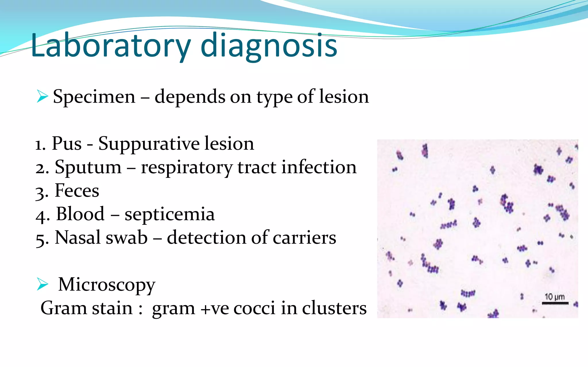
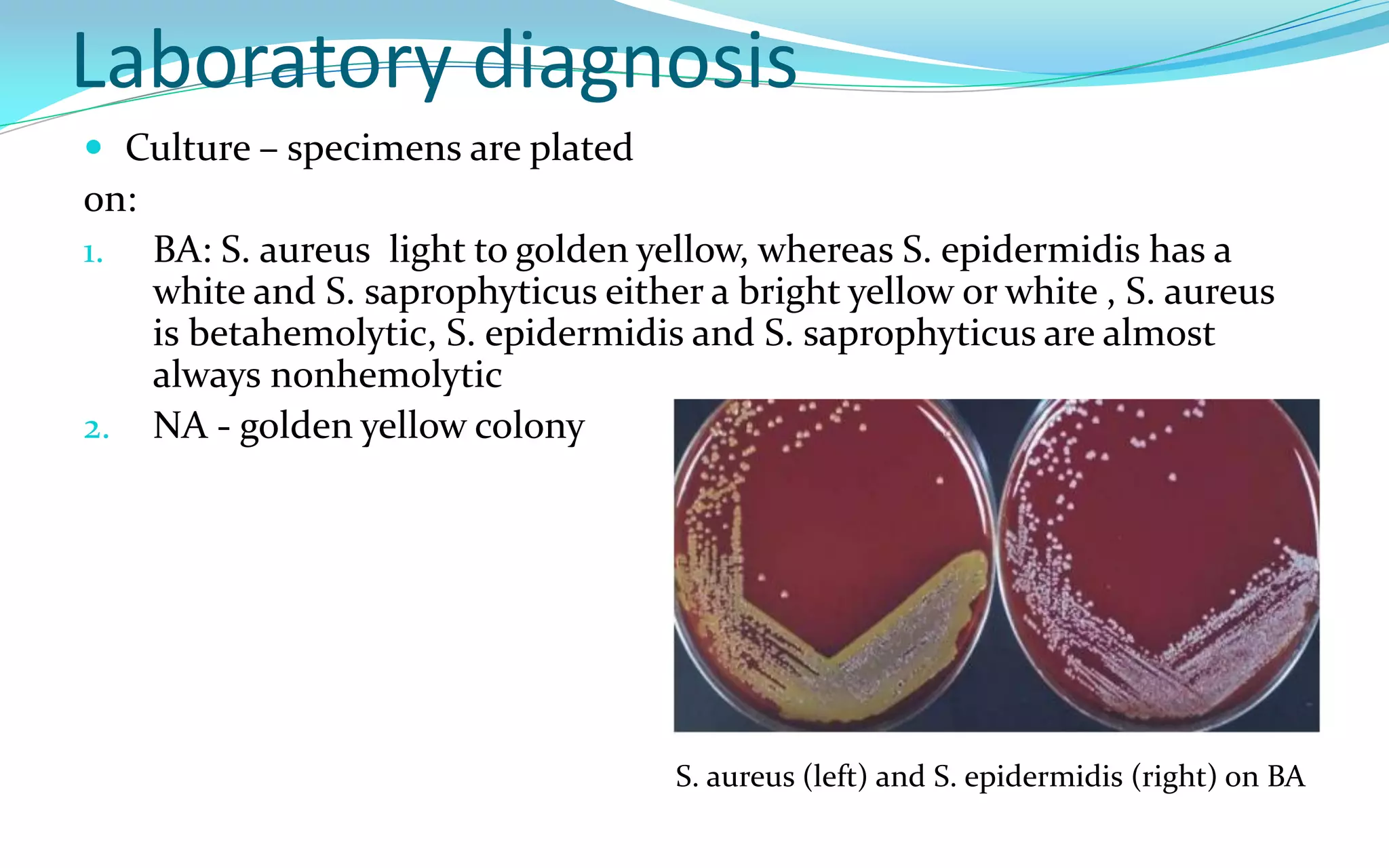
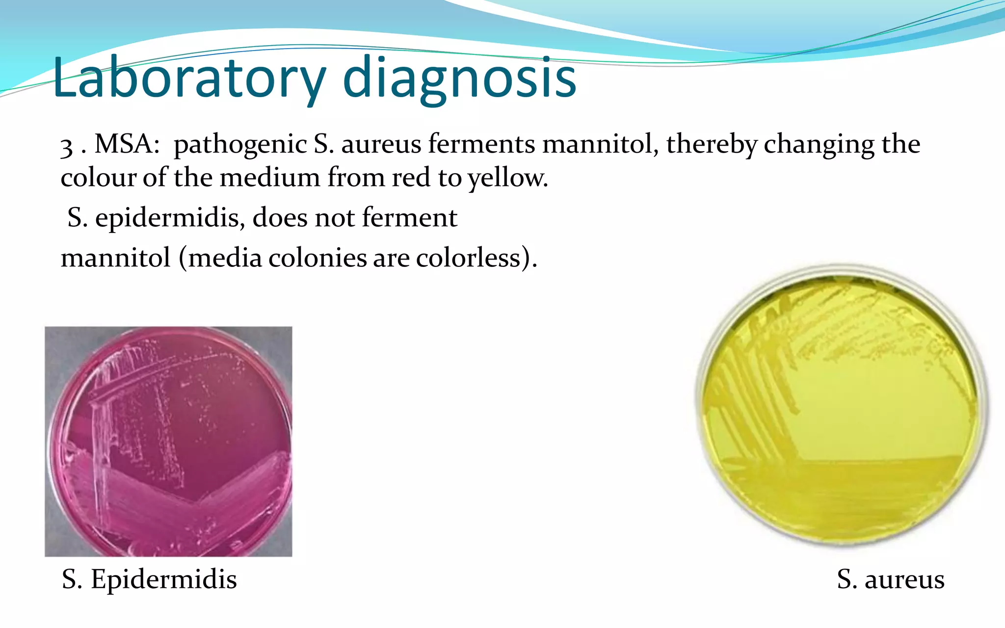
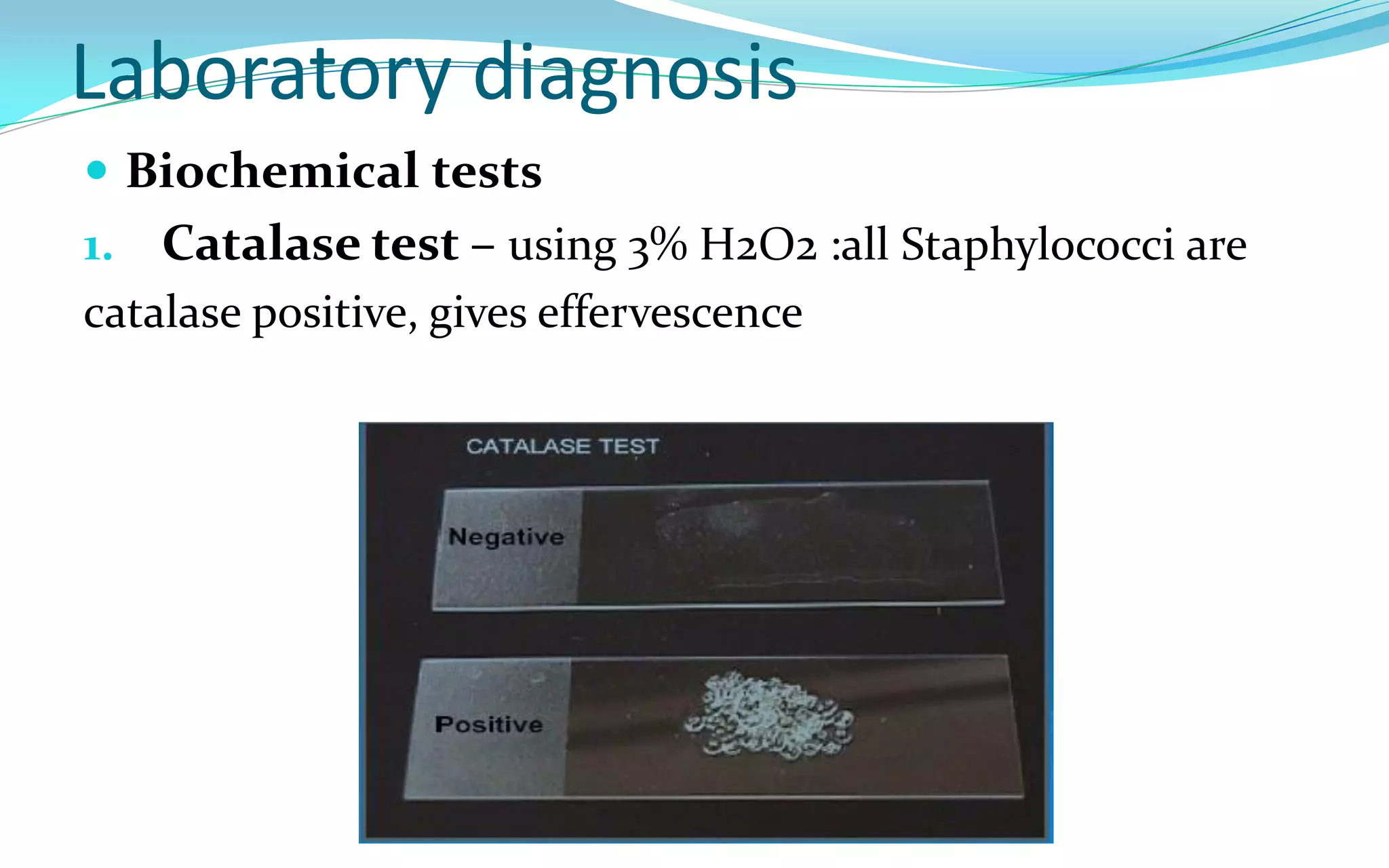
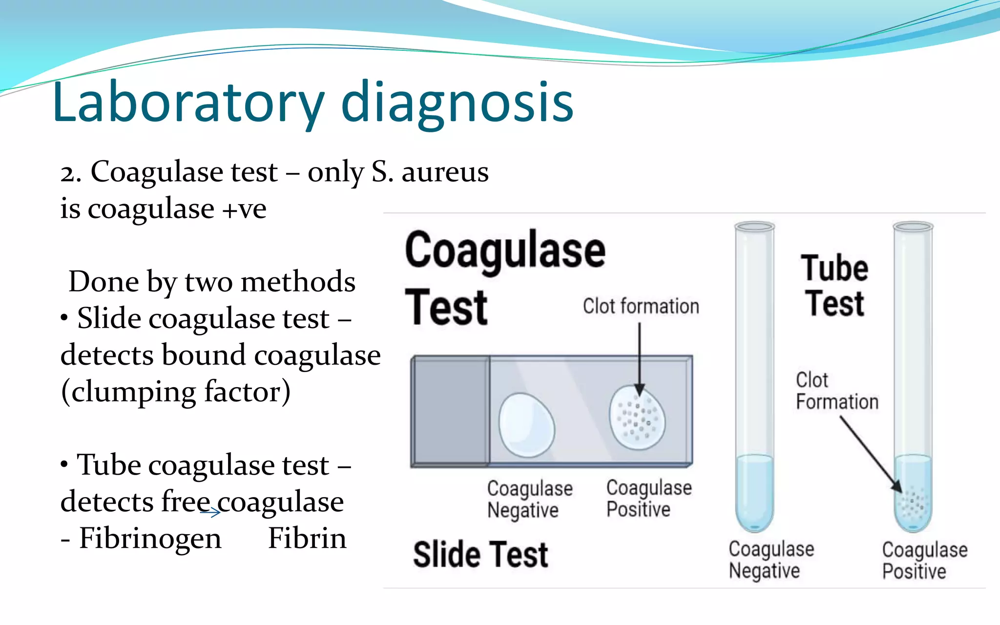
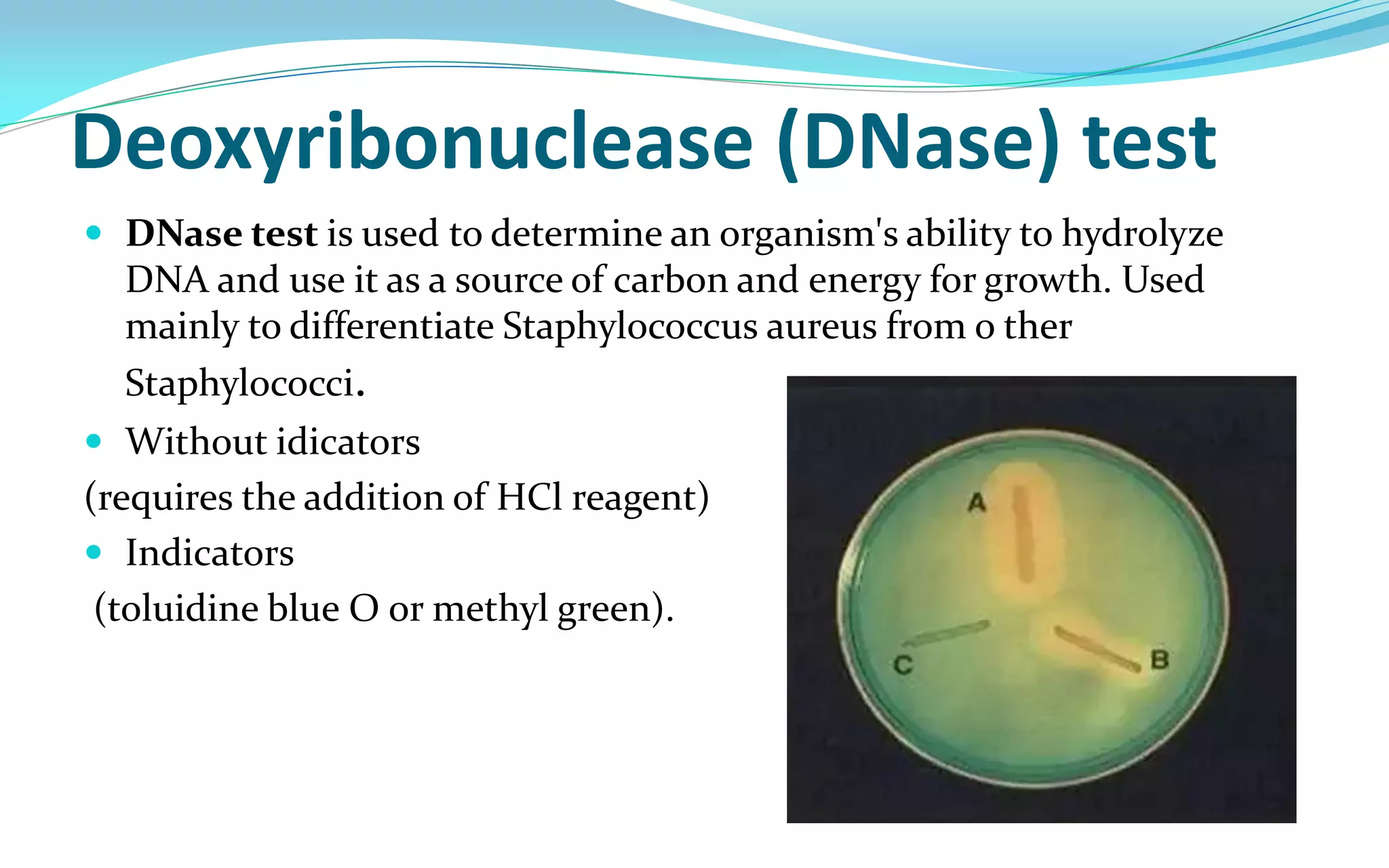
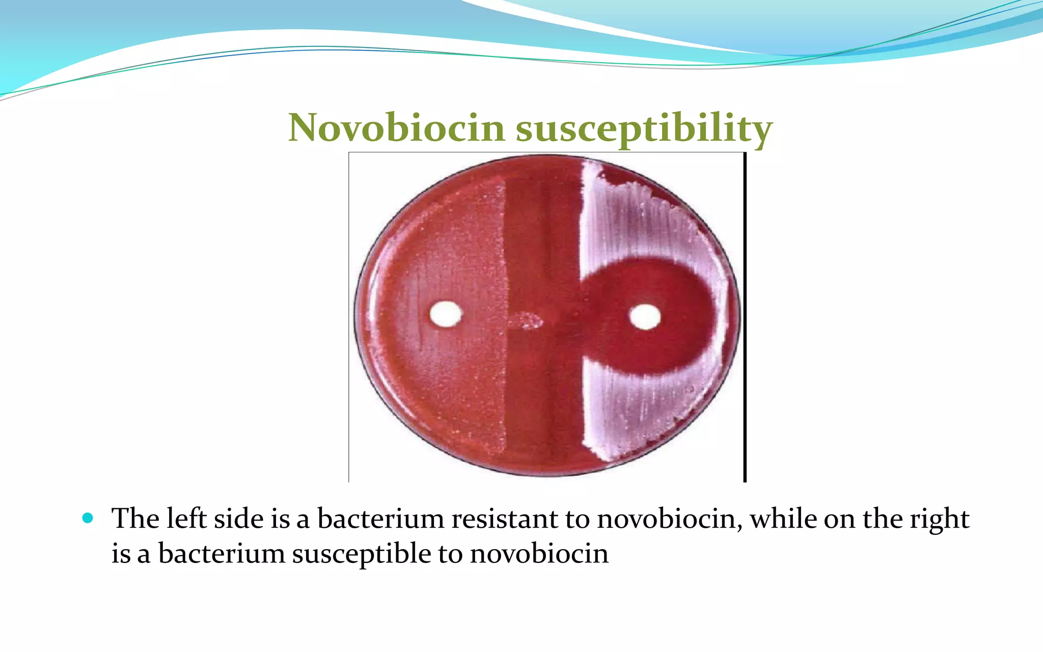
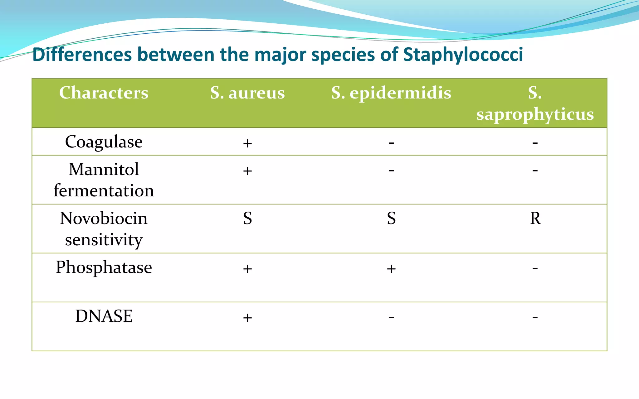
By
M.S.C. Akram A . Alqragholi
Staphylococci – General features
 Gram +ve cocci arranged in grape like cluster
 Facultative anaerobes
 Temperature range for growth is 10- 42 oc, with an optimum of
35 – 37c Produce catalase
 Oxidase negative
 Non motile
 Major component of normal flora in humans
 Skin
 Nose
 Mucosal membranes
Overview
 Many species are medically important
1. S. aureus –
• most virulent species
• Most common cause of bacterial infections, furunculosis ,food
poisoning & toxic shock syndrome
2. S. epidermidis – important cause of prosthetic implant
infections
3. S. saprophyticus – UTI, especially cystitis in women
S. aureus – General features
Coagulase positive
 Ferments mannitol
 Beta hemolytic colonies on BA
Produces phospahatse
 Highly resistant non sporing bacteria
 Can grow in the presence of 10 – 15% NaCl.
 Resistance to Penicillin
Virulence Factors
The virulence of S. aureus depends on a number of
factors. These can be:
1. Cell associated polymers and surface proteins,
2. Enzymes
3. Toxins – cytolytic & superantigen exotoxins
Virulence Factors
1. Cell associated polymers & surface proteins
 Teichoic acid:- promotes colonization of host tissues
 Capsule (slime layer) :- inhibits phagocytosis
 Protein A Protein A – binds to the Fc region of IgG and
inhibits phagocytosis by preventing opsonization
 Clumping factor:-
 Fibronectin binding protein (FnBP) – binding to
 mucosa & tissue
Virulence Factors
2. Enzymes
 Coagulase: Clotting of human blood or plasma
 Staphylokinase (Fibrinolysin) – lysis fibrin clots, spread of
infection
 Hyaluronidase – breaks down the connective tissue, initition &
spread of infection
 Lipase – infection of skin & subcutaneous tissue
 Nuclease – degrades DNA
 Catalase –
enhance their survival in phagocytes by inactivating
toxic H2O2 & free radicals released after the ingestion
of staphylococci.
Virulence Factors
3. Toxins
 Hemolysins : lysis RBCs
 Leukocidin: lysis neutrophils and macrophage
 Enterotoxin: causes food poisoning
 Toxic shock syndrome toxin (TSST):
 Exfoliatin (epidermolytic toxin):
 Pecillinase :
Laboratory diagnosis
 Specimen – depends on type of lesion
1. Pus - Suppurative lesion
2. Sputum – respiratory tract infection
3. Feces
4. Blood – septicemia
5. Nasal swab – detection of carriers
 Microscopy
Gram stain : gram +ve cocci in clusters
Laboratory diagnosis
 Culture – specimens are plated
on:
1. BA: S. aureus light to golden yellow, whereas S. epidermidis has a
white and S. saprophyticus either a bright yellow or white , S. aureus
is betahemolytic, S. epidermidis and S. saprophyticus are almost
always nonhemolytic
2. NA - golden yellow colony
S. aureus (left) and S. epidermidis (right) on BA
Laboratory diagnosis
3 . MSA: pathogenic S. aureus ferments mannitol, thereby changing the
colour of the medium from red to yellow.
S. epidermidis, does not ferment
mannitol (media colonies are colorless).
S. Epidermidis S. aureus
Laboratory diagnosis
 Biochemical tests
1. Catalase test – using 3% H2O2 :all Staphylococci are
catalase positive, gives effervescence
Laboratory diagnosis
2. Coagulase test – only S. aureus
is coagulase +ve
Done by two methods
• Slide coagulase test –
detects bound coagulase
(clumping factor)
• Tube coagulase test –
detects free coagulase
- Fibrinogen Fibrin
Deoxyribonuclease (DNase) test
 DNase test is used to determine an organism's ability to hydrolyze
DNA and use it as a source of carbon and energy for growth. Used
mainly to differentiate Staphylococcus aureus from o ther
Staphylococci.
 Without idicators
(requires the addition of HCl reagent)
 Indicators
(toluidine blue O or methyl green).
Novobiocin susceptibility
 The left side is a bacterium resistant to novobiocin, while on the right
is a bacterium susceptible to novobiocin
Differences between the major species of Staphylococci
Characters S. aureus S. epidermidis S.
saprophyticus
Coagulase + - -
Mannitol
fermentation
+ - -
Novobiocin
sensitivity
S S R
Phosphatase + + -
DNASE + - -

3 staphylococcus.pdf

  • 1.
    By M.S.C. Akram A. Alqragholi
  • 2.
    Staphylococci – Generalfeatures  Gram +ve cocci arranged in grape like cluster  Facultative anaerobes  Temperature range for growth is 10- 42 oc, with an optimum of 35 – 37c Produce catalase  Oxidase negative  Non motile  Major component of normal flora in humans  Skin  Nose  Mucosal membranes
  • 3.
    Overview  Many speciesare medically important 1. S. aureus – • most virulent species • Most common cause of bacterial infections, furunculosis ,food poisoning & toxic shock syndrome 2. S. epidermidis – important cause of prosthetic implant infections 3. S. saprophyticus – UTI, especially cystitis in women
  • 4.
    S. aureus –General features Coagulase positive  Ferments mannitol  Beta hemolytic colonies on BA Produces phospahatse  Highly resistant non sporing bacteria  Can grow in the presence of 10 – 15% NaCl.  Resistance to Penicillin
  • 5.
    Virulence Factors The virulenceof S. aureus depends on a number of factors. These can be: 1. Cell associated polymers and surface proteins, 2. Enzymes 3. Toxins – cytolytic & superantigen exotoxins
  • 6.
    Virulence Factors 1. Cellassociated polymers & surface proteins  Teichoic acid:- promotes colonization of host tissues  Capsule (slime layer) :- inhibits phagocytosis  Protein A Protein A – binds to the Fc region of IgG and inhibits phagocytosis by preventing opsonization  Clumping factor:-  Fibronectin binding protein (FnBP) – binding to  mucosa & tissue
  • 7.
    Virulence Factors 2. Enzymes Coagulase: Clotting of human blood or plasma  Staphylokinase (Fibrinolysin) – lysis fibrin clots, spread of infection  Hyaluronidase – breaks down the connective tissue, initition & spread of infection  Lipase – infection of skin & subcutaneous tissue  Nuclease – degrades DNA  Catalase – enhance their survival in phagocytes by inactivating toxic H2O2 & free radicals released after the ingestion of staphylococci.
  • 8.
    Virulence Factors 3. Toxins Hemolysins : lysis RBCs  Leukocidin: lysis neutrophils and macrophage  Enterotoxin: causes food poisoning  Toxic shock syndrome toxin (TSST):  Exfoliatin (epidermolytic toxin):  Pecillinase :
  • 9.
    Laboratory diagnosis  Specimen– depends on type of lesion 1. Pus - Suppurative lesion 2. Sputum – respiratory tract infection 3. Feces 4. Blood – septicemia 5. Nasal swab – detection of carriers  Microscopy Gram stain : gram +ve cocci in clusters
  • 10.
    Laboratory diagnosis  Culture– specimens are plated on: 1. BA: S. aureus light to golden yellow, whereas S. epidermidis has a white and S. saprophyticus either a bright yellow or white , S. aureus is betahemolytic, S. epidermidis and S. saprophyticus are almost always nonhemolytic 2. NA - golden yellow colony S. aureus (left) and S. epidermidis (right) on BA
  • 11.
    Laboratory diagnosis 3 .MSA: pathogenic S. aureus ferments mannitol, thereby changing the colour of the medium from red to yellow. S. epidermidis, does not ferment mannitol (media colonies are colorless). S. Epidermidis S. aureus
  • 12.
    Laboratory diagnosis  Biochemicaltests 1. Catalase test – using 3% H2O2 :all Staphylococci are catalase positive, gives effervescence
  • 13.
    Laboratory diagnosis 2. Coagulasetest – only S. aureus is coagulase +ve Done by two methods • Slide coagulase test – detects bound coagulase (clumping factor) • Tube coagulase test – detects free coagulase - Fibrinogen Fibrin
  • 14.
    Deoxyribonuclease (DNase) test DNase test is used to determine an organism's ability to hydrolyze DNA and use it as a source of carbon and energy for growth. Used mainly to differentiate Staphylococcus aureus from o ther Staphylococci.  Without idicators (requires the addition of HCl reagent)  Indicators (toluidine blue O or methyl green).
  • 15.
    Novobiocin susceptibility  Theleft side is a bacterium resistant to novobiocin, while on the right is a bacterium susceptible to novobiocin
  • 16.
    Differences between themajor species of Staphylococci Characters S. aureus S. epidermidis S. saprophyticus Coagulase + - - Mannitol fermentation + - - Novobiocin sensitivity S S R Phosphatase + + - DNASE + - -