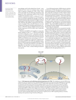
More Related Content
Similar to 3 moléculas acessórias de toll
Similar to 3 moléculas acessórias de toll (20)
More from ufamimunologia (13)
3 moléculas acessórias de toll
- 1. REVIEWS
Accessory molecules for Toll-like
receptors and their function
Clarissa C. Lee*, Ana M. Avalos* and Hidde L. Ploegh
Abstract | Toll-like receptors (TLRs) are essential components of the innate immune system.
Accessory proteins are required for the biosynthesis and activation of TLRs. Here, we
summarize recent findings on TLR accessory proteins that are required for cell-surface
and endosomal TLR function, and we classify these proteins based on their function as
ligand-recognition and delivery cofactors, chaperones and trafficking proteins. Because
of their essential roles in TLR function, targeting of such accessory proteins may benefit
strategies aimed at manipulating TLR activation for therapeutic applications.
Pattern-recognition receptors (PRRs) are germline- the recruitment of signalling adaptor molecules. TLRs
Alarmins
Endogenous mediators that encoded innate immune receptors that were originally form heterodimers or homodimers as a means of trig-
are released by necrotic cells in reported as sensors for pathogen-associated molecular gering a signal. Most TLRs form homodimers, with a few
response to infection or injury patterns (PAMPs)1. PRRs can also recognize endogenous exceptions. For example, TLR2 forms heterodimers with
and that interact with pattern- molecules that are released in response to stress or tissue TLR1 or TLR6, which enables differential recognition of
recognition receptors to
activate innate immune cells.
damage, thus behaving as sensors of alarmins. PRRs there- lipopeptides: TLR1–TLR2 recognizes triacylated lipo-
fore sense PAMPs and alarmins, which together consti- peptides, whereas TLR2–TLR6 responds to diacylated
tute damage-associated molecular patterns (DAMPs)2. lipopeptides (TABLE 1).
PRR engagement promotes the activation of innate and Extracellular and endosomal TLRs have similar
adaptive immune responses1. Members of the Toll-like ectodomain sequences, a feature that is in sharp con-
receptor (TLR) family are PRRs that recognize pathogen- trast with the diversity of the ligands that they recog-
derived macromolecules, ranging from bacterial and nize. One mode of ligand discrimination relies on the
yeast cell wall components to viral and bacterial nucleic differences in the residues present in the ectodomains
acids. TLR ligation leads to the activation of nuclear of distinct TLRs. The leucine-rich repeat modules
factor-κB (NF-κB) and interferon-regulatory factors located in the ectodomains of TLRs are each com-
(IRFs), and these transcription factors then induce the posed of 20–30 amino acids and contain the consensus
production of pro-inflammatory cytokines and type I sequence LxxLxLxxN. TLRs have different amino acid
interferons (IFNs), respectively. compositions within these modules, leading to varia-
Humans express ten functional TLRs (TLR1 to tions in structural conformation that allow for ligand
TLR10), whereas twelve TLRs (TLR1 to TLR9 and interaction3. Amino acid variations and the formation
TLR11 to TLR13) have been identified in mice. Ligands of heterodimers can only provide a limited platform
have been determined for all TLRs except for human for the recognition of the varied set of TLR ligands.
TLR10, mouse TLR12 and mouse TLR13. TLR1, TLR2, Thus, another mechanism that reflects the complexity
TLR4, TLR5, TLR6 and TLR11 reside at the plasma and diversity of TLR ligand composition must exist
membrane, where they recognize molecular compo- to ensure proper detection of PAMPs and discrimi-
Whitehead Institute for nents located on the surface of pathogens. By contrast, nation between self and non-self. Specific accessory
Biomedical Research, TLR3, TLR7, TLR8 and TLR9 are found intracellularly, proteins or cofactors can fulfil that role. A given TLR
Massachusetts Institute of
where they mediate the recognition of nucleic acids. dimer can bind to co actors that deliver molecules of
f
Technology, Cambridge,
Massachusetts 02142, USA. Thus, the subcellular distribution of TLRs correlates, a particular composition while avoiding other ligands.
Correspondence to H.L.P. to a substantial extent, with the compartments in which These cofactors can also have roles in ensuring proper
e-mail: ploegh@wi.mit.edu their ligands are found (TABLE 1). TLR folding in the endoplasmic reticulum (ER),
*These authors contributed TLRs are type I transmembrane proteins composed localization to the appropriate subcellular compart-
equally to this work.
doi:10.1038/nri3151
of an ectodomain that contains leucine-rich repeats, ment and protein processing, all of which ensure that
Published online a single transmembrane domain and a cyto lasmic p TLRs reach their assigned subcellular compartments
3 February 2012 Toll/IL‑1 receptor (TIR) domain that is involved in to bind to ligands and initiate signalling. Thus, given
168 | MARCH 2012 | VOLUME 12 www.nature.com/reviews/immunol
© 2012 Macmillan Publishers Limited. All rights reserved
- 2. REVIEWS
Table 1 | Localization and ligands of TLRs
TLR Subcellular Physiological ligands Synthetic ligands
localization
TLR1– Plasma Triacylated lipopeptides Pam3CSK4
TLR2 membrane
TLR2 Plasma Peptidoglycan, phospholipomannan, tGPI-mucins, ND
membrane haemagglutinin, porins, lipoarabinomannan,
glucuronoxylomannan, HMGB1
TLR2– Plasma Diacylated lipopeptides, LTA, zymosan FSL1, MALP2, Pam2CSK4
TLR6 membrane
TLR3 Endosome dsRNA PolyI:C
TLR4 Plasma LPS, VSV glycoprotein G, RSV fusion protein, MMTV ND
membrane envelope protein, mannan, glucuronoxylomannan,
glycosylinositolphospholipids, HSP60, HSP70,
fibrinogen, nickel, HMGB1
TLR4– Plasma OxLDL, amyloid‑β fibrils ND
TLR6 membrane
TLR5 Plasma Flagellin ND
membrane
TLR7 Endosome ssRNA Imidazoquinoline compounds:
imiquimod, resiquimod,
loxoribine
TLR8 Endosome ssRNA Resiquimod
TLR9 Endosome DNA, haemozoin CpG‑A, CpG‑B and CpG‑C ODNs
TLR11 Plasma Profilin ND
(mouse) membrane
dsRNA, double-stranded RNA; FSL1, S‑(2,3-bispalmitoyloxypropyl)-CGDPKHSPKSF; HMGB1, high-mobility group box 1 protein;
HSP, heat-shock protein; LPS, lipopolysaccharide; LTA, lipoteichoic acid; MALP2, macrophage-activating lipopeptide of 2 kDa;
MMTV, mouse mammary tumour virus; ND, not determined; ODN, oligodeoxynucleotide; oxLDL, oxidized low-density lipoprotein;
polyI:C, polyinosinic–polycytidylic acid; RSV, respiratory syncytial virus; ssRNA, single-stranded RNA; tGPI-mucin, Trypanosoma
cruzi glycosylphosphatidylinositol-anchored mucin-like glycoprotein; TLR, Toll-like receptor; VSV, vesicular stomatitis virus.
the observed differences in ligand binding and signal- Cofactors for surface TLRs
ling outcomes among TLRs, accessory proteins may LBP in ligand delivery to surface TLRs. LPS-binding
modulate various aspects of TLR function. protein (LBP) is a 481‑amino-acid acute-phase protein
For the purpose of this Review, we use the terms that binds with high affinity to lipopolysaccharide (LPS)
‘accessory protein’ and ‘cofactor’ synonymously, and we derived from the outer membrane of Gram-negative
define molecules as such when they fulfil the following bacteria12. This interaction facilitates the disaggregation
roles: they are required for TLR function; they interact of LPS and its presentation to CD14, an accessory protein
with a TLR or a TLR ligand; and their ability to facilitate that, among other functions, mediates TLR4 responsive-
the interaction of a TLR with a ligand has been experi- ness to LPS12,13 (FIG. 1). LBP can also bind to lipoteichoic
mentally confirmed. This definition aims to focus the acid (LTA), peptidoglycan and lipopeptides and transfer
Review on bona fide TLR cofactors, thereby excluding them to CD14, suggesting that LBP may assist not only
scaffolding or adaptor proteins required for signalling in the function of TLR4, but also in the function of TLR1,
(such as myeloid differentiation primary-response pro- TLR2 and TLR6 (REFS 14–16). Lbp–/– mice are highly sus-
tein 88 (MYD88) and TIR domain-containing adaptor ceptible to infection with the Gram-negative bacterium
protein inducing IFNβ (TRIF); reviewed in REFS 4,5), as Salmonella enterica subsp. enterica serovar Typhimurium
well as molecules involved in crosstalk between signal- compared with Lbp+/– mice17. LBP also has a role in the
Acute-phase protein
ling pathways (reviewed in REF. 6) and negative regulators in vivo immune response to Gram-positive pneumococci,
A member of a group of (reviewed in REFS 4,7). We also discuss certain molecules as leukocyte influx into the cerebrospinal fluid (the hall-
proteins — including C‑reactive that are essential for TLR functions (although their roles as mark of bacterial meningitis) following challenge with
protein, serum amyloid A, cofactors are less well defined than those of the other acces- pneumococci was drastically reduced in Lbp–/– mice
fibrinogen and α1‑acid
sory molecules that we describe) and that may also have compared with that in Lbp+/– mice18. LBP thus mediates
glycoprotein — that are
secreted into the blood in roles in signalling crosstalk (BOX 1). A separate category innate immune responses to PAMPs derived from both
increased or decreased comprises receptors that can interact with TLRs and may Gram-negative and Gram-positive bacteria.
quantities by hepatocytes in passively modulate TLR functions (BOX 2). Owing to space
response to trauma, constraints, we do not discuss the sequence and structure MD2 in ligand recognition by TLR4. MD2 (also known
inflammation or disease. These
proteins can be inhibitors or
of TLRs (which are reviewed in REF. 3), the possible cross- as LY96) is a 160‑amino-acid glycosylated soluble
mediators of inflammatory talk between TLRs and cytosolic innate immune receptors, protein that associates with the extracellular domain
processes. or TLR signalling (which are reviewed in REFS 4,5,8–11). of TLR4 and is required for TLR4 expression on the
NATURE REVIEWS | IMMUNOLOGY VOLUME 12 | MARCH 2012 | 169
© 2012 Macmillan Publishers Limited. All rights reserved
- 3. REVIEWS
Box 1 | Proteins with both TLR crosstalk and cofactor function
Certain proteins have been suggested to have roles both as cofactors and as molecules involved in crosstalk of Toll-like
receptor (TLR)-dependent signalling pathways.
Vitronectin
Vitronectin is a glycoprotein present in the extracellular matrix that binds to bacterial lipopeptides. Vitronectin enhances
TLR2‑mediated responses to lipopeptides and Staphylococcus aureus through interaction with its receptor, integrin β3
(REF. 86). Vitronectin also enhances responses to TLR4 ligands86, and integrins have been shown to facilitate TLR4
signalling by recruiting the adaptor protein TIRAP to the plasma membrane87.
Dectin 1
The signalling pathways triggered by dectin 1 (also known as CLEC7A) — which is a β‑glucan receptor involved in the
phagocytosis of yeast by macrophages — have been proposed to crosstalk with TLR2 signalling induced by zymosan
and β‑glucan88. Thus, dectin 1 and TLR2 may collaborate in the response to fungal pathogens.
RP105
RP105 (also known as CD180) is a lipopolysaccharide (LPS) sensor89. Its expression at the cell surface requires
association with MD1 (also known as LY86). The role of RP105 MD1 in TLR4‑mediated responses to LPS seems to vary
–
with the cell type. Whereas RP105 is required for full responsiveness to LPS in B cells, the expression of RP105–MD1 by
dendritic cells and macrophages negatively regulates TLR4 responses to LPS90. In addition, RP105 positively regulates
a TLR2‑dependent response to Mycobacterium tuberculosis lipoproteins in macrophages91. Thus, RP105 is unique in its
role in both enhancing and suppressing TLR responses in different cell types.
cell surface19,20. MD2 is necessary for TLR4‑dependent CD36 also mediates inflammatory responses to oxi-
responses to LPS in vivo 20, and Md2–/– B cells, macro dized low-density lipoprotein (oxLDL) and amyloid‑β
phages and dendritic cells (DCs) are hypo esponsive
r fibrils through the assembly of a TLR4–TLR6 hetero
to LPS. Md2 –/– mice were shown to survive LPS- dimer 26. Tlr4–/–, Tlr6–/– and Cd36–/– macrophages and
induced endotoxin shock and were more susceptible to microglial cells failed to upregulate inflammatory
S. Typhimurium than wild-type mice, thereby demon- mediators in response to oxLDL and fibrillar amyloid‑β
strating a phenotype identical to that of Tlr4–/– mice20. peptide (Aβ 1–42), respectively 26. Human embryonic
The crystal structure of TLR4–MD2 in complex with kidney 293 (HEK293) cells expressing TLR4, TLR6
Escherichia coli LPS shows how MD2 facilitates TLR4 and CD36 induced a higher level of expression of an
function21: LPS buries five of its six lipid chains into NF‑κB–luciferase reporter gene in response to oxLDL
the hydrophobic pocket of MD2. Two MD2–LPS com- or Aβ1–42 than HEK293 cells lacking CD36, suggest-
plexes are essential for bridging two TLR4 molecules21 ing that TLR4, TLR6 and CD36 function together to
(FIG. 1). Of the TLRs whose structures have been deter- mediate responses to oxLDL and amyloid‑β26. Indeed,
mined in complex with a ligand (namely, TLR1–TLR2, stimulation of THP1 monocytes with oxLDL or Aβ1–42
TLR2–TLR6, TLR3 and TLR4), TLR4 is unique in that enhanced the association of TLR4–TLR6 with CD36
it requires an accessory molecule for ligand binding 3. As (REF. 26) . Whether the TLR4–TLR6–CD36 complex
the two molecules of TLR4 in the TLR4–MD2 hetero recognizes and responds to PAMPs in addition to
dimer have limited direct interaction, MD2 is essen- endogenous ligands remains to be determined.
tial for both the ligand binding and the dimerization How CD36 mediates the function of TLR2–TLR6
of TLR4 (REF. 21). and TLR4–TLR6 is not completely understood, but the
carboxyl terminus of CD36 seems to have an impor-
Ligand di scr imination by CD36. CD36 is a tant role. A CD36 mutant that has a substitution of
472‑amino-acid double-spanning membrane glyco- tyrosine 463 (CD36Y463F) failed to induce the activation
protein of the scavenger receptor class B family that of NF‑κB or to mediate TLR4–TLR6 dimerization in
is found in lipid rafts22,23. CD36 was first implicated response to oxLDL26. An interaction between CD36
in the function of TLR2–TLR6 heterodimers by a and the tyrosine kinase LYN was shown to require
genetic screen that generated a mouse homozygous residues 460–463 of CD36, and inhibition of LYN
for a loss-of-function allele of Cd36 (Cd36 obl/obl) 24. kinase activity impaired the association of CD36
Cd36obl/obl macrophages showed an impaired produc- with TLR4–TLR6 and blocked NF‑κB activation
Lipid rafts tion of tumour necrosis factor (TNF) in response in response to oxLDL26. Recruitment of LYN to the
Structures that are proposed to to two TLR2–TLR6 ligands — namely, LTA and the C‑terminus of CD36 is thus important for the form
arise from phase separation of R‑stereoisomer of the diacylated lipopeptide MALP2 ation of a functional TLR4–TLR6–CD36 signalling
different plasma membrane
(R-MALP2) — but not to Pam 2CSK 4, Pam 3CSK 4, complex 26. The Y463F mutation in CD36 also abro-
lipids as a result of the selective
coalescence of certain lipids on LPS, peptidoglycan, zymosan A, resiquimod, gated CD36‑mediated NF‑κB activation in response
the basis of their physical polyinosinic–polycytidylic acid (polyI:C) or CpG to the ligand LTA 25. Therefore, the recruitment of
properties. This results in the DNA24. Thus, CD36 enhances immune responses to LYN to CD36 may also be important for the forma-
formation of distinct and stable some TLR2–TLR6 ligands but not to others (FIG. 1). tion of a signalling-competent TLR2–TLR6–CD36
lipid domains in membranes
that might provide a platform
In vivo, deficiency in CD36 results in an increased sus- complex. A C464S mutation in CD36 also abrogates
for membrane-associated ceptibility to infection by the Gram-positive bacterium NF‑κB activation in response to LTA25. Given that
protein organization. Staphylococcus aureus 24,25. CD36 undergoes palmitoylation on residue 464, this
170 | MARCH 2012 | VOLUME 12 www.nature.com/reviews/immunol
© 2012 Macmillan Publishers Limited. All rights reserved
- 4. REVIEWS
Box 2 | Receptor-modulators of TLR responses
In contrast to the accessory proteins described in the main text, ‘passive’ receptor cofactors of Toll-like receptor (TLR)
responses are membrane bound and do not necessarily interact with TLRs or their ligands. These receptors modulate
TLR functions by passively delivering TLR agonists to their receptors as a result of intracellular trafficking.
B cell receptor
Antigen recognition through the B cell receptor (BCR) triggers the B cell responses required for an adaptive immune
response. Immune complexes comprised of IgG and chromatin extracts induce the proliferation of autoreactive
B cells that express a BCR specific for IgG; this B cell activation was shown to be dependent on myeloid differentiation
primary-response protein 88 (MYD88) and was blocked by TLR9 inhibitors51. In addition, immune complexes containing
CG‑rich mammalian DNA sequences activated autoreactive B cells in a TLR9‑dependent manner92,93. Furthermore,
RNA-containing immune complexes can trigger TLR7 activation through a similar mechanism94. BCR ligation has
been shown to affect the trafficking of TLR9. BCR stimulation leads to TLR9 trafficking to an autophagosome-like
compartment95, and TLR9 activation by ligands delivered by the BCR results in a different cytokine profile from that
induced by CpG DNA-mediated TLR9 stimulation in the absence of a BCR stimulus96. Future studies should aim to
clarify the crosstalk pathways that link BCR activation with the trafficking and/or activation of TLRs in B cells.
RAGE
Receptor for advanced glycation end-products (RAGE) was the first reported receptor for high-mobility group box 1
protein (HMGB1)97. RAGE was originally characterized as a receptor for adducts of proteins, lipids and nucleic acids that
are produced non-enzymatically in highly oxidative environments. RAGE ligation leads to nuclear factor-κB (NF-κB)
activation and pro-inflammatory cytokine production, which is self-sustained and therefore dysregulated98. RAGE
was shown to modulate TLR9 functions in plasmacytoid dendritic cells, in which the addition of HMGB1 potentiated
CpG-induced production of interferon-α in a RAGE-dependent manner49. Whether HMGB1 loaded with RNA can
trigger TLR3 or TLR7 responses through RAGE remains to be determined.
signalling defect may be attributed to the inability of TRIF-dependent signalling and, at low doses of LPS, for
CD36C464S to be properly targeted to lipid rafts27. Thus, MYD88‑dependent signalling 13,34. It has been shown
fine-tuning of CD36‑mediated TLR assembly and that CD14 can chaperone LPS from LBP to TLR4–MD2
responses to ligands depend on the localization of at the cell surface, and new evidence demonstrates that
CD36 to plasma membrane microdomains, where it CD14 also mediates the LPS-induced endocytosis of
can interact with downstream components. TLR4, resulting in the delivery of TLR4 to a compart-
ment from which it can engage TRIF-dependent signal-
CD14: a cofactor for several TLRs. CD14 is a ling 34–37. Thus, for TLR4 activation, CD14 facilitates both
375‑amino-acid glycoprotein composed of leucine-rich ligand delivery and TLR4 endocytosis.
repeats that is present in a soluble form in the blood or CD14 also enhances immune responses to the
as a glycosylphosphatidylinositol (GPI)-anchored mem- endosomal TLR ligands polyI:C, imiquimod and CpG
brane protein on myeloid cells. CD14 interacts with DNA28,30. CD14 probably promotes the general internali-
multiple TLR ligands and enhances their ability to acti- zation of nucleic acids, as the addition of soluble CD14
vate TLRs (FIG. 1). Direct binding studies using recom- increased the internalization of polyI:C by Chinese
binant CD14 show that CD14 has the unusual ability hamster ovary (CHO) cells, and Cd14–/– macrophages
to bind to a variety of microbial products (and their internalized less CpG DNA than wild-type macro
synthetic analogues), including LPS, peptidoglycan, phages28,30. However, responses to polyI:C, imiquimod
Pam3CSK4, polyI:C and CpG DNA14,28–31. The crystal and CpG DNA are not completely abrogated in the
structure of CD14 shows it to be a dimer, and the two absence of CD14, suggesting the existence of additional
subunits together form a horseshoe-shaped structure factors that can mediate their delivery 28,30. Although
that is reminiscent of the structure of the ectodomains of CD14 has been shown to associate with TLR3, TLR7 and
TLRs. The amino terminus of each subunit is equipped TLR9 (REFS 28,30), it is unclear whether CD14 mediates
with a hydrophobic pocket, which is the principal com- the trafficking of these endosomal TLRs, as it does for
ponent of the LPS-binding site in CD14 (REF. 32). The TLR4, or whether it just mediates ligand trafficking.
CD14 binding sites for different TLR ligands appear to Reflecting its ability to bind to diverse ligands, CD14
overlap, as LPS can compete with DNA and peptidogly- also mediates TNF production in response to the TLR2–
can, LTA can compete with peptidoglycan, and double- TLR6 ligands MALP2, LTA, zymosan A and Pam2CSK4.
stranded DNA can (partially) compete with polyI:C for Moreover, CD14 participates in TLR-mediated immune
CD14 binding 14,28,30. How CD14 can bind to ligands with responses to various viruses, including respiratory
such different molecular compositions remains to be syncytial virus (RSV; which activates CD14 through its
established, and crystal structures of CD14 in complex fusion protein), vesicular stomatitis virus (VSV; which
with ligands would help to address this question. activates CD14 through its glycoprotein G), human
CD14 was first implicated in TLR4‑mediated immune cytomegalovirus (HCMV) and influenza A virus13,28,38–40.
responses. At doses of LPS or Gram-negative bacte- How exactly CD14 is involved in these processes remains
ria that kill wild-type mice, Cd14–/– mice survive and to be determined but, as shown for LPS and nucleic
produce negligible amounts of TNF and interleukin‑6 acids, CD14 may mediate the interaction of ligands with
(IL‑6)33. In response to LPS, CD14 is required for several TLRs.
NATURE REVIEWS | IMMUNOLOGY VOLUME 12 | MARCH 2012 | 171
© 2012 Macmillan Publishers Limited. All rights reserved
- 5. REVIEWS
LPS Zymosan LTA, Amyloid-β, DNA delivery RNA delivery
diacylated oxLDL HMGB1, HMGB2, HMGB1,
lipopeptides CD14, granulin, HMGB3,
LBP LL37 CD14, LL37
CD14
LPS
MD2 CD36 Cell
membrane
LYN
TLR4 TLR2–TLR6 TLR4–TLR6 Endolysosome
Endocytosis
TLR9 TLR3 or TLR7
Figure 1 | Accessory molecules mediate ligand binding and delivery to surface and endosomal TLRs.
The Toll-like receptor 2 (TLR2)–TLR6 heterodimer uses CD14 to respond to zymosan and both CD14 and CD36 to respond
Nature Reviews | Immunology
to lipoteichoic acid (LTA) and diacylated lipopeptides. LPS-binding protein (LBP) binds to lipopolysaccharide (LPS) and
presents it to CD14, which is required for TIR domain-containing adaptor protein inducing IFNβ (TRIF)-dependent
signalling in response to LPS and, at low doses of LPS, for myeloid differentiation primary-response protein 88
(MYD88)-dependent signalling. MD2 is necessary for TLR4 to bind to LPS and homodimerize. CD36 is needed for the
TLR4–TLR6 heterodimer to respond to the altered self components amyloid‑β and oxidized low-density lipoprotein
(oxLDL). Endosomal TLRs also use cofactors for nucleic acid delivery. CD14 and high-mobility group box (HMGB) proteins
bind to double-stranded RNA, single-stranded RNA and DNA, and mediate the delivery of these nucleic acids to TLR3,
TLR7 and TLR9, respectively. LL37 binds to both RNA and DNA and delivers them to TLR7 and TLR8 (in the case of RNA)
or TLR9 (for DNA). Granulin binds only to DNA and mediates DNA delivery to TLR9. Signalling from TLRs culminates in
the activation of transcription factors — namely, activator protein 1, nuclear factor-κB and interferon-regulatory factors
— and the production of pro-inflammatory cytokines and type I interferons (not shown).
Delivery of TLR ligands by TRIL. TLR4 interactor with response to synthetic oligodeoxynucleotides (ODNs) of
leucine-rich repeats (TRIL) is a type I transmembrane the CpG‑B and CpG‑C classes, and mice deficient in pro-
protein of 811 amino acids that contains 12 predicted granulin (Grn–/– mice) had a defect in TNF production45.
leucine-rich repeats in its extracellular domain. TRIL is Grn–/– bone marrow-derived macrophages (BMDMs)
highly expressed in the brain and its expression can be were less able to bind to CpG DNA than their wild-type
induced by LPS and polyI:C41,42. TRIL colocalizes with counterparts, and this defect could be corrected by the
early endosomal markers in the human astrocytoma addition of exogenous progranulin. Progranulin binds
cell line U373 but was found on the surface of HEK293 not only to CpG ODNs, but also to ODNs with inhibi-
cells, suggesting that TRIL may have cell type-specific tory activity, suggesting that granulin binds to ODNs in
localization patterns 41,42. Knockdown experiments a sequence-independent manner. The binding of CpG
demonstrated that TRIL mediates TLR3 and TLR4 ODNs to the C‑terminus of TLR9 was impaired in Grn–/–
signalling, but not TLR2 or TLR9 signalling 41,42. TRIL mice; thus, granulin may facilitate the delivery of CpG
co-immunoprecipitates with LPS, TLR3 and TLR4, sug- DNA to lysosomal compartments (FIG. 1). The inhibi-
gesting an involvement for TRIL in ligand delivery 41,42. tion of elastase activity reduced TNF responses to CpG
Future studies should clarify whether TRIL, like CD14, is DNA, suggesting that the processing of progranulin into
involved in mediating ligand delivery to TLR3 and TLR4 its fragments is required for its contribution to TLR9
and whether TRIL has a specialized function in the brain. signalling 45. Taken together, these results demonstrate
that granulin helps to deliver CpG DNA to the appropri-
Ligand delivery to endosomal TLRs ate compartment to promote TLR9 responses. It is still
Granulin delivers CpG DNA to TLR9. Granulin is a unclear whether granulins interact with a surface recep-
cysteine-rich glycosylated multifunctional protein that tor, whether they can bind to double-stranded DNA, and
is produced as a result of proteolytic processing of its what determines the enhancement of TLR9 responses to
593‑amino-acid precursor, progranulin, by the serine CpG‑B and CpG‑C but not CpG‑A ODNs. Future stud-
proteases elastase and proteinase 3 (also known as myelo ies should aim to clarify these issues and also determine
blastin)43,44. Multiple cell types constitutively secrete pro- which forms of granulin are required for TLR9 activation.
granulin, and it is present at high levels in serum43–45.
Granulin fragments were shown to interact with full- HMGB1‑mediated delivery of RNA and DNA.
length TLR9 by immunoprecipitation from RAW264.7 Members of the high-mobility group box (HMGB)
macrophages treated with the broad cysteine protease family are nuclear proteins associated with chroma-
inhibitor Z‑FA-FMK. Addition of exogenous progranulin tin that are involved in making DNA available for
enhanced TNF secretion by RAW264.7 macrophages in the regulation of transcription46. The most-studied
172 | MARCH 2012 | VOLUME 12 www.nature.com/reviews/immunol
© 2012 Macmillan Publishers Limited. All rights reserved
- 6. REVIEWS
member of this family is HMGB1, which mediates its LL37‑mediated ligand delivery to endosomal TLRs.
pro-inflammatory functions through interactions with LL37 has been reported to be a TLR9 cofactor and has
its receptors — TLR2, TLR4 and receptor for advanced been implicated in the delivery of self DNA to TLR9 in
glycation end-products (RAGE) — and has a role in plasmacytoid DCs (pDCs)54. LL37 is a 37‑amino-acid
sterile inflammation (injury) and infection 47. HMGB1 amphipathic peptide that is activated through the cleavage
is a 215‑amino-acid soluble protein composed of two of its precursor, cathelicidin antimicrobial peptide, by a
DNA-binding domains containing basic amino acids serine protease55,56. LL37–DNA complexes are resistant to
(the A and B boxes) and an acidic tail, and it binds degradation by DNases; they are internalized by pDCs and
to DNA in a sequence-independent manner. HMGB1 subsequently localize to early endosomes, from where they
displays pro-inflammatory functions once secreted mediate TLR9‑dependent IFNα production54. Patients
by the cell, and this activity led to the exploration of with psoriasis — a skin autoimmune disease character-
whether HMGB1 could deliver ligands to TLR9 and ized by local activation of DCs and T cells — have an infil-
other endosomal TLRs. tration of pDCs in the skin and high expression levels of
HMGB1 was described as a TLR9 cofactor based on LL37 in keratinocytes55. LL37 drives pDC activation and
its ability to bind to CpG DNA, to interact with TLR9 IFNα production in psoriasis by binding to DNA that is
and to enhance the delivery of TLR9 to endosomal released by injured cells. There is no evidence for a direct
compartments in response to CpG DNA. Exogenous interaction between LL37 and TLR9, suggesting that
addition of HMGB1 enhances the production of IFNα LL37 may serve mostly as a DNA-delivery molecule in
and pro-inflammatory cytokines in response to CpG situations of cell injury. LL37 has also been shown to form
DNA in DCs and macrophages48,49. IFNα secretion was complexes with self RNA and to deliver these complexes
dependent on the interaction of HMGB1 with RAGE49, to pDCs to initiate TLR7- and TLR8‑dependent IFN
which is a ‘passive’ receptor cofactor of TLR9 (BOX 2). An production. LL37–self RNA complexes are also found in
absence of HMGB1 decreases the ability of CpG DNA to psoriatic skin lesions57. Thus, LL37 — like HMGB pro-
upregulate the expression of type I IFNs and the secre- teins — can bind to self nucleic acids to mediate their
tion of pro-inflammatory cytokines (such as IL‑6, TNF delivery to endosomal TLRs. Whether LL37 is important
and IL‑12p40) by DCs48. In patients with lupus, HMGB1 for host defence in the context of TLR-driven responses
forms complexes with nucleosomes that circulate in the remains to be established.
blood as a result of increased apoptosis. Such HMGB1–
nucleosome complexes induce pro-inflammatory TLR chaperones and trafficking factors
cytokine production by peripheral blood mononuclear TLR folding by GRP94 and PRAT4A. Glucose-
cells50. Immune complexes containing mammalian regulated protein of 94 kDa (GRP94; also known
DNA, HMGB1 and IgG can activate autoreactive B cells as gp96 and endoplasmin) is an 803‑amino-acid ER
in a TLR9‑dependent manner 49,51. HMGB1 binds to paralogue of the heat-shock protein 90 (HSP90)
both CG‑rich and CG‑poor DNA, but only the delivery chaperone, which mediates protein folding. GRP94 is
by the B cell receptor of CG‑rich mammalian DNA — ubiquitously expressed and exists as an obligate solu-
in the form of HMGB1–DNA–IgG immune complexes ble homodimer, with each subunit composed of an
— promotes TLR9‑dependent B cell activation52 (BOX 2). N‑terminal ATP-binding domain, a highly charged
Therefore, HMGB1 is required for TLR9 responses to middle domain and a C‑terminal dimerization domain58.
CpG DNA and may exacerbate autoimmune disease The viability of B cells, macrophages and embryonic
owing to its ability to bind DNA. stem cells does not require GRP94; thus, GRP94 is not
Sterile inflammation
HMGB proteins may be universal mediators of innate essential for global protein quality control in the ER59–62.
An inflammatory response immune responses to nucleic acids. HMGB1 binds to So far, a limited number of targets have been identi-
triggered by tissue damage both DNA and RNA, and the closely related proteins fied for GRP94, including integrins, platelet glycoprotein
in the absence of infection. HMGB2 and HMGB3 bind to DNA and RNA, respec- complexes and TLRs58–60,62,63. GRP94 is necessary for
tively. HMGB proteins are required for type I IFN and the function of TLR1, TLR2, TLR4, TLR5, TLR7 and
Amphipathic peptide
A peptide that contains pro-inflammatory cytokine production in response to TLR9, but not that of TLR3 (REFS 59,60,62). GRP94
hydrophilic and hydrophobic RNA (through TLR3 and TLR7) and DNA (through co-immunoprecipitates with TLR1, TLR2, TLR4 and
domains, which allow the TLR9)53. Although direct binding of HMGB proteins TLR9; moreover, it is required for the surface expression
peptide to interact both with to these TLRs was not demonstrated in this study, an of TLR1, TLR2 and TLR4 and for the maturation and
charged residues and with
lipophilic structures.
absence of HMGB protein function decreased responses cleavage of TLR9 (REFS 60,62,64). Thus, GRP94 medi-
to DNA and RNA. HMGB proteins are thus required ates the folding and maturation of TLRs to allow them
Endosomes for normal inflammatory immune responses to nucleic to exit the ER. Exactly at what stage GRP94 intervenes
Vesicles of the endocytic acids53. However, it is not clear how HMGB proteins dis- in TLR folding, and how, is not known. Until recently, it
pathway that transport
tinguish between DNA and RNA. Furthermore, how can was unclear whether the function of GRP94 — like that
proteins from the plasma
membrane and the Golgi these proteins resist degradation once outside of the cell? of HSP90 — required co-chaperones. Recent evidence
compartment and have a Is their binding to nucleic acids regulated? If HMGB pro- suggests that PRAT4A (protein associated with TLR4 A)
mildly acidic pH. teins are implicated in ‘promiscuous sensing’ of nucleic may fulfil that role.
acids, then what prevents immune responses to self PRAT4A is a ubiquitous and highly conserved soluble
Paralogue
A homologous gene that
DNA and self RNA? Future studies should address these 276‑amino-acid protein found in the ER lumen that was
resulted from a gene questions to further clarify the role of HMGB proteins identified as a protein that co-immuno recipitates with
p
duplication event. in responses to nucleic acids. TLR4 (REF. 65). Bone marrow-derived DCs (BMDCs),
NATURE REVIEWS | IMMUNOLOGY VOLUME 12 | MARCH 2012 | 173
© 2012 Macmillan Publishers Limited. All rights reserved
- 7. REVIEWS
Small hairpin RNA macrophages and B cells isolated from Prat4a–/– mice As an ER luminal protein, GRP94 interacts with the
One of the two most produced lower levels of cytokines than control ectodomains of TLRs; this has been confirmed by co-
common forms of short cells in response to ligands for TLR1, TLR2, TLR4, immunoprecipitation of GRP94 with fusion proteins
(usually 21‑base-pairs long) TLR7 and TLR9, but not following stimulation with that contain the ectodomain of TLR4, TLR9 or TLR11
double-stranded RNAs used
for gene silencing. The other
ligands for TLR3, a phenotype similar to that of Grp94 fused to the transmembrane domain of the platelet-
form is known as small conditional-knockout mice 66 . Small hairpin RNA derived growth factor receptor 64. Such fusion proteins
interfering RNA (siRNA). (shRNA)-mediated knockdown of PRAT4A expres- of TLR4, TLR9 and TLR11 were expressed on the cell
sion in B cell lines impeded the passage of TLR1 and surface of wild-type pre‑B cells but not GRP94‑deficient
TLR4 through the Golgi and prevented ligand-induced pre‑B cells64. Similar results were found when meas-
trafficking of TLR9 from the ER to endolysosomes66. uring the surface expression of TLR fusion proteins
Thus, PRAT4A, like GRP94, is important for the matu- on RAW264.7 cells transduced with Prat4a‑targeted
ration of multiple TLRs in the ER. However, it is not shRNA64. Thus, both GRP94 and PRAT4A are required
a chaperone for general membrane glycoprotein syn- to mediate the proper folding of TLR ectodomains, so
thesis, as PRAT4A‑deficient BMDCs showed normal that the TLRs can exit the ER.
surface expression of CD14, MHC class I molecules Both GRP94 and PRAT4A are essential for the fold-
and CD11c66. ing of several TLRs, but many questions remain. What
PRAT4A and GRP94 work together to ensure the features of the folding, dimerization and/or stability
proper folding of TLRs (FIG. 2). PRAT4A and GRP94 of TLRs (except TLR3) dictate a need for GRP94 and
can interact directly in vitro 64, and amino acid sub- PRAT4A? Notwithstanding its structural similarity to
stitutions in GRP94 (E103A) and PRAT4A (M145K) the other TLRs, does TLR3 require a different set of spe-
that prevent the exit of TLRs from the ER also prevent cialized chaperones in place of GRP94 and PRAT4A,
GRP94 and PRAT4A from associating in vivo 60,64,67. or is it inherently less dependent on chaperones? The
Knockdown of PRAT4A expression impaired the structural motif in TLRs that is recognized by GRP94
association of GRP94 with TLR9 and, similarly, knock- and PRAT4A remains to be molecularly defined, and
down of GRP94 expression disrupted PRAT4A–TLR9 this leaves open the possibility of identifying addi-
binding 64, indicating that PRAT4A and GRP94 are tional client proteins through a search for proteins that
dependent on each other for their interactions with contain the relevant motif(s). Together, these results
TLR9. For other TLRs, a similar folding mechanism suggest that TLRs have unique folding requirements
may operate. compared with other glycoproteins (BOX 3).
DNA or RNA
Endolysosome Lysosome or LRO
TLR9
AP3
Protease
TLR7 UNC93B1 TLR9
Protein folding or TLR9
PRAT4A
GRP94 UNC93B1
ER
TLR1, TLR2, TLR7 NF-κB
TLR4, TLR7 or TLR9
p50 p65 Pro-inflammatory Type I
or TLR9 IRF7
genes IFN genes
Figure 2 | ER chaperones and trafficking and processing factors for TLRs. The endoplasmic reticulum (ER) luminal
chaperones glucose-regulated protein of 94 kDa (GRP94) and protein associated with TLR4 A (PRAT4A) are| responsible
Nature Reviews Immunology
for the proper folding and function of Toll-like receptor 1 (TLR1), TLR2, TLR4, TLR7 and TLR9, but not TLR3. The ER
membrane protein uncoordinated 93 homolog B1 (UNC93B1) is required for the translocation of TLR7 and TLR9 to
endolysosomes, where these TLRs are cleaved by cathepsins and asparagine endopeptidase. The cleaved TLRs bind
to their ligands (RNA or DNA), which trigger the recruitment of signalling components, leading to nuclear factor-κB
(NF‑κB)-dependent production of pro-inflammatory cytokines. The adaptor protein 3 (AP3) complex mediates
the translocation of TLR9 to lysosome-associated membrane glycoprotein 2 (LAMP2)-expressing lysosomes or
lysosome-related organelles (LROs), where the interferon-regulatory factor 7 (IRF7) signalling pathway is initiated,
leading to the expression of type I interferon (IFN) genes.
174 | MARCH 2012 | VOLUME 12 www.nature.com/reviews/immunol
© 2012 Macmillan Publishers Limited. All rights reserved
- 8. REVIEWS
Lysosomes En d o s o m a l T L R t r a f f i c k i ng b y U N C 9 3 B 1 . the Unc93b1D34A allele die prematurely owing to sys-
Organelles involved in protein Uncoordinated 93 homolog B1 (UNC93B1) is a temic inflammation73. TLR7 is responsible for the
degradation that have a low pH 598‑amino-acid ER‑resident glycoprotein that is pre- pathologies of the Unc93b1 D34A/D34A mice, because
and correspond to the last step dicted to span the membrane 12 times68. Mice homo Unc93b1D34A/D34ATlr7–/– mice showed normal survival and
of the endocytic pathway.
zygous for a missense mutation (H412R) located in the splenic cell numbers. Increased trafficking of TLR7 to
Lysosome-related organelles ninth predicted transmembrane domain of UNC93B1 endolysosomes in the absence of ligand was observed
(LROs). Cell type-specific (known as 3d mice) have impaired signalling via in Unc93b1D34A/D34A stem cell-derived DCs compared
compartments that share TLR3, TLR7 and TLR9 and show an increased sus- with the levels in wild-type DCs73. The D34A muta-
properties with lysosomes
ceptibility to various viral and bacterial pathogens68. tion in UNC93B1 thus leads to aberrant trafficking and
but have specialized functions.
LROs include melanosomes,
Similarly, cells from human patients with mutations activation of TLR7.
lytic granules, MHC class II that result in truncated UNC93B1 transcripts have The role of UNC93B1 in TLR biology is intriguing.
compartments, platelet-dense defective TLR3, TLR7, TLR8 and TLR9 signalling 69. How the cell perceives and processes the signals that
granules, basophil granules Thus, UNC93B1 is required for endosomal TLR are initially required for the trafficking of UNC93B1–
and azurophil granules.
responses (FIG. 2). TLR complexes from the ER to their endolyso omals
Co-immunoprecipitation experiments show that destination remains unknown. Are there small
UNC93B1 interacts with TLR3, TLR7, TLR8, TLR9 numbers of functional nucleic acid-sensing TLRs at
and TLR13, but not with TLR4, and its interaction the cell surface that could transmit this signal? Are
with TLRs is eliminated by the H412R mutation 70. additional nucleic acid sensors involved? It is also
Replacement of the transmembrane domain of TLR3 becoming apparent that the intracellular distribution
or TLR9 with that of TLR4 resulted in a TLR chimaera of nucleic acid-sensing TLRs mediated by UNC93B1
that was unable to interact with UNC93B1, whereas may differ between cell types. For example, in B cells
replacement of the transmembrane domain of TLR4 from transgenic mice that express green fluorescent
with that of TLR3 or TLR9 resulted in chimeric pro- protein-linked TLR9, TLR9 was shown to preferen-
teins that could interact with UNC93B1. The trans- tially localize to an endolysosomal compartment even
membrane domain of endosomal TLRs thus controls in the absence of any obvious stimulation (A.M.A.,
association with UNC93B1 (REF. 70). Ligand-induced M. M. Brinkmann and H.L.P., unpublished obser-
trafficking of UNC93B1, TLR7 and TLR9 to CpG- vations). Also, how TLR–UNC93B1 oligomeric
containing endolysosomal compartments is defec- structures assemble in the ER remains a ‘black box’.
tive in BMDCs from 3d mice 71. Therefore, nucleic
acid-sensing TLRs must interact with UNC93B1 via Divergence of TLR9 responses by AP3. Adaptor
their transmembrane domains so that UNC93B1 can protein 3 (AP3) is a required component of the traf-
mediate their delivery to endolysosomes, where they ficking machinery for TLR9. Members of the adaptor
can bind and respond to their respective ligands. protein family are tetrameric complexes that mediate
UNC93B1 discriminates between various nucleic the sorting of membrane proteins in the secretory and
acid-sensing TLRs. A D34A missense mutation in endocytic pathways74. AP3 — which consists of the
UNC93B1 renders TLR7 hyperresponsive and TLR9 subunits δ, μ3A, β3A and σ3 — recruits cargo pro-
hyporesponsive, whereas TLR3 is unaffected. This is teins into endosomes for delivery to lysosomes and
due to a stronger association between this UNC93B1 lysosome-related organelles74. Compared with control
mutant and TLR7 (REF. 72) . Mice homozygous for cells, BMDMs lacking the β3A subunit of AP3 (Ap3b1–/–
BMDMs) had reduced IFN expression in response
to CpG‑A ODNs complexed with the cationic lipid
Box 3 | Unique folding of TLRs in the ER DOTAP (DOTAP–CpG‑A), polyI:C or LPS 75. pDCs
Several proteins associate with Toll-like receptors (TLRs) early in the course of their from mice with mutations in Ap3b1 show a similar
biosynthesis. These include chaperones — such as glucose-regulated protein of defect in IFN production in response to CpG‑A ODNs,
94 kDa (GRP94) and protein associated with TLR4 A (PRAT4A), which are soluble VSV or influenza virus75,76. Following treatment of
proteins that reside in the lumen of the endoplasmic reticulum (ER) — as well as AP3‑deficient BMDMs with DOTAP–CpG‑A, TLR9
ER membrane proteins that possess multiple membrane-spanning segments was not observed in lysosome-associated membrane
(such as uncoordinated 93 homolog B1 (UNC93B1)). Some of these proteins, glycoprotein 2 (LAMP2)-expressing compartments at
including GRP94, clearly assist in the biogenesis of glycoproteins other than TLRs. 6 hours post-stimulation, suggesting a role for AP3
Others, such as UNC93B1, appear to be far more selective for TLRs. Little is known in recruiting TLR9 to lysosomes or lysosome-related
about the oligomeric states of these TLR-associated proteins themselves, but the
organelles75. Furthermore, the recruitment of IRF7 to
current incomplete picture that has emerged is that of a highly complex launching
pad that prepares TLRs for their release from the ER in a properly assembled form.
lysosomes containing CpG‑A ODNs was impaired in
Other accessory molecules may not associate with TLRs until they have reached AP3‑deficient BMDMs75. Thus, the failure of TLR9 to
their final destination. reach LAMP2+ compartments and the lack of IRF7
The proper assembly of multiprotein complexes such as the T cell receptor is a recruitment are responsible for the defective IFN
prerequisite for their exit from the ER, a concept referred to as architectural editing. response in AP3‑deficient BMDMs.
This means that the absence of a single subunit compromises assembly, egress from These findings contrast with observations made at
the ER and hence surface display. A similar concept may apply to the formation of earlier time points (such as 90 minutes post-stimulation),
signalling-competent TLR assemblies. However, we have little mechanistic information when CpG‑A ODNs are observed in endosomes of
on how each of the proteins discussed in this article participates in the generation of pDCs 77,78 . DOTAP-mediated retention of CpG‑A
active TLRs. This, clearly, is an area in need of further exploration.
ODNs in endosomes of macrophages, which do not
NATURE REVIEWS | IMMUNOLOGY VOLUME 12 | MARCH 2012 | 175
© 2012 Macmillan Publishers Limited. All rights reserved
- 9. REVIEWS
normally produce IFNs, promoted the recruitment trafficking of TLR9 to LAMP2 + compartments is
of IRF7 and MYD88 to CpG‑A ODN-containing necessary for IFN production by BMDMs stimulated
endosomes and the expression of IFNs 78. Thus, the with DOTAP–CpG‑A. Further trafficking studies thus
endosomal compartment was thought to be crucial need to be conducted to clarify this point and to deter-
BPI1 for BPI2 production. It is necessary to reconcile these
IFN mine whether AP3 is also involved in the trafficking of
BPI1
BPI1 BPI2
BPI2
findings with the observation that AP3‑mediated TLR3, TLR4 or TLR7.
BPI1 BPI1 BPI2 BPI2
BPI1 MLBPI2 ML
BPI1ML BPI2
Table 2 | AccessoryMLBPI1ML BPI2 TLR function
moleculesBPI2
BPI1 for
Name ML domain structure
Protein ML Localization Interacting TLR Interacting ligand Refs
BPI1 BPI2
ML
Mediators of ligand deliveryML BPI2 recognition
BPI1 and/or
BPI1 BPI2
LBP BPI1
ML
BPI2 Secreted None demonstrated LPS 12
ML
MD2 ML Plasma membrane TLR4 LPS 19–21
LRR1 ML LRR11 LRR11
CD36 LRR1
LRR1 LRR11 Plasma membrane, Golgi TLR2, TLR4, TLR6 FSL1, LTA, oxLDL, 23,26,
LRR1 LRR1 LRR1
LRR11 LRR11 amyloid-β fibrils 99–101
LRRNT LRR1
LRR1
LRR1 LRRNT
LRRNT LRR1 LRR11
LRR1
LRRNTLRRNT LRR1LRR11
LRR1
LRR1 LRR1 LRR11
CD14 LRRNTLRR12 LRR1 LRR11 Secreted, plasma TLR2, TLR3, TLR4, LPS, peptidoglycan, 14,23,29–31,
LRRNT
LRR12
LRR12
LRRCT LRR1
LRR1 membrane (GPI-linked), TLR7, TLR8, TLR9 Pam3CSK4, polyI:C, CpG 102,103
LRRNT
LRR1
LRR12LRRNT
LRRCT LRR11
LRRCT
LRR1LRR12 LRR11 endolysosomes DNA
LRR1
LRRCTLRRCT
LRR12 LRR1 LRR11
TRIL LRR1LRR12
LRRNT LRR1 LRR11
Plasma membrane, early TLR3, TLR4 LPS 41,42
LRRCTLRRNT
LRR12 LRR1 endosomes
LRRCT
LRR12
LRRNT
LRRCT
LRRCT LRR1
LRRNT
LRR12
P G LRR12 A C D E
F B
FLRR12
G LRRCT
P Granulin A C D E
F B
P G LRRCT A C D E
B
LRR12
F GLRRCT
P GGranulin F B A C D E
Granulin A C D E
P B
domain
domain
Granulin F B A C D E
G LRRCT
Pdomain
Granulin
domainP G F B A C D E
domain
Granulin
ProgranulinHMG boxPA GF HMGA AboxD DE Acidic tail
HMGGranulinFBHMG C C B
G
P box
domain A B E Secreted, TLR9 CpG-A, CpG-B, CpG-C 45
GranulinHMG box B Acidic tail
HMG box Adomain
Granulin box B Acidic tail endolysosomes and inhibitory ODNs
HMG box Adomain box B box B Acidic tail
HMG
HMG box G F HMGA Acidic tail
P A
domain B C D E
HMG box A G HMGB A B C Acidic tail E
Granulin B A C D E
P F box
HMGB1 HMG boxG F HMG box B D Acidic tail
P A
Granulin Nucleus, cytoplasm, can TLR9, possibly TLR3 CpG-A ODNs, CpG-B 48,49,53
domain
Granulin
AmphipathicHMG CB D EAcidic tail be secreted following
HMG box G AF HMGA box B Acidic tail
P A box
HMGdomain Bhelix-bend-helix
box
domain
and TLR7 ODNs, DNA, RNA
Amphipathic helix-bend-helix
Granulin
Amphipathic helix-bend-helix TLR ligation
LL37 HMGdomain HMG box
Amphipathic helix-bend-helixB Acidic tail Early endosomes
box A
Amphipathic HMG box B Acidic tail
helix-bend-helix Possibly TLR7 and Mammalian DNA, 54,57
HMG box A
Amphipathic helix-bend-helix Acidic tail
HMG box A HMG box B TLR9 mammalian RNA
AmphipathicCharged
ATP-binding helix-bend-helix
Dimerization
ATP-binding A Chargedbox B Dimerization
HMG box Charged HMG
Amphipathic domain
helix-bend-helixdomaintail
Acidic
Chaperones domain Charged helix-bend-helix
ATP-binding
domain
ATP-binding
Amphipathic Dimerization
domain
domain Dimerization
domain
ATP-binding domain
Charged domain
Dimerization
GRP94 domain domain Amphipathic domaindomain
ATP-binding domain helix-bend-helix
Charged domain
Dimerization ER TLR1, TLR2, TLR4, None demonstrated 58,60,62,64
Amphipathic helix-bend-helix
ATP-binding domain Saposin Dimerization
Charged domain
domain Amphipathic helix-bend-helix TLR9
domain
ATP-binding
ATP-binding Chargeddomain
Saposin
B-type
Saposin domain
Charged Dimerization
Dimerization
PRAT4A domain Amphipathicdomain
domain Saposin helix-bend-helix
domain
B-type
B-type domain
domain ER TLR1, TLR2, TLR4, None demonstrated 64–67
Saposin
B-typeB-type
ATP-binding Saposin Charged Dimerization TLR9
ATP-binding
domain Charged
Saposin
domain Dimerization
domain
Molecules that ATP-binding B-type
facilitate Charged
domain
B-type
Saposin
Dimerization
domain the trafficking of endosomal TLRs
domain
domain
ATP-binding Saposin Dimerization
domain
Charged domain
N B-type
B-type C
UNC93B1 domain domain C domain ER, endolysosomes TLR3, TLR7, TLR8, None demonstrated 68,70,71
N
N Saposin C
Saposin C
TLR9, TLR13
N N C
B-type
Saposin
B-type
N B-type C
AP3 N Saposin C TGN, endolysosomes, TLR9 None demonstrated 75
NN B-type C LROs
µ3A C
δ
µ3A β3A
δ µ3A β3A
δ σ3β3A
N µ3A β3A µ3A C
Nδ δ σ3 β3A
σ3 C
N σ3µ3A C
TLR-processing enzymes δ β3A
µ3A
σ3
N δ β3A C
σ3µ3A µ3A C1
Cathepsins δ δ σ3 β3A
Peptidase β3A Endosomes, lysosomes TLR9, possibly TLR3 None demonstrated 79,80,84
Peptidase C1
Peptidase C1
σ3 σ3 and TLR7
Peptidase C1µ3AC1
Peptidase β3A
AEP δ µ3A
Peptidase C13 Endosomes, lysosomes TLR9, possibly TLR3 None demonstrated 83,84
δ C13
Peptidaseµ3A β3A
PeptidaseC1
Peptidaseσ3 β3A
δ C13
Peptidase C1
Peptidase C13σ3C13 and TLR7
µ3A
Peptidase C1
δ σ3
Peptidase β3A
Peptidase C13 C1
Peptidase
AEP, asparagine endopeptidase; AP3, adaptor protein 3; BPI1, BPI/LBP/CETP N-terminal domain; BPI2, BPI/LBP/CETP C-terminal domain; ER, endoplasmic reticulum;
σ3
Nature Reviews | Immunology
Peptidase C13
FSL1, S-(2,3-bispalmitoyloxypropyl)-CGDPKHSPKSF; GPI, glycosylphosphatidylinositol; GRP94, glucose-regulated protein of 94 kDa; HMGB1, high-mobility group
box 1 protein; LBP, LPS-bindingReviewsLPS,C1
NaturePeptidase||C13
Nature Reviews Immunology
protein; Immunology
PeptidaseC13
Peptidase C1 lipopolysaccharide; LRO, lysosome-related organelle; LRR, leucine-rich repeat; LRRCT, LRR C-terminal domain;
Peptidase
Nature Reviews Immunology
LRRNT, LRR N-terminal domain; LTA,| lipoteichoic acid; ML, MD2-related lipid-recognition domain; ODN, oligodeoxynucleotide; oxLDL, oxidized low-density
Nature Reviews | Immunology
Peptidase C1acid; PRAT4A, protein associated with TLR4 A; TGN, trans-Golgi network; TLR, Toll-like receptor; TRIL, TLR4
lipoprotein; polyI:C, polyinosinic–polycytidylic
Nature Reviews | Immunology
NaturePeptidase | C1
PeptidaseC13
Reviews Immunology
interactor with leucine-rich repeats; UNC93B1, uncoordinated 93 homolog B1.
Peptidase C13
Nature Peptidase| C13
Reviews Immunology
Nature Reviews | Immunology
Peptidase C13
176 | MARCH 2012 | VOLUME 12 Reviews | Immunology
Nature www.nature.com/reviews/immunol
Nature Reviews | Immunology
© 2012 Macmillan Publishers Limited. All rights reserved
Nature Reviews | Immunology
- 10. REVIEWS
TLR processing factors Many questions still remain regarding the role of cofactors
TLR9 undergoes proteolytic processing following its in different aspects of TLR biosynthesis, trafficking,
arrival at endolysosomal compartments, and possibly ligand recognition and activation.
also in early endosomes that are endowed with a low pH The involvement of UNC93B1 in the trafficking
and proteases79,80. Proteases that process TLRs must inter- of signalling-competent TLR3, TLR7 and TLR9 and
act with them, at least transiently, and are thus considered how this polytopic protein regulates its interactions
cofactors. The endosomal–lysosomal complement of with its client TLRs is incompletely understood. The
proteases is composed mostly of cathepsins. Cathepsins emerging picture is that the role of UNC93B1 is more
were first implicated in TLR9 function when it was complex than merely serving as a delivery platform for
found that inhibition of cathepsin K ameliorated disease endo omal TLRs. Much remains to be learnt about the
s
in an adjuvant-induced mouse model of arthritis, and assembly of UNC93B1–TLR complexes in the ER, their
cathepsin K deficiency resulted in a decreased BMDC exit sites and their targeting to organelles for proper
cytokine response to CpG DNA but not to TLR3, TLR7, TLR function. Trafficking factors such as AP3 are
TLR8 or TLR2–TLR6 ligands81. By functional cDNA important for compartment-specific regulation of TLR
cloning, cathepsin B, cathepsin L, cathepsin S and signalling. But are all cells subject to this differential
cathepsin F were identified as factors associated with TLR distribution? With the discovery of AP3 as a cell-
TLR9 function in a B cell line. Furthermore, the inhi- and compartment-specific cofactor — together with the
bition of these cathepsins by small molecules blocked recent identification of viperin (also known as RSAD2)
TLR3‑, TLR7- and TLR9‑mediated responses in pri- as a possible TLR7 and TLR9 signalling adaptor mol-
mary B cells82. The combined action of cathepsin L ecule specific for the production of IFNα85 — this area
and cathepsin S results in cleavage of TLR9 (REF. 80), of TLR biology is coming into focus.
an event required for signalling 79,80. Inhibitors for The ability of surface TLRs to sense a wide variety of
individual cathepsins failed to fully inhibit cleavage diverse ligands contrasts with the restricted specificity
and TLR-driven responses79,80; the activity of multiple of endosomal TLRs (TABLE 1). Is this due to the presence
cathepsins is therefore required for full TLR9 activity. of more surface TLR cofactors for ligand discrimina-
Asparagine endopeptidase (AEP; also known as legu- tion or to the variety of extracellular PAMPs found in
main) is a lysosomal protease that cleaves C‑terminal nature? The more-restricted pattern of endosomal TLR
to asparagine residues. AEP can cleave TLR9 and thus ligand recognition probably evolved as an adaptation to
mediates its activation in DCs83. Therefore, AEP and prevent the recognition of self nucleic acids that mimic
cathepsins appear to have redundant and/or sequen- those of microbial origin. The use of cofactors for ligand
tial roles in the cleavage of TLR9 in different types of recognition makes a special case for TLR7 and TLR8:
antigen-presenting cells84. their activation by small molecules such as the imidazo
Proteolytic processing has also been reported for TLR3 quinolines is not easily reconciled with the mode of
and TLR7 (REFS 79,84), and cleavage may therefore be natural ligand binding to other TLRs, and the existance
a general occurrence for endosomal TLR activation. of a TLR7- or TLR8‑associated cofactor would be an
However, the partial inhibition of TLR cleavage and obvious solution to this conundrum. Importantly, it
activation that is achieved by inhibitors of cathepsin also remains to be determined how ubiquitous cofactors
and AEP suggests that proteolysis, although important, (such as granulin and HMGB1, which can potentially
may not be essential for the activation of TLR3 and carry pro-inflammatory endogenous nucleic acids)
TLR7 (REF. 84). A differential requirement for either enter cells and reach TLR-containing compartments.
AEP or cathepsins in the proteolytic conversion and
activation of TLR9 probably reflects variations in Conclusions and future perspectives
protease expression in different tissues and cell types. The TLR field has rapidly evolved since the initial dis-
A better understanding of the regulation of TLR3, covery of receptors that recognize widely different
TLR7 and TLR9 cleavage and function demands a cell PAMPs but contain structurally conserved ectodomains.
type-specific exploration of the proteases required Many molecules that contribute to ligand discrimination
for activation. However, it remains to be established and receptor signalling have been identified, and such
whether these results can be generalized to other molecules have different roles, for example as cofac-
species, including humans. tors, signalling adaptors and molecules, and regulators
of TLR function. The final result of TLR specificity
Cofactors and advances in TLR biology and activation must stem from a combination of such
The identification of new TLR accessory molecules and mediators, resulting in complex signalling platforms.
the elucidation of their mechanisms of action have led to Because of their contributions to TLR function, the
a greater understanding of TLR biology. From a biological study of cofactors that help to activate TLRs yields the
perspective, ligand discrimination by different TLRs can obvious dividend of a better understanding of TLR path-
be accomplished through the use of different cofactors ways that control innate and adaptive immunity. Whether
that aid in the specificity of ligand recognition. However, such knowledge can be applied to devise new therapies
many accessory proteins (exemplified by UNC93B1 and is impossible to gauge, but the elucidation of additional
CD14) appear to be used by several TLRs, suggesting means of manipulating TLRs remains a highly desirable
an additional layer of complexity in the mechanisms by goal. We may thus anticipate important advances in our
which TLRs distinguish one ligand from another (TABLE 2). understanding of the roles of TLR accessory proteins.
NATURE REVIEWS | IMMUNOLOGY VOLUME 12 | MARCH 2012 | 177
© 2012 Macmillan Publishers Limited. All rights reserved