More Related Content
Similar to 1 células dendríticas - primeira apresentação
Similar to 1 células dendríticas - primeira apresentação (20)
More from ufamimunologia (18)
1 células dendríticas - primeira apresentação
- 1. REVIEWS
Transcriptional programming of the
dendritic cell network
Gabrielle T. Belz and Stephen L. Nutt
Abstract | Specialized subsets of dendritic cells (DCs) provide a crucial link between the
innate and adaptive immune responses. The genetic programme that coordinates these
distinct DC subsets is controlled by both cytokines and transcription factors. The initial steps
in DC specification occur in the bone marrow and result in the generation of precursors
committed to either the plasmacytoid or conventional DC pathways. DCs undergo further
differentiation and lineage diversification in peripheral organs in response to local
environmental cues. In this Review, we discuss new evidence regarding the coordination
of the specification and commitment of precursor cells to different DC subsets and
highlight the ensemble of transcription factors that control these processes.
Dendritic cells (DCs) are essential for antigen presen- Unravelling the complexity of the DC network
tation and the initiation of protective T cell responses DCs are a heterogeneous group of cells that have been
and, thus, constitute a front-line defence against invad- divided into different subsets. This segregation was
ing pathogens. DCs are located throughout the body initially based on their distinct patterns of cell-surface
and form a sophisticated and complex network that molecule expression. The four major categories of DCs
allows them to communicate with different popula- are conventional DCs, which predominate in the steady
tions of lymphocytes, thereby forming an interface state; Langerhans cells; plasmacytoid DCs (pDCs);
between the external environment and the adaptive and monocyte-derived DCs, which are induced in
immune system. To provide this protection, different response to inflammation.
subsets of DCs have evolved, and these DC subsets
are specialized to exist in distinct locations, where Conventional DCs. Conventional DCs are special-
they acquire antigens and transport them to draining ized for antigen processing and presentation. They
lymph nodes for T cell priming. The DC network is can be grouped into two main classes based on their
programmed by a group of transcription factors that localization in tissues and their migratory pathways
determine the specification and differentiation of the as they circulate in the body (FIG. 1a; TABLE 1). The first
different subsets of DCs. Recently, it has been shown category of conventional DCs is generally referred to
that defects in transcription factor expression under- as the migratory DCs. These DCs develop from early
pin developmental defects in DCs and other immune precursors in the peripheral tissues, where they act
cells, and these defects result in severe immuno as antigen-sampling sentinels. From the peripheral
deficiencies and enhanced susceptibility to bacterial, tissues, they migrate to the regional lymph nodes via
fungal and viral infections in humans1–3. Thus, disrup- afferent lymphatics, a process that is accelerated in
tion of transcription factor expression and selective loss response to danger signals, such as those that occur
of DC subsets is likely to have important implications during pathogen infection. Migratory DCs are not
for human disease. found in the spleen and are restricted to the lymph
In this Review, we focus on the role of transcription nodes4, where they constitute a variable proportion of
Division of Molecular
Immunology, Walter and factors in generating different DC subsets and highlight the steady-state DC population; this proportion
Eliza Hall Institute of Medical the synergistic functions of cytokines in shaping DC depends on the specific tissues that are drained by the
Research, 1G Royal Parade, fate decisions. Furthermore, we discuss the molecular lymph node5 (FIG. 1). Migratory DCs can be broadly
Melbourne, Victoria 3052, pathways that may allow plasticity in DC fate decisions divided into CD11b+ DCs (also known as dermal or
Australia.
e-mails: belz@wehi.edu.au;
and that enable the rapid recruitment and differen- interstitial DCs) and CD11b– DCs6, which have more
nutt@wehi.edu.au tiation of DCs in response to diverse environmental recently been shown to express CD103 (also known as
doi:10.1038/nri3149 stimuli. integrin αE)4,7.
NATURE REVIEWS | IMMUNOLOGY VOLUME 12 | FEBRUARY 2012 | 101
© 2012 Macmillan Publishers Limited. All rights reserved
- 2. REVIEWS
a Lymph nodes
Spleen
Blood-derived DCs Lymphoid tissue-resident DCs Migratory DCs
Monocyte- pDCs CD4–CD8α– CD4+ DCs CD8α+ DCs CD103+ CD11b+ Langerhans
derived DCs (DN) DCs DCs (interstitial or cells
dermal) DCs
Inflammation Steady state
b
pDC DN DC CD8α+ DC
Bone marrow
CD103+ CD11b+ Langerhans
Non-lymphoid tissue DC DC cell Skin
CD11b+ DC-SIGN+ Lymph node Langerhans cell
monocyte-derived DC
Epidermis
CD103+ CD11b+
DC DC Dermis
HSC
CD103+ DC CD11b+ DC
FLT3+
CMP
MDP
Pre-DC pDC
Monocyte Blood
CDP
Pre-DC
DN DC
CD4+ DC CD8α+ DC
CD11b+ DC-SIGN+
monocyte-derived DC Spleen
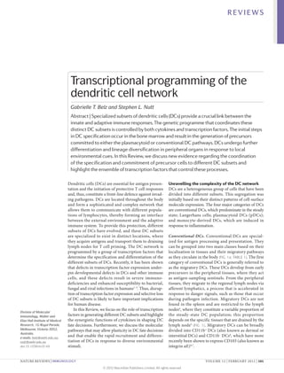
Figure 1 | Differentiation and trafficking of DC subsets. a | The figure shows the organization of the dendritic cell (DC)
network, and includes the key surface phenotype markers of different DC subsets, which are delineated on the basis of their
localization in secondary lymphoid tissues. Gut-associated DCs that express both CD103 and CD11b have been included in
Nature Reviews | Immunology
the CD11b+ DC subset. Inflammatory monocyte-derived DCs are rapidly recruited to sites of inflammation, whereas other
DC subsets are normally present in the steady state. The relationship between inflammatory and steady-state DCs remains
an open issue. Moreover, it is unclear whether monocyte-derived DCs can arise through in situ proliferation in addition to
arriving at tissues via the circulation. b | In the mouse bone marrow, haematopoietic stem cells (HSCs) differentiate into
common myeloid progenitors (CMPs), a fraction of which express FMS-related tyrosine kinase 3 (FLT3) and differentiate
into more-restricted macrophage and DC progenitors (MDPs). MDPs appear to be the direct precursor to common DC
progenitors (CDPs), which give rise to the DC lineages. CDPs produce precursor DCs (pre-DCs) and plasmacytoid DCs
(pDCs) that exit the bone marrow and travel through the blood to secondary lymphoid organs and non-haematopoietic
tissues. A small proportion of DCs may also be derived from CLPs in the bone marrow and from early T cell progenitors in the
thymus. Under steady-state conditions, lymphoid tissue-resident DCs that arise from pre-DCs are the only subsets found in
the spleen. This population is comprised of three conventional DC subsets, namely CD4+ DCs, CD8α+ DCs and CD8α–CD4–
double-negative (DN) DCs. Peripheral lymph nodes contain CD8α+ and CD8α– DC populations but are also populated by
two groups of migratory DCs. Langerhans cells develop in the epidermis and migrate through the basement membrane to
the draining lymph nodes via terminal lymphatic vessels that arise in the dermis. The dermal DC population is broadly
composed of CD11b+ and CD103+ DCs, and these cells migrate through the lymphatics to the lymph node. Monocytes
arrive at tissues from the blood. In response to inflammation, they can develop into monocyte-derived DCs, which adopt
many of the characteristics of conventional DCs. DC-SIGN, DC-specific ICAM3‑grabbing non-integrin.
102 | FEBRUARY 2012 | VOLUME 12 www.nature.com/reviews/immunol
© 2012 Macmillan Publishers Limited. All rights reserved
- 3. REVIEWS
Table 1 | Phenotypic markers of DC subsets
DC subset DC type CD8α CD103 CD205 EPCAM CD11b B220 or DC-SIGN Langerin Antigen Major
(CD326) CD45RA (CD207) presentation cytokine
produced
pDCs Lymphoid- +/– – – – – + ++ – Poor IFNα
resident DCs
CD8α+ DCs Lymphoid- + low + – + – – +/– Cross- IL‑12p70,
resident DCs presentation IFNλ
on MHC class I;
expression of
cystatin C
CD4+ DCs Lymphoid- – – – – + – – – Presentation on
resident DCs MHC class II
DN DCs Lymphoid- – – – – + – – – Presentation on
resident DCs MHC class II
CD11b+ Migratory – +/– + – + – ND – Presentation on
DCs DCs MHC class II
CD103+ DCs Migratory Cross-
• Lung DCs – + ++ +/– – – – + presentation on
• Intestine – + – – + – – – MHC class I
Langerhans Migratory – – ++ + + – – ++ Presentation IL‑10
cells DCs of self antigens
for tolerance
induction
Monocyte- Induced by – – – – + – + – Cross- TNF
derived inflammation presentation
DCs
DC, dendritic cell; DC-SIGN, DC-specific ICAM3-grabbing non-integrin; DN, double-negative; EPCAM, epithelial cell adhesion molecule; IFN, interferon;
IL, interleukin; ND, not determined; pDC, plasmacytoid DC.
The second major category of conventional DCs that are present early in embryonic development and
is the lymphoid tissue-resident DCs that are found in that undergo a proliferative burst in the epidermis in
the major lymphoid organs, such as the lymph nodes, the first few days after birth24.
spleen and thymus. These DCs can be further classified
by their expression of the surface markers CD4 and pDCs. pDCs are quiescent cells that are broadly distrib-
CD8α into CD4+ DCs, CD8α+ DCs and CD4–CD8α– uted in the body. They are characterized by their ability
DCs (typically referred to as double-negative DCs)8,9 to rapidly produce large amounts of type I interferons
(TABLE 1). CD8α + DCs are noted for their capacity to (IFNs)25,26, a feature most evident during viral infection.
cross-present antigens10 and for their major role in pDCs express several characteristic markers, includ-
priming cytotoxic CD8+ T cell responses11–16 (BOX 1). ing sialic acid-binding immunoglobulin-like lectin H
CD4 + DCs and CD4 –CD8α – DCs can also present (SIGLEC‑H) and bone marrow stromal antigen 2 (BST2)
MHC class I‑restricted antigens in some settings 15,17, in mice and blood DC antigen 2 (BDCA2; also known
but appear to be more efficient at presenting MHC as CLEC4C) and leukocyte immunoglobulin-like recep-
class II-associated antigens to CD4 + T cells 18–20 . tor, subfamily A, member 4 (LILRA4; also known as
Lymphoid tissue-resident DCs do not traffic from ILT7) in humans. In addition, both mouse and human
other tissues but develop from precursor DCs found pDCs express CD45RA27. pDCs have poor antigen-
in the lymphoid tissues themselves21. In the absence of presenting capacity, and their precise contribution to
infection, they exist in an immature state (which is char- immune responses is still unclear 28.
acterized by a high endocytic capacity and lower MHC
class II expression compared with activated DCs), and Monocyte-derived DCs. Under inflammatory con-
their residency in lymphoid tissues makes them ideally ditions, circulating blood monocytes can be rapidly
placed to sense antigens or pathogens that are transported mobilized and can differentiate into cells that possess
in the blood12,22,23. many prototypical features of DCs21,29–32 (FIG. 1). In the
steady state, monocytes express the macrophage colony-
Langerhans cells. Langerhans cells are resident in the stimulating factor receptor (M-CSFR; also known as
skin and, like migratory DCs, migrate to the lymph CD115), which is essential for their development, as well
nodes to present antigens (FIG. 1). However, unlike con- as other markers, such as LY6C and CX3C-chemokine
ventional DCs, which arise from a bone marrow precur- receptor 1 (CX3CR1). In response to growth factors such as
sor cell, Langerhans cells are derived from a local LY6C+ granulocyte–macrophage colony-stimulating factor
myelomonocytic precursor cell population in the skin. (GM-CSF) in vitro or to Toll-like receptor 4 (TLR4) ligands
This precursor population originates from macrophages or bacteria in vivo, fully differentiated monocyte-derived
NATURE REVIEWS | IMMUNOLOGY VOLUME 12 | FEBRUARY 2012 | 103
© 2012 Macmillan Publishers Limited. All rights reserved
- 4. REVIEWS
Box 1 | Direct presentation, cross-presentation and cross-dressing
Cytokines regulate DC development
The differentiation of DCs from haematopoietic
Efficient presentation of antigens to CD8+ T cells depends on the generation of progenitor cells relies on the activity of cytokines,
peptides for loading into MHC class I complexes. Several pathways have been in particular FMS-related tyrosine kinase 3 ligand
uncovered to achieve this activation of CD8+ T cells. Direct antigen presentation by (FLT3L), M‑CSF and GM‑CSF. These cytokines con-
infected or malignant cells ensures the destruction of these cells by cytotoxic CD8+
trol the initial production and lineage diversification
T cells (CTLs), without harming adjacent healthy cells. The restriction of antigen
presentation to directly infected cells, however, is not sufficient to ensure the activation
of DCs, although the factors that regulate the expres-
of CD8+ T cells, particularly when the pathogen does not infect professional sion of the receptors for these key cytokines and the
antigen-presenting cells, such as dendritic cells (DCs). In this case, CD8+ T cells can be downstream transcriptional programmes instigated by
activated by DCs that present extracellular antigens on their MHC class I molecules via FLT3L, M‑CSF and GM‑CSF are only now emerging.
the process of cross-presentation. This pathway can result in the generation of CTLs The ability of these cytokines to stimulate the differ-
that are reactive to foreign antigens, or in the induction of tolerance through the entiation of DCs in vitro (BOX 2) provides a tractable
deletion of autoreactive CD8+ T cells following the cross-presentation of self antigens. model system to address the influence of extrinsic
In mice, most focus has been on the cross-presenting capacities of CD8α+ DCs, and factors on the DC transcriptional network.
more recently CD103+ DCs6,7,10,138, although other populations also possess this ability. FLT3L and FLT3 constitute the best-characterized
For example, fully differentiated monocyte-derived DCs that express DC-specific
growth factor–receptor axis for DCs, as mouse haemato
ICAM3‑grabbing non-integrin (DC‑SIGN) are potent cross-presenting cells33.
An alternative pathway for the acquisition of peptide–MHC complexes is known as
poietic progenitor cells cultured with FLT3L generate a
‘cross-dressing’139,140 (originally known as trogocytosis141). Through this pathway, DCs diverse array of conventional DC subsets and pDCs36,37.
acquire preformed peptide–MHC class I complexes from infected cells. Complexes In agreement with the important role of FLT3L in DC
acquired in this way can drive the activation of memory, but not naive, CD8+ T cells differentiation, DCs can be generated from essentially
during viral infection, perhaps owing to the lower activation threshold of memory cells. any FLT3+ progenitor cell either in vitro or following
The molecular mechanisms involved in cross-presentation and cross-dressing are only adoptive transfer in vivo38–40 (FIG. 2). In addition, enforced
beginning to be unravelled. expression of FLT3 in megakaryocyte–erythrocyte
progenitors (MEPs), which are normally FLT3–, results
in the acquisition of DC potential41. An instructive role
DCs emerge. Similarly to conventional DCs, monocyte- for FLT3 in DC development is further supported by the
derived DCs express CD11c, MHC class II molecules, finding that in all cell lineages except the DC lineage,
CD24 and SIRPα (also known as CD172a), and they upreg- FLT3 is downregulated following differentiation and,
ulate their expression of DC-specific ICAM3‑grabbing at least in the case of B cells, this repression of FLT3 is
non-integrin (DC‑SIGN; also known as CD209a) but essential for further development 42.
lose expression of both M‑CSFR and LY6C33 (TABLE 1). Mice deficient for FLT3L or signal transducer and
Monocyte-derived DCs also express the macrophage activator of transcription 3 (STAT3; a signalling mol-
marker MAC3 (also known as CD107b and LAMP2)21,32. ecule downstream of FLT3) have markedly reduced
In addition, these cells acquire potent antigen-presenting numbers of lymphoid-resident conventional DCs and
capacity, including the ability to cross-present antigens33–35 pDCs, whereas mice lacking FLT3 have a milder pheno-
(BOX 1). Thus, it is emerging that monocyte-derived DCs type, which suggests the presence of a second ligand for
are a crucial reservoir of professional antigen-presenting this receptor 43–46. FLT3 signalling is also crucial for the
cells (APCs) that are recruited into immune responses development of migratory DCs, as the numbers of both
to certain microorganisms and potentially have an pre-DCs and CD103+ DCs were found to be reduced in
emergency back-up role in cases of acute inflammation. a range of tissues from Flt3l–/– mice compared with the
Box 2 | In vitro models for investigating DC development and behaviour
The establishment of well-defined cell culture systems that allow the generation of large numbers of dendritic cells (DCs)
from bone marrow has been instrumental for understanding DC biology. Recent refinement of the tools and surface
markers used to analyse cultures now allows the resolution of DC precursors equivalent to those found in vivo, together
with fully differentiated DC subsets, in the tissue culture flask.
Generation of steady-state DC subsets in vitro
The development of steady-state DCs depends on signalling through FMS-related tyrosine kinase 3 (FLT3), which is
expressed on the surface of DC precursors. Bone marrow precursors cultured with FLT3 ligand (FLT3L) give rise to
plasmacytoid DCs (pDCs) and multiple lymphoid tissue-resident conventional DC subsets36,37,89. Intriguingly,
in vitro-generated DCs do not express CD4 or CD8, but their patterns of expression of the markers CD103, CD11b,
CD172a and CD24 indicate the presence of conventional DC subsets in addition to DC precursors.
Generation of monocyte-derived DCs in vitro
Perhaps the most commonly used approach to generate DCs involves the culture of bone marrow precursors in a medium
supplemented with granulocyte–macrophage colony-stimulating factor (GM-CSF). As these DCs can also be produced by
culturing monocytes in GM‑CSF and interleukin‑4 (IL‑4), they are referred to as monocyte-derived DCs and correspond
to the dominant inflammatory DC type that is mobilized during some bacterial infections33.
These highly refined culture systems allow comparative studies between different DC subsets. Furthermore, the
ability to generate large numbers of DCs in vitro should greatly facilitate molecular exploration of the genomic and
transcriptional machinery that leads to the generation of different DC subsets.
104 | FEBRUARY 2012 | VOLUME 12 www.nature.com/reviews/immunol
© 2012 Macmillan Publishers Limited. All rights reserved
- 5. REVIEWS
BATF3
FLT3L
Transitional CD103+ DC
Lymphocytes pre-DC
ID2 E4BP4 BATF3
CLP
FLT3L
IRF8 Pre-CD8α+ DC PU.1hi
IRF2 CD8α+ DC ID2hi
PU.1 GFI1 PU.1 RELB IRF4 Ikaros (null)
? FLT3+
LMPP MDP CDP Pre-DC
CMP GM-CSF
M-CSF
FLT3L CD11b+ DC
Monocyte
Immature pDC Loss
M-CSF GM-CSF of E2-2
E2-2 Ikaros (L) IRF8
PU.1low
ID2–
E2-2hi
Macrophage Monocyte- Mature pDC
derived DC
Figure 2 | Growth factors and transcription factors that regulate DC differentiation. The developmental pathways
from myeloid and lymphoid progenitors to precursor dendritic cells (pre-DCs) in the bone marrow and Reviews | Immunology
Nature the peripheral
diversification of DC subsets are shown (see FIG. 1 legend for details). The approximate points at which key transcription
factors are first required for DC development are indicated by vertical lines. Stages at which key growth factors have been
determined to be essential are indicated. The development of both DCs and monocytes depends on high concentrations
of PU.1, which regulates the expression of the cytokine receptors FMS-related tyrosine kinase 3 (FLT3), macrophage
colony-stimulating factor receptor (M-CSFR) and granulocyte–macrophage colony-stimulating factor receptor (GM-CSFR).
The development of CD8α+ and CD103+ DCs relies on the stepwise activity of interferon-regulatory factor 8 (IRF8), inhibitor
of DNA binding 2 (ID2), E4 promoter-binding protein 4 (E4BP4) and basic leucine zipper transcription factor, ATF-like 3
(BATF3), as well as on FLT3 signalling. CD11b+ DCs depend on a unique set of transcription factors, including RELB, IRF2, IRF4
and Ikaros, and to some extent on the cytokines M‑CSF and GM‑CSF. The plasmacytoid DC (pDC) lineage requires IRF8, a low
level of PU.1 and the absence of ID2. The differentiation of pDCs from an immature precursor requires E2‑2 and Ikaros, with
induced loss of E2‑2 converting pDCs into cells that closely resemble CD8α+ conventional DCs. CDP, common DC progenitor;
CLP, common lymphoid progenitor; CMP, common myeloid progenitor; FLT3L, FLT3 ligand; GFI1, growth factor
independent 1; LMPP, lymphoid-primed multipotent progenitor; MDP, macrophage and DC progenitor.
numbers in wild-type mice. By contrast, the development that resemble monocyte-derived DCs, while repress-
of CD11b+ DCs and Langerhans cells is largely independ- ing the development of pDCs in a STAT5‑dependent
ent of FLT3L47. Thus, FLT3L has two distinct roles in DC manner 46,51. This finding has generally been interpreted
biology: it is required for the early development of DCs to show that GM‑CSF has a greater role in the produc-
from haematopoietic progenitors; and later it functions tion of monocyte-derived DCs than in the generation
to maintain DC homeostasis by promoting limited levels of other DC subsets21,52, although the importance of this
of proliferation of DCs in peripheral tissues43. process in vivo is still to be established.
In addition to FLT3L, GM‑CSF has long been known M-CSF is the major cytokine involved in the produc-
to stimulate DC differentiation in culture (BOX 2) . tion of monocytes and macrophages53. A role for this
However, GM‑CSF is not essential for DC differentia- cytokine in DC biology was suggested by the expression
tion in the steady state, as mice that lack the GM‑CSF of M‑CSFR by DCs54,55 but came to prominence with the
receptor (GM-CSFR) have only mildly reduced num- finding that M‑CSFR-deficient mice lack Langerhans
bers of DCs48. Nonetheless, GM‑CSF is not completely cells56. Surprisingly, mice lacking M‑CSF (op/op mice)
redundant in DC production, as mice deficient in both have normal numbers of Langerhans cells, a quandary
GM‑CSF and FLT3L have a greater loss of DCs than either that was resolved by the identification of interleukin‑34
single-knockout strain44. Moreover, other recent studies (IL‑34) as a second ligand for M‑CSFR57. The relatively
have demonstrated that GM‑CSF is necessary for there normal DC numbers in mice lacking M‑CSF, despite
to be normal numbers of CD103+CD11b+ DCs in the the profound reduction in monocytes, demonstrates
lamina propria49,50. The addition of GM‑CSF to cultures that the monocytic system is not the major source of
of bone marrow cells promotes the development of cells steady-state DCs. M‑CSF is, however, required for the
NATURE REVIEWS | IMMUNOLOGY VOLUME 12 | FEBRUARY 2012 | 105
© 2012 Macmillan Publishers Limited. All rights reserved
- 6. REVIEWS
normal development of CD103–CD11b+ DCs in non- that specifies the DC lineage in more-immature
lymphoid tissues49 and is able to support conventional pro enitors and then drives differentiation into the DC
g
DC and pDC differentiation in cell culture in the subsets. Three transcription factors — PU.1 (encoded by
absence of FLT3 (REF. 58). Sfpi1), Ikaros and growth factor independent 1 (GFI1)
— appear to be prime candidates for DC-specifying fac-
Stages of programming DC identity tors. In addition, the signalling and transcription factors
DC ontogeny. Although it is well established that all STAT3 and STAT5 are known to have a role in DC differ-
DCs, with the exception of Langerhans cells, are derived entiation, as they mediate the signals transduced through
from bone marrow-resident haematopoietic stem cells FLT3 and GM‑CSFR, respectively (TABLE 2).
(HSCs), mapping the origins of the DC lineages has PU.1 belongs to the ETS family of transcription
proven to be both difficult and controversial. Early factors, which has multiple context-specific roles in
transfer experiments led to the surprising conclusion haematopoiesis. PU.1 is an attractive candidate for being
that DCs can develop with approximately equal effi- a crucial regulator of the DC lineages, as it is expressed by
ciency from both lymphoid and myeloid progenitors59,60, all DCs and by CDPs66–68. A role for PU.1 in DC develop-
whereas in vitro cultures with GM‑CSF show that DCs ment was initially suggested by the analysis of mice with
can arise from monocytic precursors, as mentioned a germline deficiency of PU.1. Indeed, one such study
above. However, monocytes are not likely to be a major concluded that PU.1 was necessary for all embryonic DC
source of steady-state DCs in lymphoid organs, as development 69, although a second study reported that
lineage-tracing experiments have shown that monocytic Sfpi1–/– fetal thymi could generate DCs70. However, these
cells give rise to neutrophils and macrophages but not approaches could not distinguish between the require-
DCs5. More recently, the adoption of a ‘FLT3‑centric’ ments for PU.1 in multipotent progenitors and the role
view of haematopoiesis38–40,61 has established that most of PU.1 specifically in the DC lineages. Moreover, the
steady-state DCs arise from FLT3+ progenitors (FIG. 2). impact of enforced expression of PU.1 in haematopoietic
The pathway of DC differentiation from primitive progenitors suggests an instructive and concentration-
bone marrow progenitors has been extensively reviewed dependent role for PU.1 in promoting macrophage and
elsewhere4,53,62 and is only briefly summarized here. FLT3 DC development71–73.
expression is first induced in a subset of the HSC com- A recent study used conditional gene deletion in
partment that has only short-term pan-haematopoietic defined haematopoietic progenitors and CDPs to show
repopulating activity, and the expression of this recep- that PU.1 is absolutely essential for the generation of
tor is then maintained in lymphoid-primed multipotent all conventional DCs and pDCs both in vivo and in
progenitors (LMPPs)63 and in a subpopulation of com- FLT3L‑containing cultures in vitro 66. Moreover, in
mon myeloid progenitors (CMPs)38. CMPs are thought line with its established role in regulating GM‑CSFR
to differentiate into macrophage and DC progenitors expression, PU.1 is required for GM‑CSF-induced DC
(MDPs)64, which appear to be the direct precursors of differentiation from early haematopoietic progenitors.
common DC progenitors (CDPs)40,61. Both MDPs and Among the many genes that are potentially regulated
CDPs are proliferating cells that reside in the bone mar- by PU.1, Flt3 was demonstrated by molecular studies
row and express FLT3 and M‑CSFR. CDPs differentiate to be directly regulated by PU.1 in DCs and haemato
directly into pDCs and into the precursors of conven- poietic progenitors66. This regulation occurred in a
tional DC subsets, termed pre-DCs, but they lack the concentration-dependent manner, as Sfpi1+/– cells had
potential to give rise to macrophages21,65. Pre-DCs then reduced FLT3 expression and an impaired ability to
leave the bone marrow and are found in blood, second- generate conventional DCs. Interestingly, previous stud-
ary lymphoid organs and some tissues21,49,52,65, where ies have shown that FLT3 signalling is able to activate
they mature into the conventional DC subsets (FIG. 1). PU.1 expression in MEPs, suggesting a self-reinforcing
Differentiation into different conventional DC subsets loop between PU.1 and FLT3 in DCs41. Whether PU.1
appears to be a late step in DC development that is per- is required for monocyte-derived DC formation in vivo
haps important in maintaining the stability or plasticity remains to be determined.
of the peripheral DC compartment. The key features and Ikaros is a zinc-finger transcription factor that has
mechanisms involved in the plasticity of the DC network important roles in haematopoiesis74. Expression of a
are likely to include the short lifespan of mature conven- dominant-negative form of Ikaros that also impairs the
tional DCs (5–7 days, although up to 25 days in some cir- function of other Ikaros family members, such as Aiolos,
cumstances4,47); the rapid recruitment and proliferation of resulted in a complete loss of all conventional DC sub-
pre-DCs and their capacity to respond to extrinsic signals sets. By contrast, a null mutation in the gene encoding
(such as TLR ligands and pro-inflammatory cytokines); Ikaros led to the selective loss of CD11b+ DCs, with some
and the active expression by DCs of transcription factors CD8α+ DCs being retained75. Whether Ikaros directly
such as E2‑2 (also known as TCF4). regulates DC differentiation, as opposed to having a role
in early myeloid progenitors, is at present unclear 76, as
Initiating the DC programme in haematopoietic progeni- mice homozygous for a severely hypomorphic allele of
tors. The information outlined above demonstrates the the Ikaros gene (IkL/L mice) lack mature pDCs but con-
rapid progress that is being made in understanding the tain relatively normal numbers of conventional DCs77.
developmental stages and cell biology of the DC lineages. DC‑specific conditional mutagenesis is now required to
Much less is known about the transcriptional programme decipher the exact function of Ikaros in DCs (FIG. 2).
106 | FEBRUARY 2012 | VOLUME 12 www.nature.com/reviews/immunol
© 2012 Macmillan Publishers Limited. All rights reserved
- 7. REVIEWS
Table 2 | Transcription factors guiding steady-state DC subset development
Transcription Transcription factor family Function Refs
factor
PU.1 (SFPI1, ETS-domain transcription factor; Required for the development of all DC subsets 66,
SPI1) binds to PU box sequences 69,70
IRF2 Interferon-regulatory factor; inhibits Alters pDC ratios; in its absence the numbers of 122
the IRF1-mediated transcription of CD8α– DCs and Langerhans cells are reduced
type I IFNs
IRF4 Interferon-regulatory factor Required for non-CD8α+ DC lineage development 126,127
IRF8 (ICSBP) Interferon-regulatory factor Required for the development of pDCs and most 90,92,
conventional DCs 93,104
GFI1 Zinc-finger protein; transcriptional GFI1 deficiency results in a 50% reduction in the 79
repressor numbers of conventional DCs and pDCs and
increased numbers of Langerhans cells
ID2 Inhibitor of DNA binding family Required for the development of CD103+ DCs and 47,
protein containing HLH domains CD8α+ DCs in PLNs and spleen; not required for 87,89
DCs in MLNs
E4BP4 (NFIL3) PAR-related bZIP transcription factor Required for the development of CD8α+ DCs 108
E2‑2 (TCF4) E protein containing bHLH domains Required for the development and maintenance 80,84
of pDCs
STAT3 Signal transducer and activator of STAT3 deficiency results in a substantial reduction 46
transcription in conventional DC numbers
STAT5A and Signal transducer and activator of Inhibit pDC development by interacting with IRF8; 51
STAT5B transcription deficiency results in reduced conventional DC and
increased pDC numbers
Ikaros (IKZF1) Zinc-finger DNA-binding protein Ikaros deficiency results in the absence of most 75,77
DCs; a hypomorphic mutation leads a specific loss
of pDCs
BATF3 bZIP family; heterodimerizes with BATF3-deficient mice fail to develop CD103+ DCs 13,
JUN and show impaired survival of precursor CD8α+ DCs 89,111
RELB REL-homology domain family; RELB deficiency results in the loss of CD8α– DCs 129,130
interacts with NF‑κB family members
SPIB ETS-domain transcription factor Required for human pDC differentiation 91
BATF3, basic leucine zipper transcription factor, ATF-like 3; bHLH, basic HLH; bZIP, basic leucine zipper; DC, dendritic cell; E4BP4,
E4 promoter-binding protein 4; GFI1, growth factor independent 1; HLH, helix-loop-helix; ID2, inhibitor of DNA binding 2; IFN,
interferon; IRF, interferon-regulatory factor; MLN, mesenteric lymph node; NF‑κB, nuclear factor‑κB; PAR, proline- and acidic-rich
region; pDC, plasmacytoid DC; PLN, peripheral lymph node; STAT, signal transducer and activator of transcription.
GFI1 is a small, zinc-finger-containing transcriptional it is surprising how little we actually understand about
repressor that is important for early haemato oiesis78.
p this process. Two developmental systems appear to be
GFI1 is expressed in DC precursors, and Gfi1–/– mice in place to separate pDCs and conventional DCs (FIG. 2).
have reduced numbers of all lymphoid-resident DC First, pDCs absolutely rely on the expression of the
subsets, whereas Langerhans cell numbers were actu- E protein E2‑2 (REF. 80) and the absence of the E protein
ally increased79. Interestingly, GFI1‑deficient haemato antagonist inhibitor of DNA binding 2 (ID2)81. Second,
poietic progenitor cells were unable to develop into DCs pDCs have a uniquely low level of PU.1 (REF. 68) and an
in vitro in the presence of either FLT3L or GM‑CSF and extremely high concentration of interferon-regulatory
instead differentiated into macrophages, suggesting that factor 8 (IRF8), a transcription factor that can form
GFI1 is a crucial modulator of DC versus macrophage a complex with PU.1 on a class of ‘composite’ DNA
development (FIG. 2). elements82.
One model to explain the pDC versus conventional
Establishing pDC and conventional DC identity. DC lineage split is to assume that the conventional DC
E protein
Restriction of the developmental programme of DC is the default setting and that progenitors have to be
The E proteins (including E12,
E47, HEB and E2‑2) have progenitors to the conventional DC and pDC lineages diverted to the pDC lineage83. E2‑2 fits the bill for a fac-
emerged as key regulators of occurs at the CDP stage40,61 (FIG. 2). Conventional DCs tor that could control this diversion, as it is abundantly
the immune system. They are a and pDCs differ markedly in their appearance, functions expressed by pDCs and is required for pDC lineage
family of basic helix-loop-helix and transcriptional programmes, and thus how a CDP specification80. Once progenitors have committed to a
factors that work together with
their antagonists, the ID
is influenced to develop into either a pDC or a conven- pDC fate, it appears that continuous expression of E2‑2
proteins (ID1–ID4), to regulate tional DC is a question of major importance for under- is essential to maintain the mature pDC phenotype84.
lymphocyte development. standing and manipulating DC biology. In this context, In mice, a specific deletion of the gene encoding E2‑2
NATURE REVIEWS | IMMUNOLOGY VOLUME 12 | FEBRUARY 2012 | 107
© 2012 Macmillan Publishers Limited. All rights reserved
- 8. REVIEWS
in pDCs led to the expansion of a population of DCs they have been shown to share a few targets, such as
that exhibit many characteristics of conventional DCs. Ciita94, Tlr9 (REF. 95) and Ifna96). However, BXH2 mice,
In the in vivo setting, however, it is particularly dif- which harbour a spontaneous point mutation in Irf8,
ficult to dissect whether E2‑2‑deficient pDCs undergo have defects in CD8α + DC development but not in
phenotypic conversion to conventional DCs owing to a pDC generation. This mutation is thought to ablate
reduction in E2‑2‑mediated repression of ID2, as pro- the interaction of PU.1 and IRF8, raising the possibil-
posed, or whether normal conventional DC numbers ity that the PU.1–IRF8 complex is not as crucial for
are increased in the absence of a full pDC compart- the differentiation of pDCs as for the development
ment, as occurs in other settings. Nevertheless, pDCs of conventional DCs93. There are, as yet, no genome-
are particularly sensitive to E2‑2 concentration, as both wide DNA-binding datasets for IRF8 and PU.1 in
E2‑2‑deficient mice and patients with a rare mono- DCs, although similar data from macrophages sug-
allelic loss of E2‑2 (Pitt–Hopkins syndrome) show gest that PU.1 might occupy most of the active regula-
impaired pDC formation and function80. E2‑2 binds tory regions in DCs97,98 and that IRF8 might bind to
directly to the promoters of several pDC-expressed a subset of these sites99. Importantly, in macrophages
genes, including BDCA2, LILRA4, IRF7, the pre-TCR and myeloid progenitors, PU.1 is directly involved in
α-chain gene, IRF8 and SPIB (which encodes a close nucleosome remodelling, and this leads to the generation
relative of PU.1 that is expressed by human and mouse of an open chromatin conformation and histone modi‑
pDCs)80. The reliance of pDCs on an E protein such as fications, suggesting that PU.1 can directly programme
E2‑2 may explain the observation that pDCs express the fate of myeloid cells97,98.
many lymphocyte-associated transcripts (including The ETS-family transcription factor SPIB — the
SPIB, RAG1, IL7R and TDT), as E proteins are central closest homologue of PU.1 in the mammalian genome
to many aspects of lymphopoiesis27. — is expressed, within the DC lineages, specifically by
ID proteins are direct inhibitors of DNA bind- pDCs100. Knockdown of the expression of either SPIB
ing by E proteins. ID2 is the predominant ID protein or PU.1 in human haematopoietic progenitors strongly
expressed in the DC lineage and is also involved in inhibits pDC formation, suggesting that both factors
the development of multiple lineages during haemato function in human pDCs91. The extent of any functional
poiesis, particularly that of lymphoid tissue-inducer redundancy between PU.1 and SPIB in pDCs has so far
cells (LTi cells) and natural killer (NK) cells85–88. ID2 not been addressed in mice.
expression is extremely low in CDPs, pre-DCs and In summary, the separation of pDC and conven-
pDCs, whereas all conventional DC populations tional DC lineages represents the first major division
Lymphoid tissue-inducer express high levels of ID2 (REF. 89) (FIG. 3). In line with in the DC pathway and requires the concerted action of
cells
(LTi cells). A cell type that is
this expression pattern, ID2‑deficient mice have a both E proteins and the PU.1–IRF8 complex (FIGS 2,3).
present in developing lymph profoundly altered conventional DC compartment Although the activation of STAT5 inhibits pDC for-
nodes, Peyer’s patches and but still produce pDCs (see below)87. This leads to a mation in vitro 51, the exact signals that initiate this
nasopharynx-associated model whereby the acquisition of high levels of ID2 process are not known. In addition, the ways in which
lymphoid tissue (NALT).
and subsequent suppression of E2‑2 activity blocks the E2‑2–ID2 and PU.1–IRF8 axes interact are still
LTi cells are required for
the development of these pDC development and allows progression along the unclear, as are the identities of most of the genes tar-
lymphoid organs. The inductive conventional DC pathway. This model is clearly an geted by these transcription factors. pDCs represent the
capacity of these cells for the oversimplification, as E2‑2‑deficient mice still produce end point of their lineage; however, the production of
generation of Peyer’s patches some pDC progenitors and ID2 is not essential for the conventional DCs is only the first step in their further
and NALT has been shown by
adoptive transfer, and it is
differentiation of all conventional DCs. diversification, which is outlined in the next section.
generally assumed that they The development of pDCs also depends on PU.1,
have a similar function in the IRF8 and potentially SPIB 66,90,91. pDCs are absent Genetic programming of conventional DC subsets
formation of lymph nodes. in PU.1- or IRF8‑deficient mice 66,90,92, as well as in Although it is fairly clear that conventional DCs, pDCs,
humans with a mutation in IRF8 (REF. 3), although it Langerhans cells and monocyte-derived DCs represent
Nucleosome remodelling
Changes in the nucleosome remains to be proven whether these factors function developmentally distinct lineages, the relationships
structure are mediated by specifically in the pDC lineage or have a role in CDPs between the various anatomically, phenotypically and
dedicated nuclear enzymes (FIG. 2) . However, circumstantial evidence favours a functionally distinct conventional DC populations
(for example, ATP-dependent specific function for these factors in determining pDC remain to be fully elucidated. Conventional DCs are
nucleosome-remodelling
enzymes) that change the
versus conventional DC fate. PU.1 is expressed at a thought to be derived from circulating pre-DCs21,65.
accessibility of DNA and uniformly high level in CDPs, but at a lower level in Although it remains possible that multiple develop-
the expression of genes. pDCs, with the timing of downregulation coinciding mentally distinct types of pre-DC exist and act as the
with pDC formation66, whereas IRF8 is expressed at precursors for individual conventional DC lineages,
Histone modifications
very high levels in both CDPs and pDCs (FIG. 3). The we favour a model that considers all the populations
Histones are essential to
maintain DNA organization stoichiometric relationship between PU.1 and IRF8 is of conventional DCs as related subsets derived from a
and may be modified by likely to be important for the pDC versus conventional single pre-DC population. In this model, the conven-
methylation and acetylation — DC branch point, as both of these factors are known to tional DC subsets are induced by the environmental
changes that are thought to function in a dose-dependent manner 66,93 and can bind milieu that they reside in, and thus it is the interaction
keep genes active or silent,
respectively — thereby altering
to distinct DNA sequences both individually and in a of these extrinsic signals with the core transcriptional
the genetic code read by complex 82. The extent to which PU.1 and IRF8 share programme of conventional DCs that dictates the
transcriptional regulators. target genes in pDCs is currently unknown (although outcome of DC terminal differentiation.
108 | FEBRUARY 2012 | VOLUME 12 www.nature.com/reviews/immunol
© 2012 Macmillan Publishers Limited. All rights reserved
- 9. REVIEWS
BATF3 +++ clearly has a key role in the function of conventional DCs,
IRF8 +++ Irf8–/– mice develop a myeloproliferative syndrome that is
ID2 +++ characterized by the overproduction of granulocytes105.
IRF4 ++
E4BP4 ++ This implies that IRF8 may also be required for the gen-
CD103+ DC
PU.1 +++ eration or maintenance of MDPs, which can give rise
E2-2 + to monocytes, conventional DCs and pDCs. The exact
Transitional developmental stage at which IRF8 exerts its activity on
pre-DC Pre-CD8α+ DC BATF3 ++ conventional DCs remains to be fully determined.
IRF8 +++
ID2 +++ E4BP4 is a mammalian basic leucine zipper (bZIP)
IRF4 + transcription factor that is required for the development
E4BP4 ++ of NK cells but not of other lymphocyte lineages106,107.
IRF8 +++ IRF8 +++ PU.1 +++
ID2 +/– ID2 ++ CD8α+ DC E2-2 + In NK cells, E4BP4 acts in a dose-dependent man-
PU.1 +++ PU.1 +++ ner downstream of the IL‑15 receptor to regulate ID2
CDP Pre-DC BATF3 ++ expression106,107. More recently, the induction of E4BP4
IRF8 + has been shown to be important for the development of
ID2 ++ CD8α+ DCs108. E4BP4‑deficient CDPs had lower levels
IRF4 +++
E4BP4 ++ of BATF3 expression than control CDPs, and enforced
IRF8 +++ IRF8 +++
CD11b+
PU.1 +++ expression of BATF3 in the mutant cells rescued CD8α+
ID2 +/– ID2 +/– E2-2 + DC development in vitro108. Thus, E4BP4 is emerging as
PU.1 +++ PU.1 +++ DC
a key regulator of conventional DC diversity. Whether
Immature pDC BATF3 +
IRF8 +++ E4BP4 also acts through ID2 to mediate these effects
ID2 +/– in DCs has not yet been addressed.
IRF4 ++ ID2 is expressed by all conventional DC subsets,
E4BP4 ++
IRF8 +++ Mature PU.1 + with the highest levels of expression in CD8α + and
ID2 +/– pDC E2-2 +++ CD103+CD11b– DCs47,89. Loss of ID2 prevents the devel-
PU.1 + opment of these two subsets in the skin-draining lymph
E2-2 +
nodes and spleen. However, CD103+CD11b+ DCs in the
Figure 3 | Differential expression of transcription factors regulating DC mesenteric lymph nodes, together with CD4+ and CD4–
differentiation. The stages of differentiation of conventional dendritic cells Immunology
Nature Reviews | (DCs) and CD8α– DCs in lymphoid tissues, appear to develop nor-
plasmacytoid DCs (pDCs) from the common DC progenitor (CDP) are shown, together mally in the absence of ID2 (REFS 13,47,87). Although it
with the relative levels of expression of key transcription factors in each cell type indicated
is at present unclear which E protein (or E proteins) —
on an arbitrary scale. The expression pattern of some of the factors has not been
determined at the earliest stages of DC ontogeny. –, no expression; +, low expression; E2A (also known as TCF3), E2‑2 or HEB (also known as
++, intermediate expression; +++, maximal expression; BATF3, basic leucine zipper TCF12) — is the crucial target of ID2 in conventional DCs,
transcription factor, ATF-like 3; E4BP4, E4 promoter-binding protein 4; ID2, inhibitor selective deletion of the gene encoding E2‑2 in mature
of DNA binding 2; IRF, interferon-regulatory factor; pre-DC, precursor DC. Figure is pDCs results in the spontaneous differentiation of pDCs
modified, with permission, from REF. 89 © Macmillan Publishers Ltd. All rights reserved. into cells that exhibit conventional DC properties, perhaps
through the induction of ID2 (REF. 84). Progress in dis-
cerning the key target genes of E proteins in conventional
Transcriptional regulators of CD8α + and CD103 + DCs will be required to understand why ID2 has such
DCs. CD8α+ and CD103+ DCs have gained consider- an important role in CD8α+ and CD103+CD11b– DCs.
NFAT
(Nuclear factor of activated
able attention owing to their specialized roles as induc- BATF3, which is also known as JUN-dimerization
T cells). A family of ers of MHC class I‑restricted immune responses to protein p21SNFT, is a bZIP transcription factor that
transcription factors that are pathogens. Moreover, a human BDCA3+ DC subset has acts to repress the activity of NFAT –AP1 complexes
regulated by calcium signalling recently been identified that shares features with both by competing with FOS for JUN dimerization 109,110.
and expressed by a variety of
of these mouse DC subsets; such features include cross- BATF3 was the first transcription factor that appeared
immune cells.
presenting capacity and the expression of XC-chemokine to have an exclusive role in the development of the
AP1 receptor 1 (XCR1)101–103. The differentiation of DC pre- CD8α+ DCs111, although it has since been shown to
(Activator protein 1). cursors into the CD8α+ and CD103+ DC lineages appears be involved in the development of CD103+CD11b–
A heterodimeric transcription to depend on the integration of four key transcription DCs in peripheral lymphoid tissues13, but not that of
factor that is composed of
proteins belonging to the FOS,
factors — namely, IRF8, E4BP4 (E4 promoter-binding CD103+CD11b+ DCs isolated from gut lymphoid tis-
JUN and JUN-dimerization protein 4; also known as NFIL3), ID2 and BATF3 (basic sues112. More recently, detailed analyses of Batf3–/– mice
protein families. AP1 controls leucine zipper transcription factor, ATF-like 3). have shown that despite the reduction in the frequency
various cellular processes, IRF8 is highly expressed in CDPs, pDCs (dis- of CD8α+ DCs, particularly in the spleen, CD8α+ DCs
including differentiation,
cussed in detail above) and conventional DCs, par- are still present in the absence of BATF3 (REFS 89,113).
proliferation and apoptosis.
ticularly the CD8α+ and CD103+ DC subsets (FIG. 3). Nevertheless, Batf3–/– mice exhibit severe defects in their
Cross-priming IRF8‑deficient mice lack many mature DC subsets, capacity to respond to pathogen infections — includ-
A mechanism by which including Langerhans cells90,104. In addition to regulating ing West Nile virus, influenza virus and Toxoplasma
immunogenic CD8+ T cells are the generation of pDCs and conventional DC subsets, gondii infections111,114–116 — and an impaired ability to
activated by the presentation
of an antigen that was not
IRF8 controls various functional features of DCs, such mediate cross-priming. This suggests that the main role
synthesized by the as the expression of TLR9 and IFNα by pDCs and the of BATF3 may be in regulating the cross-presentation of
antigen-presenting cell itself. production of IL‑12 by CD8α+ DCs93. Although IRF8 exogenous antigens to CD8+ T cells (BOX 1).
NATURE REVIEWS | IMMUNOLOGY VOLUME 12 | FEBRUARY 2012 | 109
© 2012 Macmillan Publishers Limited. All rights reserved
- 10. REVIEWS
The transcriptional network in CD8α+ and CD103+ IRF2 acts as a transcriptional repressor of genes
DCs. As outlined above, a deficiency of IRF8, E4BP4, encoding type I IFNs (IFNα and IFNβ) and thus limits
ID2 or BATF3 results in a lack of both the CD8α+ and inflammation120,121. Mice lacking IRF2 exhibit a selective,
CD103+ DC subsets. This dependency on the same cell-autonomous loss of CD4+ DCs in the spleen and epi-
transcriptional regulators, as well as similar func- dermis, and these subsets are restored when type I IFN
tional and localization characteristics13,47,111, suggests signalling is eliminated122,123. Precisely how IRF2 is regu-
that CD8α+ and CD103+ DCs represent a single sub- lated during DC development is not clear, but one role of
set. However, similar developmental requirements IRF2 may be to protect developing DCs from maturation
do not necessarily imply close lineage relationships; arrest when the levels of type I IFNs are elevated. IRF2
for example, PU.1 is required for the development of has also been reported to form complexes with IRF8
multiple distinct lineages, including macrophages, and to act cooperatively with this factor in regulating
granulo ytes and DCs, but very different mechanisms
c the expression of IL12 (REF. 124).
are involved66,117. IRF4, a crucial regulator of many aspects of lympho-
One approach used to investigate the relative con- cyte differentiation125, is most highly expressed in CD4+
tributions of IRF8, ID2 and BATF3 to conventional DCs (in which IRF8 expression is lowest). In line with
DC differentiation has been to engineer a fluorescent this expression profile, CD4+ DCs are largely absent from
reporter into the Id2 locus (to generate Id2gfp mice)89. mice lacking IRF4, but these mice also show defects in
Analyses of Id2gfp mice showed that neither CDPs nor pDCs in the spleen126,127. Moreover, DCs generated in vitro
pre-DCs expressed ID2. Thus, it is more likely that ID2 through the stimulation of bone marrow precursors with
drives the terminal differentiation of different DC sub- FLT3L appear to rely on IRF8 rather than IRF4, but those
sets rather than influencing early lineage-commitment generated in the presence of GM‑CSF depend on IRF4
decisions. Using Irf8–/– or Batf3–/– mice crossed with (REFS 126,127). Thus, it has been proposed that the main
Id2gfp mice, it became clear that IRF8 is required for action of IRF4 is to coordinate signals from GM‑CSF
the generation of DC precursors from a very early stimulation through the nuclear factor-κB (NF‑κB) path-
time point, whereas BATF3 has a role later in conven- way 128. The extent to which IRF4 and IRF8 regulate simi-
tional DC development, downstream of ID2 (REF. 89). lar or distinct sets of target genes, with or without PU.1,
BATF3‑deficient progenitors gave rise to precursors of remains an open question that needs to be addressed.
SIRPα– DCs (which are the precursors of CD8α+ and RELB is a member of the NF-κB family and can func-
CD103+ DCs) in FLT3L‑containing cultures and to a tion either as an activator or as a repressor of transcrip-
lesser extent in vivo, although both the frequency of tion by forming heterodimers with the p50 and p52
these DCs and their expression of CD8α were reduced NF‑κB family members. RELB is most highly expressed
compared with wild-type CD8α+ DCs89 (FIG. 2). Thus, in the CD8α– and CD11b+ DC subsets, and these DC
it is clear that IRF8, ID2 and BATF3 each functions at subsets are absent in RELB-deficient mice129,130. To date,
a distinct point in the differentiation of CD103+ and there have been no studies addressing the mechanism
CD8α+ DCs. What is lacking, however, is an under- by which RELB controls DC differentiation. It is pos-
standing of how the programmes that are activated by sible that IRF4 — the deletion of which also results
each of these transcription factors interact to give rise in a substantial loss of CD4+ DCs127 — may be a key
to the diversity of DC subsets with unique functions. At target of the RELB pathway. RELB has a crucial role in
present, the major approaches for dissecting this net- the upregulation of the signalling molecule CD40, which
work are the deletion of individual transcription factor is required to induce immunogenic DCs and for the
genes from the entire haematopoietic compartment, induction of IFNα131,132. Loss of IFNα results in impaired
and/or limited sampling of tissues and inference with the cross-priming of exogenous antigens132,133. CD40 acts
on/off regulatory switches. But these approaches provide through TNF receptor-associated factor 6 (TRAF6) to
only ‘black and white snapshots’ of transcription factor activate the NF‑κB cascade134. Strikingly, Traf6–/– mice
involvement in DC development. The establishment lack CD8α– DCs and have an impairment in DC matu-
of the identity of DC subsets, their differentiation and ration similar to that observed in Relb–/– mice. It seems
their maintenance are unlikely to be so simple and may likely that TRAF6 regulates CD4+ DC development in a
require combinatorial interactions between transcrip- RELB-dependent manner.
tion factors to guide fate decisions118,119. Genome-wide Much attention has been focused on delineating the
DNA-binding data will be crucial for the precise eluci- development of CD8α+ and CD103+ DCs. However,
dation of the manner in which different transcription despite the crucial role of CD8α – DCs in immune
factors work together to define different DC subsets. responses — particularly in the activation of CD4 +
T cells18,19 and possibly of follicular helper T cells135–137
Transcriptional regulation of CD8α– DCs. Much less is — a detailed understanding of the transcription factor
known about the transcription factors that regulate the networks that drive CD8α– DC development is lacking.
differentiation decisions of CD8α– conventional DC lin- Furthermore, transcription factors normally associ-
eages, despite the dominant role of these cells in present- ated with CD8α+ and CD103+ DC lineages, such as ID2
ing antigens to CD4+ T cells. The transcription factors (REF. 89) and IRF8, are differentially expressed among
IRF2, IRF4 and RELB have been shown to be important CD8α– DCs, suggesting that they may have additional
in the development of these subsets and their subsequent roles in the generation of fully matured DCs that do not
maturation (FIG. 2; TABLE 2). express CD8α.
110 | FEBRUARY 2012 | VOLUME 12 www.nature.com/reviews/immunol
© 2012 Macmillan Publishers Limited. All rights reserved