• Adenocarcinoma stomachhas been one of the leading causes of cancer
death lately.
• Ranks 4th according to recent GLOBOCAN data, with estimated 9,68,784
new cases diagnosed annually and estimated 6,60,175 deaths worldwide.
• It ranks 6th according to GLOBOCAN, wrt total cases in India.
• More common in males, and have genetic predisposition.
• Stomach cancer incidence rates are highly variable by region, being
highest in Eastern and Central Asia and in western parts of Latin
America.
• Notably, the global incidence of gastric cancer has declined rapidly over
the past few decades, partly due to recognition of certain risk factors such
as H. pylori and other dietary and environmental risk factors.
4
5.
• In Asia(mainly China), the decline has been less dramatic with
increase in incidence of early-onset gastric cancer.
• Gastric adenocarcinomas are primarily classified
topographically as cardia (proximal) or non—cardia (distal).
• Despite the overall decline, there has been increase in in
incidence of cancer of the cardia.
• The shift from distal to proximal may be in part due to decline
in distal gastric cancer, however it has also been proposed that
cardia cancer is different entity from rest of gastric carcinoma.
5
6.
• The proximaltumors share demographic and pathologic features with
Barrett’s-associated esophageal adenocarcinoma & are more likely to occur
in males, which parallels the male predominance in the increasing
incidence of the carcinoma of the lower third of the esophagus.
• The proximal tumors are not associated with severe gastritis characterized
by atrophic and/or intestinal metaplasia.
• These tend to be more aggressive.
• Environmental risk factors such as cigarette and alcohol are more strongly
associated with cardia carcinomas.
• Also there has been rise in incidence (of both, cardia and non-cardia
gastric cancers) among young adults.
6
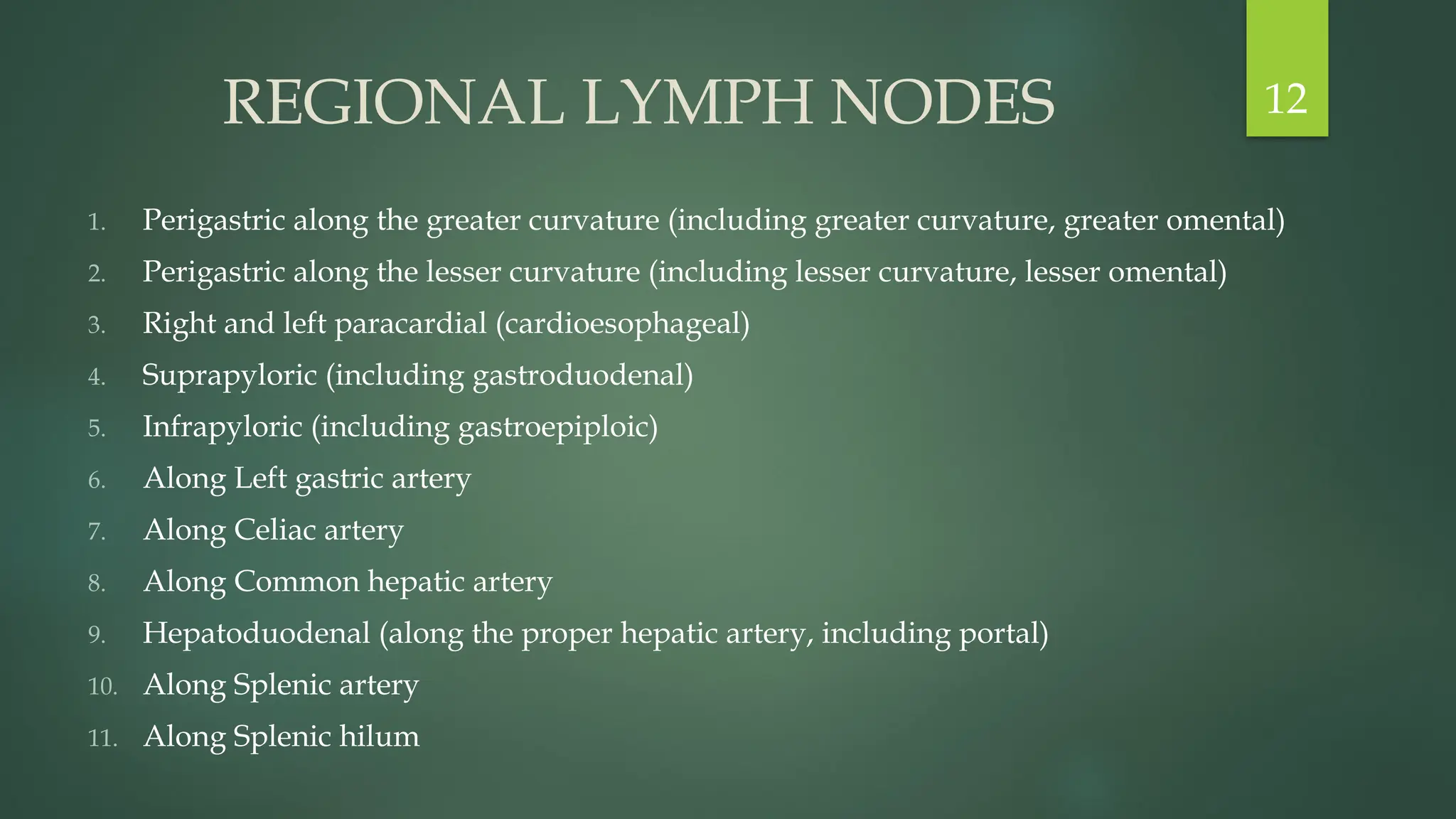
REGIONAL LYMPH NODES
1.Perigastric along the greater curvature (including greater curvature, greater omental)
2. Perigastric along the lesser curvature (including lesser curvature, lesser omental)
3. Right and left paracardial (cardioesophageal)
4. Suprapyloric (including gastroduodenal)
5. Infrapyloric (including gastroepiploic)
6. Along Left gastric artery
7. Along Celiac artery
8. Along Common hepatic artery
9. Hepatoduodenal (along the proper hepatic artery, including portal)
10. Along Splenic artery
11. Along Splenic hilum
12
SIEWERT CLASSIFICATION
• Donein all patients with adenocarcinomas involving the EGJ.
1. Siewert Type I: adenocarcinoma of the lower esophagus (often associated with
Barrett esophagus) with the epicenter located within 1 cm to 5 cm above the
anatomic EGJ.
2. Siewert Type II: true carcinoma of the cardia at the EGJ, with the tumor epicenter
within 1 cm above and 2 cm below the EGJ.
3. Siewert Type III: subcardial carcinoma with the tumor epicenter between 2 cm and 5
cm below the EGJ, which infiltrates the EGJ and lower esophagus from below.
• The treatment of Siewert types I and II is as described in the NCCN Guidelines for
Esophageal and Esophagogastric Junction Cancers.
• Siewert type III lesions are considered gastric cancers, and thus should be treated as
described in the NCCN Guidelines for Gastric Cancer.
17
1. Anorexia
2. Earlysatiety
3. Abdominal pain & discomfort (usually epigastric, vague, mild in early
disease but more severe and constant in advanced disease)
4. Unintentional weight loss
5. Dysphagia (notably if GE junction or proximal stomach involvement)
6. Anemia-related weakness (occult GI bleed with or without IDA)
7. Nausea and vomiting
8. Tarry stools
9. Duration of symptoms is <3 months in almost 40% of patients and >1
year in 20%.
22
23.
10. M/C sitesof metastasis are liver, peritoneal surfaces, and the non
regional or distant lymph nodes; less commonly in ovary (Krukenberg
Tumor), CNS, bone.
• Physical examination can reveal advanced disease, for which the
presentation may include-
1. an abdominal mass (epigastric or liver mass as well as a periumbilical
node [i.e., Sister Mary Joseph node])
2. palpable left supraclavicular nodes (i.e., Virchow node)
3. an enlarged left axillary lymph node (Irish node)
4. rectal shelf (representing peritoneal seeding [i.e., Blumer shelf]).
23
24.
• Paraneoplastic manifestationsinclude-
1. Dermatologic findings may include
sudden appearance of diffuse
seborrheic keratoses ( sign of Leser-
Trélat ) or Acanthosis nigrans.
2. MAHA ( microangipathic hemolytic
anemia )
3. Membranous nephropathy
4. Hypercoagulable state ( Trousseau’s
syndrome)
5. Polyarteritis nodosa
24
• Approximately 10%of gastric cancers are linked to genetic syndromes.
• The most common being hereditary diffuse gastric cancer (HDGC), which is characterized
by an AD inheritance, pertaining a 60% to 80% increased risk of gastric cancer.
• Some of these cases are believed to be due to mutations in E-cadherin (CDH1). The
presence of CDH1 mutations portends a lifetime risk of gastric cancer development of 70%
to 80%. Prophylactic gastrectomy is often recommended once patients are >20 years of age
in these patients.
• Other syndromes associated with increased risk include
1. Lynch syndrome (DNA mismatch repair gene mutation),
2. Familial adenomatous polyposis (APC mutation),
3. Peutz-Jeghers syndrome (STK11 mutation),
4. Juvenile polyposis syndrome (SMAD4 mutation),
5. Hereditary breast and ovarian cancer syndrome, and
6. Li-Fraumeni syndrome (TP53 mutation)
26
• Tis orT1a tumors may be candidates for EMR or ESD.
• T1b–T3: Adequate gastric resection to achieve negative microscopic
margins along with lymphadenectomy.
1. Distal gastrectomy
2. Subtotal gastrectomy
3. Total gastrectomy
• T4b tumors require en bloc resection of involved structures.
• Gastric resection should include the regional lymphatics—perigastric
lymph nodes (D1) and those along the named vessels of the celiac axis
(D2), with a goal of examining at least 16 or greater lymph nodes.
36
37.
EMR (ENDOSCOPIC MUCOSALRESECTION),
ESD (ENDOSCOPIC SUBMUCOSAL DISSECTION)
EMR or ESD of early-stage gastric cancer can be considered
adequate therapy when
the lesion is ≤2 cm in diameter
is shown on histopathology to be well or moderately well
differentiated
does not penetrate beyond the superficial submucosa
does not exhibit LVI, and
has clear lateral and deep margins.
37
38.
• R0 resection:no tumor microscopically at the surgical margin.
• R1 resection: tumor microscopically at the surgical margin.
• R2 resection: tumor macroscopically at the surgical margin.
• D0 dissection: dissection of some of the perigastric lymph nodes.
• D1 dissection: dissection of all perigastric lymph nodes.
• D2 dissection: dissection of perigastric + celiac axis lymph nodes.
(Celiac axis lymph nodes: The celiac trunk, left gastric artery, common hepatic
artery, and lymph nodes around the splenic artery.)
• D3 dissection: D2 + hepatoduodenal, peripancreatic, and mesentery root
lymph nodes dissection.
• D4 dissection: D3 + para-aortic lymph nodes dissection.
38
• Regimens shouldbe chosen in the context of performance status (PS),
medical comorbidities, and toxicity profile.
• Trastuzumab should be added to first-line chemotherapy for advanced
HER2 overexpression positive adenocarcinoma.
• Two-drug regimens are preferred for patients with advanced disease
because of lower toxicity. The use of three cytotoxic drugs in a regimen
should be reserved for patients who are medically fit with excellent PS
and easy access to frequent toxicity evaluations.
• PERIOPERATIVE CT (4 cycles preoperative and 4 cycles postoperative)
1. FLOT (Fluorouracil, leucovorin, oxaliplatin, and docetaxel)
2. Fluoropyrimidine + Oxaliplatin
42
43.
CAPECITABINE
• Antimetabolite.
• Inactiveas a prodrug, converted to 5-FU.
• Converted to 5-FU in liver and tumor tissues by thymidine phosphorylase.
• Good oral bioavailability, hence given orally.
• 1250 mg/m2
, PO, BD, for 2 weeks with 1 week rest, or 1500mg/m2, P/O, BD, for 1 week
with 1 week off- when used as monotherapy.
• 850-1000 mg/m2
, PO, BD, for 2 weeks when used with other chemotherapeutic agents.
• Taken with a glass of water within 30 min of meal.
• Main S/E-
Hand foot syndrome/ palmo-plantar erythrodysaesthesia is a characteristic S/E.
T/t- Vitamin B6 50 mg PO, BD, or celecoxib 200 mg PO, BD, or nicotine patch, and adequate
hydration, urea-based moisturizers and lotions, etc.
43
44.
OXALIPLATIN
• Platinum analogue.
•Cell cycle non specific.
• Binds N7 position of adenine and guanine and disrupts cell division.
• IV only.
• Usual dosage- 100-130 mg/m2
IV over 2 hrs in a 3-weekly schedule.
• Main S/E-
1. Neurotoxicity. Ca+2
/Mg+2
infusions for treating neurotoxicity.
2. Nephrotoxicity
3. Anaphylaxis
44
45.
5 FLORO URACIL
•Antimetabolite.
• S phase specific.
• IV only.
• Usual dose- 450 mg/m2
IV day 1-5 every 28 days.
• Antitumor activity enhanced by leucovorin and methotrexate.
• Main S/E-
1. Hand foot syndrome
2. Myelosuppression. T/t- Vistonuridine 10 gm PO, QID for 5
days.
45
46.
FOLINIC ACID/ LEUCOVORIN
•It is a Folic acid analogue.
• Given along with 5 FU to potentiate its action.
• It acts as a ligand, gets attached to the receptor of tumor cells
and aids the binding of 5FU to the cells.
• Administered before giving 5 FU.
• It is also given along with Methotrexate.
• When given with Methotrexate, it replenishes the Folic Acid
reserves which have been decreased due to Methotrexate
toxicity.
46
47.
PACLITAXEL
• Taxane.
• Antimicrotubuleagent.
• Cell cycle specific agent (M phase), enhaces microtubule polymerization.
• Only IV.
• Usual dosage- 135-175 mg/m2
IV 3 hr infusion q3w, 80-100 mg/m2
IV 1 hr infusion
q1w .
• Potent radiosensitizer.
• Main S/E-
1. Hypersensitivity reaction
2. Neurotoxicity
3. Cardiotoxic (mild)
4. Myelosuppression
47
48.
DOCETAXEL
• Taxane, semisyntheticagent.
• Antimicrotubule agent.
• Cell cycle specific agent (M phase), enhances microtubule polymerization.
• Only IV.
• Usual dosage- 75 mg/m2
IV q3w.
• Radiosensitizer.
• Main S/E-
1. Hypersensitivity
2. Myelosuppression
3. Fluid retention syndrome
4. Maculopapular rash
5. Vesicant
48
49.
CISPLATIN
• Platinum analog.
•AKA CDDP.
• Binds to DNA at N7 position of adenine and guanine, produces cross links.
• Cell cycle non specific agent.
• IV or Intraperitoneal.
• Used mostly in squamous cell neoplasms.
• Radiosensitizer.
• Usual dosage- 50-100 mg/m2
IV as either q1w or q3w. Also 20 mg/m2
IV if used on day 1-5 q3w.
• If used with paclitaxel, administered after it.
• Main S/E-
1. Most emetogenic
2. Nephrotoxic
3. Peripheral neuropathy
4. Ototoxicity
5. SIADH
49
50.
CARBOPLATIN
• Platinum analog.
•Binds to DNA at N7 position of adenine and guanine, produces cross links.
• Cell cycle non specific agent.
• Only IV.
• Dose calculated by AUC, based on GFR.
• Calvert formula- Dose (mg) = Target AUC x (GFR+25).
• If used with paclitaxel, administered after it.
• Main S/E-
1. Myelosuppression
2. Nausea and vomiting
3. Nephrotoxic
4. Peripheral neuropathy
50
51.
TRASTUZUMAB
• AKA Herceptin.
•Anti HER 2 antibody.
• Recombinant humanized monoclonal antibody.
• Usual dose- 8 mg/kg IV over 90 min loading dose, f/b
6 mg/kg IV over 90 min maintenance dose, q3w.
• Main S/E-
1. Cardiotoxic
2. Infusion reaction
51
• Radiation therapy,usually administered with concomitant fluoropyrimidine
based chemotherapy, is indicated for locally confined gastric cancer that
either is not technically resectable or occurs in medically inoperable patients.
• Historically, 2D based radiation planning has been carried out primarily
using anatomic landmarks as well as fluoroscopic barium swallow to
determine field borders.
• Those who undergo gastric resection with incomplete tumor resection or
have truly positive margins of resection are also appropriately managed by
combined-modality therapy.
• For unresectable lesions with moderate periesophageal extension, it may not
be possible to exclude an adequate amount of the heart with AP/PA fields,
and the use of lateral or oblique fields for a portion of treatment, as well as
adopting IMRT techniques, is likely indicated.
53
54.
• Patients withlocally advanced disease that is unresectable with negative margins would
be identified preoperatively with endoscopic ultrasonography and CT staging.
• Preoperative chemoradiation then could precede an attempt at gross total resection,
alone or in combination with IORT, and maintenance chemotherapy.
• In patients with high-risk features-
1. poorly differentiated or higher-grade cancers
2. lymph vascular invasion
3. perineural invasion
4. age <50 years
5. suboptimal resection including lymph node resection,
postoperative chemoradiotherapy may be indicated.
54
55.
• RT Dosing:
45-50.4 Gy (1.8 Gy/day),
total 25-28 fractions
Higher doses may be used
for positive surgical
margins in selected cases
as a boost to that area- 55-
60 Gy.
Meticulous treatment
planning is required to
reduce unnecessary dose
to OARs.
55
56.
• POSITION: Supine
•FIELD: AP/PA parallel opposed fields
• BORDERS:-
SUPERIOR BORDER: at T10/T11
INFERIOR BORDER: at L3/L4 ,for proximal 1/3 or GE
junction: at L1/L2
LEFT LATERAL: include all remaining peri-gastric nodes,
antral/distal 1/3 lesions, splenic hilum
RIGHT LATERAL: preop location of primary tumour or
porta hepatis (whichever is further)
56
• INDICATION OFPOST-OP CT-RT-
If no pre-op CT/ CT-RT
All pT3 and pT4 less than D2 dissection
High nodal burden
All R1 and R2 resection if no pre-op RT or CT-RT.
• If Pre-op RT/ CT-RT given-
Not indicated
59
60.
• POSTOPERATIVE CT-RT-
for patients who received less than a D2 lymph node dissection
The INTERGROUP 0116 TRIAL formed the basis for postoperative adjuvant CT-RT
strategy
• REGIMEN 1 (FU + Leucovorin) :
2 cycles before and 4 cycles after CT-RT.
For cycles after CT-RT, begin CT 1 month after CT-RT.
Leucovorin 400 mg/m2
IV on day 1, Fluorouracil 400 mg/m2
IV push on day
1 ,Fluorouracil 1200 mg/m2
IV continuous infusion over 24 hours daily on days 1
and 2 cycled every 14 days.
With radiation: Fluorouracil 200–250 mg/m2
IV continuous infusion over 24 hours
daily on days 1–5 weekly for 5 weeks.
60
61.
• REGIMEN 2(Capecitabine) :
1 cycle before and 2 cycles after CT-RT.
For cycles after CT-RT, begin CT 1 month after CT-
RT.
Capecitabine 750–1000 mg/m2
PO BID on Days 1–14
cycled every 21 days, for pre and post CT-RT.
With RT: Capecitabine 625–825 mg/m2
PO BID on
days 1–5 weekly for 5 weeks.
61


![10. M/C sites of metastasis are liver, peritoneal surfaces, and the non
regional or distant lymph nodes; less commonly in ovary (Krukenberg
Tumor), CNS, bone.
• Physical examination can reveal advanced disease, for which the
presentation may include-
1. an abdominal mass (epigastric or liver mass as well as a periumbilical
node [i.e., Sister Mary Joseph node])
2. palpable left supraclavicular nodes (i.e., Virchow node)
3. an enlarged left axillary lymph node (Irish node)
4. rectal shelf (representing peritoneal seeding [i.e., Blumer shelf]).
23](https://image.slidesharecdn.com/22-250508054059-44be626b/75/22-Gastric-Cancer-powerpoint-presentation-23-2048.jpg)


