SlideShare a Scribd company logo
One Platform Comparison of Estrone and Folic Acid Anchored
Surface Engineered MWCNTs for Doxorubicin Delivery
Neelesh Kumar Mehra†,‡
and N. K. Jain*,†,‡
†
Pharmaceutics Research Laboratory, Department of Pharmaceutical Sciences, Dr. H. S. Gour University, Sagar 470 003, India
‡
Pharmaceutical Nanotechnology Research Laboratory, ISF College of Pharmacy, Moga 142 001, India
ABSTRACT: Our main aim in the present investigation was to assess and compare
the in vitro and in vivo cancer targeting propensity of doxorubicin (DOX) loaded folic
acid (FA) and estrone (ES) anchored PEGylated multiwalled carbon nanotubes
(MWCNTs) employing tumor bearing Balb/c mice. The DOX was loaded into the
developed functionalized MWCNTs after proper characterization using dialysis
diffusion method. The in vitro, ex vivo, and in vivo studies were performed on the
MCF-7 cell line for assessment of the cancer targeting propensity. Both qualitative and
quantitative cell uptake studies indicated the preferential higher uptake of estrone
anchored nanotube formulation compared to other formulations and free DOX owing
to the overexpression of estrogen receptors (ERs) on human breast MCF-7 cells.
Similarly, the pharmacokinetic and increased antitumor activities also confirmed the
elevated cancer targeting propensity of the estrone and folic acid anchored MWCNT
formulations. The DOX/ES-PEG-MWCNTs has also shown significantly longer
survival span (43 days) than free DOX (18 days) and control group (12 days). Present outcomes from the ex vivo and in vivo
studies are deemed to be of great scientific value and shall assist targeted drug delivery formulation scientists for selection of the
targeting moieties in the treatment of human breast cancer.
KEYWORDS: carbon nanotubes, drug targeting, doxorubicin, pharmacokinetic, breast cancer, estrone, folic acid, antitumor activity
1. INTRODUCTION
On recalling the “Magic bullet concept” tremendous sophisti-
cation has been visualized in targeting concepts and delivery of
bioactives. In the present scenario, carbon nanotubes (CNTs)
have attracted escalating attention after attachment of huge
targeting moieties to offer sustained/controlled release behavior
with cellular targeting and enhanced specificity.1,2
CNTs were first discovered and fully explored in 1991.3
CNTs are unique, three-dimensional sp2
hybridized carbon
nanomaterials that have attracted tremendous attention as a
valuable, promising, safe and effective alternative nanoarchitec-
ture for biomedical applications due to their unique
physicochemical properties such as biocompatibility, non-
immunogenicity, high loading efficiency, high aspect ratio,
structural flexibility, and noncytotoxic nature.2−9
Functionalized
CNTs (f-CNTs) have the capacity to readily cross the plasma
membrane via an energy-dependent, endosomally mediated
and direct translocation, which is known as tiny nanoneedle
mechanism.9,10
Due to the tiny nanoneedle tubular shape, they
pass through cell membrane without causing cell death, thus
allowing efficient tissue and tumor specific drug delivery with
minimal or no toxic effects. Compared with other nanocarrier
systems, which have low entrapment efficiency due to steric
hindrance, the f-CNTs have greater entrapment due to the
availability of high surface area.4,11,12
Despite excellent progress in using surface engineered CNTs
as targeted drug delivery carriers, more research is needed to
further optimize their ability to selectively accumulate in
diseased/targeted tissues and to release their toxic payload in a
controlled manner. To date, numerous research reports are
available considering functionalized CNTs for drug delivery
including doxorubicin hydrochloride,13−15
paclitaxel,16
docetax-
el,17
gemcitabine,18
amphotericin B,19
gliotoxin,20
sulfasala-
zine,21
oxaliplatin,22
etc.
The anthracycline antibiotic, doxorubicin hydrochloride,
chosen for the present investigation as an anticancer agent is
widely used in the treatment of cancers of the breast, prostate,
brain, and cervix and Hodgkin lymphoma. Short half-life and
severe toxicity (acute cardiotoxicity) associated with doxor-
ubicin restrict its clinical application. According to the
Biopharmaceutical Classification System (BCS) doxorubicin
hydrochloride belongs to class III, i.e., high solubility and low
permeability (BCS III). BCS is a scientific framework for
classifying substances according to their aqueous solubility and
permeability.23−25
Very recently our laboratory explored the
delivery of DOX employing D-α-tocopheryl polyethylene glycol
1000 succinate (TPGS) as targeting ligand with improved
therapeutic outcomes and antitumor targeting efficacy on the
MCF-7 cell line.4
The purpose of the present investigation was
to compare the cancer-targeting propensity of the folic acid
(FA) and estrone (ES) appended surface functionalized
Received: October 27, 2014
Revised: December 13, 2014
Accepted: December 17, 2014
Published: December 17, 2014
Article
pubs.acs.org/molecularpharmaceutics
© 2014 American Chemical Society 630 DOI: 10.1021/mp500720a
Mol. Pharmaceutics 2015, 12, 630−643
MWCNTs employing breast cancer cells on Balb/c mice. The
cancerous cells overexpress the folate receptors (FRs) and
estrogen receptors (ERs) belonging to folate and nuclear
hormone receptor superfamily, respectively.26
The MWCNTs
tethering ES and FA probably deliver DOX more efficiently
into the cancerous cells after binding selectively to their
respective receptors through endocytosis or tiny nanoneedle
mechanism. The ligand-driven MWCNTs are an established,
well-known concept, and these targeting moieties can be easily
attached to f-MWCNTs. The comparative cancer targeting
propensity of targeting ligand conjugated f-MWCNTs studied
on a single platform reported herein is expected to be of greater
scientific interest in targeted drug delivery. We have extensively
characterized the ligand (ES and FA) conjugated MWCNTs in
vitro, ex vivo, and in vivo in terms of biodistribution and
pharmacokinetics, Kaplan−Meier survival analysis, and tumor
targeting efficacy on Balb/c mice.
2. MATERIALS AND METHODS
2.1. Materials. Multiwalled carbon nanotubes (MWCNTs)
produced by chemical vapor deposition (CVD) with purity
99.3% were purchased from Sigma-Aldrich Pvt. Ltd. (St. Louis,
Missouri, USA). Doxorubicin hydrochloride was received as a
benevolent gift from M/s Sun Pharm Advanced Research
Centre (SPARC), Vadodara, India. Folic acid (FA), dimethyl
sulfoxide (DMSO), 1-ethyl-3-(3-dimethylaminopropyl)-
carbodiimide (EDC), and dialysis membrane (MWCO, 5−6
kDa) were purchased from HiMedia Pvt. Ltd. Mumbai, India.
Estrone (ES) was purchased from Sigma-Aldrich Pvt. Ltd. USA.
All the reagents and solvents were used as received.
2.2. Functionalization of MWCNTs. The procured
pristine MWCNTs were initially purified and treated with
strong oxidizing acid following a previously reported method
from our and other laboratories after minor modifica-
tions.2,4,10,13,27,28
Briefly, MWCNTs (500 mg) were treated
with concentrated nitric acid and sulfuric acid
(HNO3:H2SO4::1:3) mixture with continuous magnetic stirring
at 100 rpm (Remi, Mumbai, India) maintaining 100 ± 5 °C
temperature for 24 h. Then dispersed MWCNTs were washed
with deionized water, ultracentrifuged, vacuum-dried, and
collected. Treated MWCNTs (300 mg) were further treated
with ammonium hydroxide (NH4OH) and hydrogen peroxide
(H2O2) in 50:50 ratio in a round-bottom flask (RBF) at 80 ± 5
°C for 24 h. The treated oxidized MWCNTs (ox-MWCNTs)
were repeatedly washed to neutral pH, ultracentrifuged
(Z36HK, HERMLE LaborTchnik GmbH, Germany) at
20,000 rpm for 15 min, and finally lyophilized (Heto dry
Winner, Germany).
2.3. Conjugation of Estrone to Surface Functionalized
MWCNTs (ES-PEG-MWCNTs). The conjugation of estrone
(ES) with oxidized-MWCNTs was performed using standard 1-
ethyl-3-(3-dimethylaminopropyl)carbodiimide (EDC) chemis-
try in three steps following the previously reported methods
after minor modifications.23,27,29
2.4. Activation of Estrone. Briefly, estrone (ES; 8 mM),
succinic anhydride (SA; 10 mM), dimethylaminopyridine
(DMAP; 8 mM), and triethylamine (TEA; 8 mM) were
dissolved in dioxane and magnetically stirred overnight at 25 ±
2 °C. The dioxane was evaporated under vacuum, and the
obtained residue was dissolved in dichloromethane (DCM) and
filtered. The filtrate was further dried and precipitated using
diethyl ether to obtain the carboxylic acid derivative of estrone
(ES-COOH). The ES-COOH (0.5 mM) was dissolved in
DCM (0.5 mM) in equimolar concentration, and hydrox-
ybenzotriazole (HOBT; 0.1 mM), PEG-3000-amine, and DCC
were added to it and continually magnetically stirred at 25 ± 2
°C for 24 h until precipitate was formed. The pellet of ES-PEG-
NH2 was collected by centrifugation (Scheme 1), repeatedly
washed with deionized water, and collected.23,30
The COOH-MWCNTs (33.36 mg) and EDC (6.41 mg)
were dissolved in DMSO with continuous magnetic stirring at
100 rpm. After 6 h, ES-PEG-NH2 in DMSO (4.60 mg/mL)
solution was added and kept magnetic stirred up to 5 days.
Then the unreacted biproducts/materials were separated out,
lyophilized (Heto dry Winner, Denmark, Germany), collected,
and stored (Scheme 2).27
2.5. Synthesis of FA-PEG Conjugation to Function-
alized MWCNTs. The tert-butoxycarbonyl (t-Boc)-protected
folic acid (FA) for amine protection of FA was synthesized, and
its further activation was performed following the previously
reported well-known method to obtain t-Boc-FA-NHS.2,31
2.5.1. Conjugation of t-Boc-FA to Surface Functionalized
MWCNTs. The t-Boc-FA-NHS (16.25 mg) was mixed with
PEG-3k bis amine (Sigma-Aldrich, USA) (225.0 mg) in DMSO
in the presence of triethylamine (TEA) and magnetically stirred
for 24 h at 25 ± 2 °C. The obtained t-Boc-FA-PEG-NH2 as a
pale yellow solid was collected and dried.
The t-Boc-FA-PEG-NH2 was mixed with ox-MWCNTs in
DMSO followed by addition of EDC and continually
magnetically stirred for 5 days maintaining the dark condition
at 25 ± 2 °C (Scheme 3). The resultant conjugate was
collected, dried, and characterized by FTIR spectroscopy.2,10,32
The activation and conjugation of both ligands with
MWCNTs were extensively characterized by UV/vis spectros-
copy (Shimadzu 1601, UV−visible spectrophotometer, Shi-
madzu, Japan), and Fourier transform infrared spectroscopy
(FTIR) was performed using the KBr pellet method
(PerkinElmer 783, Pyrogon 1000 spectrophotometer, USA)
scanned in the range from 4000 to 500 cm−1
.
2.6. Characterization of the MWCNT Conjugates. The
developed MWCNT conjugates were characterized for particle
size by photon correlation spectroscopy in a Malvern Zetasizer
nano ZS90 (Malvern Instruments, Ltd., Malvern, U.K.) at room
Scheme 1. Conjugation of PEG to Activated Estrone
Molecular Pharmaceutics Article
DOI: 10.1021/mp500720a
Mol. Pharmaceutics 2015, 12, 630−643
631
temperature, atomic force microscope (AFM) using Digital
Nanoscope IV Bioscope (Veeco Innova Instruments, Santa
Barbara, CA, USA), FTIR spectroscopy using PerkinElmer
FTIR spectrophotometer (PerkinElmer 783, Pyrogon 1000
Spectrophotometer, Shelton, Connecticut), 1
H nuclear mag-
netic resonance (NMR) spectroscopy (Bruker, DRX, USA),
and Raman spectroscopy RINSHAW, inVia Raman spectro-
photometer (Renishaw, Gloucestershire, U.K.).
2.7. DOX Loading Studies. DOX was loaded into the
MWCNT dispersion using modified equilibrium dialysis
method as reported previously.10,15,25
Briefly, DOX was initially
dissolved in acetone, and aqueous triethylamine (TEA)
solution was added in a 2:1 molar ratio (DOX:TEA). The
DOX was magnetically stirred (100 rpm; Remi, India) with the
developed MWCNT conjugates and pristine MWCNTs in
phosphate buffer solution (PBS: pH 7.4) using Teflon beads
overnight at 37 ± 0.5 °C for 24 h to facilitate DOX loading.
MWCNT conjugates were then ultracentrifuged until the
nanotube dispersion became color free. The amount of
unbound DOX was determined measuring absorbance at λmax
480.2 nm spectrophotometrically (Shimadzu 1601, UV−visible
spectrophotometer, Shimadzu, Japan). The developed DOX
loaded MWCNT (DOX/MWCNTs, DOX/ox-MWCNTs,
DOX/ES-PEG-MWCNTs, and DOX/FA-PEG-MWCNTs)
conjugates were collected, dried, lyophilized (Heto dry Winner,
Denmark, Germany), and stored at 2−8 °C for further use. The
DOX loading efficiency (%) was calculated using the following
formula:
Scheme 2. Development of ES-PEG-MWCNT Conjugate
Scheme 3. Conjugation Scheme of FA-PEG and ES-PEG Conjugated MWCNTs
Molecular Pharmaceutics Article
DOI: 10.1021/mp500720a
Mol. Pharmaceutics 2015, 12, 630−643
632
=
−
×
% loading efficiency
wt of loaded DOX wt of free DOX
wt of loaded DOX
100
2.8. In Vitro Release Studies. The dispersion of DOX/
MWCNT, DOX/ox-MWCNT, DOX/ES-PEG-MWCNT, and
DOX/FA-PEG-MWCNT conjugates was studied in sodium
acetate buffer saline (pH 5.3) and phosphate buffer saline (pH
7.4) as recipient media using a modified dissolution method
with maintenance of 37 ± 0.5 °C physiological temperature.
The MWCNT conjugates were filled in pretreated dialysis
membrane (MWCO 5−6 kDa, HiMedia, Mumbai, India)
separately and kept in the releasing media under constant
magnetic stirring (100 rpm; Remi, Mumbai, India) at 37 ± 0.5
°C. At definite time points, the MWCNT samples were
withdrawn, and after each sampling, the withdrawn medium
was replenished with fresh sink solution maintaining strict sink
condition. The drug concentration was determined after proper
dilution in a UV/visible spectrophotometer at λmax 480.2 nm
(UV/vis, Shimadzu 1601, Kyoto, Japan).
2.9. Ex Vivo (Cell Line) Studies. The ex vivo (cell line)
studies were performed on Michigan Cancer Foundation
human breast cancer cells (MCF-7) cell line procured from
National Center for Cell Sciences (NCCS), Pune, India. The
MCF-7 cells were maintained in Dulbecco’s modified Eagle’s
medium (DMEM; HiMedia, Mumbai, India) containing 10%
fetal bovine serum (FBS; Sigma, St. Louis, Missouri)
supplemented with 1% antibiotic (penicillin−streptomycin)
solution grown to confluence in humidified atmosphere
containing 5% CO2 at 37 °C. Cells were seeded at a density
of 4 × 10³
cells/well and incubated for 24 h prior to
commencement of the experiments. The DMEM medium was
changed as per requirement every alternate day, and cells with
approximately 80% confluence were used for further studies.
After that, medium was decanted and washed with fresh PBS (6
mL for 25 cm2
) and cells were trypsinized using 0.25% trypsin
solution. Trypsin was removed completely, and subsequently
medium was added to split the cells.4,14
2.10. Cell Viability Assay. The cell viability assay was
performed using 3-(4,5-dimethylthiazol-2-yl)-2,5-diphenyltetra-
zolium bromide (MTT) dye reduction assay to a blue formazan
derivative by living cells to assess the cytotoxic propensity of
the nanotube formulations at different concentrations.27,28,33
Briefly, MCF-7 cells were placed onto 96-well flat-bottomed
tissue culture plates (Sigma, Germany) and allowed to adhere
for 24 h at 37 °C prior to assay. The cells in quadruplicate were
treated with free DOX, DOX/MWCNTs, DOX/ox-MWCNTs,
DOX/FA-PEG-MWCNTs, and DOX/ES-PEG-MWCNTs
with increasing concentration (0.001−100 μM) of DOX
simultaneously under controlled environment for 24 and 48 h
at 37 ± 0.5 °C in a humidified atmosphere with 5% CO2.
Thereafter, medium was decanted and 50 μL of methylthiazole
tetrazolium (MTT; 1 mg/mL) in DMEM (10 μL; 5 mg/mL in
Hanks balanced salt solution; without phenol red) was added to
each well and incubated for 4 h at 37 °C. Formazan crystals
were solubilized in 50 μL of isopropanol by shaking at rt for 10
min. The absorbance was taken using a microplate reader
(Medispec Ins. Ltd., Mumbai, India) at 570 nm wavelength,
blanked with DMSO solution, and (%) cell viability was
calculated using the following formula:
= ×cell viability (%)
[A]
[A]
100test
control
where [A]test is the absorbance of the test sample and [A]control
is the absorbance of control samples.
2.11. Cell Uptake Study. Cellular uptake studies were
carried out through flow cytometry analysis using FACS
Calibur flow cytometer (Becton, Dickinson Systems, FACS
canto, USA) on MCF-7 cell line to determine the intracellular
uptake efficiency of the developed surface engineered MWCNT
formulations.20,27,34−36
The cellular uptake of free DOX and
DOX loaded MWCNT formulations using MCF-7 cell line was
performed through flow cytometry. The cell suspension was
incubated at 25 ± 2 °C for 4 h with intermittent mixing every
20 min. The 4 × 10³
cells per well were seeded in a 12-well
plate and incubated for 24 h at 37 ± 0.5 °C with 5% CO2, and
then the medium in each well was replaced with 2 mL of
serum-free and antibiotic-free medium. Then various concen-
trations of MWCNT formulations (DOX solution, DOX/
MWCNTs, DOX/ox-MWCNTs, DOX/FA-PEG-MWCNTs,
and DOX/ES-PEG-MWCNTs) were incubated for 3 h.
Three hours post in vitro incubation, cells were washed three
times with ice-cold PBS, trypsinized (0.1%; w/v), pelletized via
centrifugation (1000g) to remove the trypsin, and finally
resuspended in PBS. Cells associated with DOX were measured
quantitatively (FACS Calibur flow cytometer; Becton, Dick-
inson Systems, FACS canto, USA) and qualitatively (fluo-
rescence microscope, Leica, Germany).
2.12. Cell Cycle Distribution Measurement. The DNA
content in the different cell cycle phases was determined by
flow cytometry for 24 h after treatment with the developed
MWCNT formulations.4,37−39
Briefly, 4 × 10³
cells were seeded
and allowed to attach overnight at 37 ± 0.5 °C; the cultured
cells were treated with developed optimized nanotube
formulation (2 nM/mL concentration of drug) under CO2
atmosphere, and the incubated cells were harvested, washed,
and fixed using 70% cold ethanol overnight. Subsequently the
cells were trypsinized, washed again, fixed, and further
resuspended in hypotonic propidium iodide solution (PI; 50
μg/mL) containing ribonuclease A (RNase free, 100 μg/mL)
and incubated for 30 min at 37 °C in dark before measurement.
The distribution of cell cycle was determined with FACS
Calibur flow cytometer and analyzed using Cell Quest software
(Becton, Dickinson Systems, FACS canto, USA).
2.13. In Vivo Studies. 2.13.1. Animals and Dosing. The
Balb/c mice of either sex (20−25 g) were used for in vivo
studies, as mice present a more sensitive model for in vivo
evaluations. The experimental design was duly approved by the
Committee for the Purpose of Control and Supervision of
Experiments on Animals (CPCSEA) [Registration No. 379/
01/ab/CPCSEA/02] of Dr. H. S. Gour Vishwavidyalaya, Sagar-
470003 India. The Balb/c mice of uniform weight (20−25 g)
were housed in ventilated plastic cages with access to water ad
libitum. The mice were acclimatized at 25 ± 2 °C by
maintaining the relative humidity (RH) 55−60% under natural
light/dark condition prior to studies, avoiding any kind of stress
on Balb/c mice.4,16,40−43
2.13.2. Antitumor Targeting Efficacy Studies. The present
antitumor targeting efficacy study was performed on tumor
bearing Balb/c mice employing tumor growth inhibition study
and Kaplan−Meier survival curve analysis. The tumor was
developed on Balb/c mice using the well-known right flank
method4
by injecting serum-free cultured MCF-7 cells in the
Molecular Pharmaceutics Article
DOI: 10.1021/mp500720a
Mol. Pharmaceutics 2015, 12, 630−643
633
right hind leg of the mice. The development of tumor was
regularly monitored by palpating the injected area with index
finger and thumb. When the tumor grew to approximately 100
mm3
in size, mice were subjected to in vivo studies and Balb/c
mice were accommodated in a pathogen-free laboratory
environment.20,42,44
The sterilized free DOX, DOX/
MWCNT, DOX/ox-MWCNT, DOX/PEG-MWCNT, DOX/
FA-PEG-MWCNT, and DOX/ES-PEG-MWCNT formulations
(equivalent dose = 5.0 mg/kg body weight) were intravenously
administered into the mice through the tail vein route. The size
of the tumor was measured using an electronic digital Vernier
caliper and calculated using the formula =1/2 × length ×
width2
. The median survival time was also recorded, and the
study was terminated 45 days post treatment.
2.13.3. Pharmacokinetic and Biodistribution Studies after
Intravenous Administration. The pharmacokinetic and bio-
distribution studies were performed on Balb/c mice after
intravenous administration of the free DOX and DOX loaded
MWCNT formulations (DOX/MWCNTs, DOX/ox-
MWCNTs, DOX/ES-PEG-MWCNTs, DOX/FA-PEG-
MWCNTs) at equivalent dose 5.0 mg/kg body weight dose.
The blood was collected in to the Hi-Anticlot blood collecting
vials (HiMedia, Mumbai, India) through retro-orbital plexus of
the animal’s eyes under mild anesthetic condition at the
different time points. The collected blood was centrifuged after
addition of 100 μL of trichloroacetic acid (TCA) in methanol
(10% w/v) to separate RBCs. Supernatant was collected,
vortexed, and ultracentrifuged (Z36HK, HERMLE LaborTch-
nik GmbH, Germany), and drug concentration was determined
using the high performance liquid chromatography (HPLC)
method. The pharmacokinetic parameters such as peak plasma
concentration (Cmax), the area under the curve (AUC0−t), area
under the first moment curve (AUMC), mean residence time
(MRT), half-life (t1/2), and half value duration (HVD) were
also calculated.4,16,18,45
The in vivo biodistribution study was also performed on
tumor bearing Balb/c mice after administration of the same
sterilized formulations. At 1, 6, 12, and 24 h time points mice
were carefully sacrificed by the decapitation method. The
different organs were separated out, washed with Ringer’s
solution for removal of any adhered debris, dried with tissue
paper, weighed, and stored frozen until used. Weighed tissues
samples were immediately homogenized (York Scientific
Instrument, New Delhi, India), vortexed, and ultracentrifuged
at 3000 rpm for 15 min (Z36HK, HERMLE LaborTchnik
GmbH, Germany). The obtained clear supernatant was
collected in HPLC vials and loaded onto the HPLC system
to determine the concentration of DOX (Shimadzu, C18,
Japan) using a mobile phase consisting of buffer pH 4.0/
acetonitrile/methanol (60:24:16; v/v/v) with 1.2 mL/min flow
rate at 102/101 bar.41,46−49
2.14. Statistical Analysis. The obtained data from the
studies were presented as mean ± standard deviation (n = 3),
and statistical analysis was performed using GraphPad InStat
software (Version 6.00, Graph Pad Software, San Diego,
California, USA) by one-way analysis of variance (ANOVA)
followed by Tukey−Kramer multiple comparisons test. The
comparison of the Kaplan−Meier survival analysis curve was
performed using the Log-rank (Mantel−Cox) test (conserva-
tive).The pharmacokinetic data analysis of the plasma
concentration time profile was conducted using the Kinetica
5.0 software (Thermo Scientific, USA), followed by non-
compartmental analysis. A probability p ≤ 0.05 was considered
significant while p ≤ 0.001 was considered as extremely
significant.
3. RESULTS AND DISCUSSION
The surface engineered smart, multifunctional carbon nano-
tubes appear to be useful in cancer chemotherapy as drug
delivery systems, owing to their unique physicochemical
properties and being continually explored in controlled and
targeted drug delivery. As-synthesized pristine MWCNTs were
procured and purified using different purification approaches
rendering them suitable for further studies. The presence of
impurities in pristine CNTs restricts their clinical application;
thus emerges functionalization that renders them more
biocompatible, safe, and effective for drug delivery. Function-
alization is a well-known approach for surface alteration of
CNTs. The f-MWCNTs used in the present investigation were
prepared after proper functionalization and conjugation of
targeting moieties, devoid of fluorescent dye, for cellular
trafficking because doxorubicin has red-autofluorescence.
The FTIR spectrum of procured unmodified (pristine)
MWCNTs depicts absorption peaks at 1626 and 2400.24 cm−1
.
The characteristic peak at 1626 cm−1
suggests the presence of
carbon residue on the CNT surface, while a clear single
characteristic peak at 2400.24 cm−1
is ascribed to the stretching
of the carbon nanotube backbone. The oxidized MWCNTs
shows characteristic peaks approximately at 1637.4 and 3425.6
cm−1
attributable to asymmetrical stretching of CO
stretching vibration mode and O−H stretching vibration,
respectively.4
The FTIR spectrum of the FA-PEG-MWCNTs showed
characteristic peaks at 1025.0, 3438.6, 2920.0, 1650.0, 1435.2,
and 1320.5 cm−1
. The peaks at 1650.0 and 1435.2 cm−1
of NH2
stretching and O−H deformation of phenyl skeleton and a
strong clear sharp characteristic peak observed at 1025.0 cm−1
of C−O stretching of ether linkage suggest the attachment of
FA-PEG to MWCNTs (Figure 1 A).
The estradiol conjugated with surface engineered MWCNTs
shows the characteristic peaks in the range of 1500−1655 cm−1
of −CO anhydride and amide bonding of estradiol
conjugated MWCNTs. A strong and sharp characteristic peak
Figure 1. FTIR spectra of (A) FA-PEG-MWCNTs and (B) ES-PEG-
MWCNTs.
Molecular Pharmaceutics Article
DOI: 10.1021/mp500720a
Mol. Pharmaceutics 2015, 12, 630−643
634
was found at 1020.04 cm−1
of C−O stretch of ether linkage due
to the presence of polyether backbone of PEG. The
characteristic peak at 1650.94 cm−1
of C−O stretching of
amide bond formation clearly indicates confirmation of
conjugation of estrone to MWCNTs (Figure 1 B).
The 1
H NMR spectra of the FA-PEG-MWCNTs and ES-
PEG-MWCNTs are shown in Figures 2A and 2B. The
characteristic signal of the pteridine ring proton and aromatic
protons of the p-aminobenzoic acid (PABA) of FA was clearly
present in the case of FA-PEG-MWCNTs (Figure 2 A).
Similarly, ES-PEG-MWCNTs represent the protons character-
istic of the phenyl ring of the steroidal moiety showing
chemical shifts as presented in the NMR spectrum (Figure 2
B).
Raman spectroscopy used for further characterization of the
developed MWCNT conjugates provides information on
hybridization state and defect site chemistry. It also gives
information on changes in electronic structure upon anchoring
of different chemical functional moieties. The MWCNTs show
the disorder related to D mode (approximately at 1330−1360
cm−1
) and high-energy mode known as tangential G mode
(approximately at 1500−1600 cm−1
).4
Raman spectra of ox-MWCNTs, FA-PEG-MWCNTs, and
ES-PEG-MWCNTs are shown in Figures 3A, 3B, and 3C. The
Raman spectrum of ox-MWCNTs shows the Raman shifts at
1584.73 and 1352.0 cm−1
, which correspond to the G band
(graphite-like mode) and D band (disorder-induced band),
respectively (Figure 3 A). The Raman spectrum of the FA-
PEG-MWCNTs shows the G band around 1565 cm−1
and D
band around 1310 cm−1
(Figure 3 B). The Raman spectrum of
the ES-PEG-MWCNTs shows the G band around 1580 cm−1
and D band around 1320 cm−1
(Figure 3 C). The shifting of G
and D band intensity toward higher intensity in the case of FA-
PEG-MWCNTs and ES-PEG-MWCNTs is mainly due to
increase in the extent of conjugation of FA/ES-PEG- with
functionalized MWCNTs, which would increase the single
bond characteristics in the functionalized systems.
Figure 2. NMR spectra of (A) FA-PEG-MWCNTs and (B) ES-PEG-MWCNTs.
Molecular Pharmaceutics Article
DOI: 10.1021/mp500720a
Mol. Pharmaceutics 2015, 12, 630−643
635
The transmission electron microscopy (TEM) clearly
revealed that the developed formulations were in nanometric
size range, even after conjugation.
The particle size and polydispersity index (PI) of the DOX/
MWCNTs, DOX/ox-MWCNTs, DOX/FA-PEG-MWCNTs,
and DOX/ES-PEG-MWCNTs were found to be 350.02 ±
5.24, 230.12 ± 6.56, 242.28 ± 5.85, and 240.26 ± 4.50 and 0.22
± 0.012, 0.32 ± 0.023, 0.55 ± 0.024, and 0.85 ± 0.034,
respectively. The loading efficiency of DOX/MWCNTs, DOX/
ox-MWCNTs, DOX/FA-PEG-MWCNTs, and DOX/ES-PEG-
MWCNTs was calculated to be 92.50 ± 2.62, 93.25 ± 3.52,
96.50 ± 2.52, and 96.80 ± 3.44, respectively, and determined
by absorption peak at 480.2 nm. A significant improvement in
the loading efficiency was observed in the case of ligand
conjugated MWCNTs through various interaction mechanisms
(such as π−π stacking, hydrogen bonding, and hydrophobic
interactions) owing to the aromatic structure of the
doxorubicin. Also large π-conjugated structures of the nano-
tubes can form π−π stacking interaction with the quinine part
of the DOX. One more possible reason is that cationically
charged DOX molecule could easily be adsorbed at lower
potential of ES-/FA-PEG-MWCNTs than pristine and ox-
MWCNTs via electrostatic interaction as well as π−π stacking
interaction that plays an important role with respect to DOX
loading.2,4,15
Huang et al. reported approximately 91% of DOX
loading efficiency in surface engineered SWCNTs.15
Similarly,
the high surface area and conjugation chemistry of MWCNTs
have been suggested to provide strong π−π stacking interaction
between CNTs and drug moieties.25
According to Niu et al.
DOX tends to strongly interact with side-walls of SWCNTs
through π−π stacking and hydrophobic interactions owing to
their aromatic nature.10
The DOX released from known amounts of DOX/FA-PEG-
MWCNTs, DOX/ES-PEG-MWCNTs, DOX/PEG-MWCNTs,
DOX/ox-MWCNTs, and DOX/MWCNTs was determined
separately in PBS (pH 7.4) and sodium acetate buffer solution
(pH 5.3) up to 200 h. The obtained release pattern exhibited a
nonlinear profile characterized by relatively faster initial release
followed by sustained and slower release at later periods in both
DOX/FA-PEG-MWCNT and DOX/ES-PEG-MWCNT for-
Figure 3. Raman spectra of (A) ox-MWCNTs, (B) ES-PEG-MWCNTs, and (C) FA-PEG-MWCNTs.
Molecular Pharmaceutics Article
DOI: 10.1021/mp500720a
Mol. Pharmaceutics 2015, 12, 630−643
636
mulations (Figure 4). Moreover DOX/ox-MWCNTs and
DOX/MWCNTs show faster release of DOX in both media.
The release of DOX from the MWCNT formulation mainly
depends on the solubility and pH of the media as well as the
interaction and cleavage of bonding; an appreciable DOX
release could be observed in an acidic pH solution, suggesting
pH-dependent release. It is clearly observed that pH-dependent
DOX release is beneficial in targeted drug delivery to cancerous
cells. However, the DOX molecules stacked on the surface
engineered MWCNT formulations are very stable in a neutral
solution (pH 7.4) at 37 °C. This pH-triggered DOX release
may be caused by a larger degree of protonation of the carboxyl
(−COOH) and amino (−NH2) groups on DOX at lower pH,
which weakens the interaction between DOX and MWCNTs.
At low pH, the hydrophilicity of DOX increases due to the
protonation of the NH2 groups native to its structure. The
increased hydrophilicity aids in overcoming the interaction
(π−π) among the DOX and the f-MWCNTs while facilitating
its detachment from the nanotubes4,14,15,51
Thus, the order of
release from MWCNT formulations at all pH ranges is as
follows:
Zhang and co-workers reported pH-triggered drug release
response from the modified nanotubes under normal
physiological conditions and release at reduced pH, typical of
microenvironments of intracellular lysosomes or endosomes
and cancerous tissue.14
Our in vitro release data of DOX are in
line with the previous reports.4,13−15,28
The MTT assay was performed to determine the cancer
targeting propensity of the DOX loaded nanotube formulations
at 0.001, 0.01, 1, 10, and 100 μM concentration for 24 and 48 h
treatment. The higher anticancer activity was achieved with
targeting ligand anchored MWCNTs most probably due to the
interaction of activated overexpressed estrone and folate
receptors on the cell surface hence higher targeting ligand−
receptor interaction affinity leading to apoptosis by intercalat-
ing DOX (anthracycline antibiotic) with DNA (Figure 5).
Figure 4. In vitro cumulative release behavior of DOX from developed carbon nanotube formulations at different pH conditions. Values represented
as mean ± SD (n = 3).
Figure 5. Percent cell viability of MCF-7 cells after treatment with free DOX, DOX/MWCNTs, DOX/ox-MWCNTs, DOX/PEG-MWCNTs,
DOX/FA-PEG-MWCNTs, and DOX/ES-PEG-MWCNTs at (A) 24 and (B) 48 h (n = 3).
Molecular Pharmaceutics Article
DOI: 10.1021/mp500720a
Mol. Pharmaceutics 2015, 12, 630−643
637
However, the nontargeted (DOX/MWCNTs and DOX/ox-
MWCNTs) and PEGylated MWCNTs (DOX/PEG-
MWCNTs) showed lesser cytotoxicity owing to low internal-
ization in cancerous cells. The obtained results of cytotoxicity
(MTT) assay are in good agreement with the previous
published reports.14,27,33
Flow cytometric analysis was used to determine the cellular
uptake of the DOX loaded MWCNT formulations by MCF-7
cells. The cellular uptake efficiency was performed on MCF-7
cells after 3 h treatment with the different MWCNT
formulations (free DOX, DOX/MWCNTs, DOX/ox-
MWCNTs, DOX/PEG-MWCNTs, DOX/FA-PEG-MWCNTs,
and DOX/ES-PEG-MWCNTs) and is shown in Figures 6 and
7. The fluorescence intensity of DOX/ES-PEG-MWCNTs,
DOX/FA-PEG-MWCNTs, DOX/PEG-MWCNTs, DOX/ox-
MWCNTs, DOX/MWCNTs, and free DOX was found to be
78.65, 78.12, 66.22, 62.46, 60.25, and 58.15%, respectively. The
higher red fluorescence intensity was achieved in the case of
targeting ligand-anchored MWCNT formulations compared to
other formulations and control group. It is well reported that
the DOX has red-autofluorescence intensity. In line with the
cytotoxicity assay, cell uptake study also showed higher cellular
uptake of the targeting ligand anchored nanotube formulations
possibly due to the targeting ligand−receptor interactions
following receptor-mediated endocytosis and tiny nanoneedle
specific mechanism and higher drug release. In spite of the high
chemotherapeutic efficacy of DOX, clinical use is restricted due
to its dose-limiting cardiotoxicity and renal and hepatic toxicity.
The overall high cellular uptake efficiency was ranked in the
following order: DOX/ES-PEG-MWCNTs > DOX/FA-PEG-
MWCNTs > DOX/PEG-MWCNTs > DOX/ox-MWCNTs >
DOX/MWCNTs > free DOX > control.
The DNA cell cycle distribution study was performed to
investigate the phases in which the developed nanotube
formulations arrest the cancerous cells. The cell cycle analysis
could be recognized by the four distinct phases in a
proliferating cell population: G1, S, G2, and M phase.50
The
DOX/ES-PEG-MWCNTs showed 78.64%, 13.52%, and
11.22% cell arrest in G1, G2, and S phase, respectively.
However, DOX/FA-PEG-MWCNTs showed 76.97%, 12.17%,
and 10.86% cell arrest in G1, G2, and S phase, respectively.
Thus, we conclude that the doxorubicin loaded MWCNT
formulations were more efficient at attacking and killing the
cancerous cells in the G1 phase, i.e., DNA synthesis by
accumulation into nucleus. As it is well reported that the
doxorubicin interacts with DNA by intercalation, thus the
functions of DNA are affected and it is believed that the
Figure 6. Fluorescent images of breast cancer MCF-7 cells of (A) control and after treatment with (B) free DOX, (C) DOX/MWCNTs, (D)DOX/
PEG-MWCNTs, (E) DOX/ox-MWCNTs, (F) DOX/FA-PEG-MWCNTs, and (G) DOX/ES-PEG-MWCNTs (20 μM concentration).
Molecular Pharmaceutics Article
DOI: 10.1021/mp500720a
Mol. Pharmaceutics 2015, 12, 630−643
638
scission of DNA is mediated by binding of DOX to DNA and
topoisomerase II enzyme.4
The pharmacokinetic parameters of DOX, DOX/ox-
MWCNTs, DOX/MWCNTs, DOX/PEG-MWCNTs, DOX/
Figure 7. Quantitative cell uptake in breast cancer MCF-7 cells of (A) control and after treatment with (B) free DOX, (C) DOX/MWCNTs,
(D)DOX/PEG-MWCNTs, (E) DOX/ox-MWCNTs, (F) DOX/FA-PEG-MWCNTs, and (G) DOX/ES-PEG-MWCNTs (20 μM concentration).
Table 1. Pharmacokinetic Parameters of Free DOX, DOX/MWCNT, DOX/ox-MWCNT, DOX/PEG-MWCNT, DOX/FA-PEG-
MWCNT, and DOX/ES-PEG-MWCNT Formulationsa
pharmacokinetic
parameters HVD (h)
AUC0−t (μg/mL·
h)
AUC0−∞ (μg/
mL·h)
AUMC0−t (μg/
mL·h2
)
AUMC0−∞ (μg/
mL·h2
) t1/2 (h) MRT (h)
free DOX 0.3564 ± 0.04 9.3066 ± 0.08 9.3292 ± 0.22 21.6860 ± 0.26 22.0079 ± 0.22 1.5648 ± 0.01 2.3590 ± 0.01
DOX/MWCNTs 0.8329 ± 0.03 22.3127 ± 1.20 22.9233 ± 0.42 131.0810 ± 4.66 149.8770 ± 3.86 4.7022 ± 0.02 6.5381 ± 0.02
DOX/ox-
MWCNTs
1.5208 ± 0.02 26.2936 ± 1.60 26.4129 ± 0.62 182.6100 ± 3.65 189.7500 ± 5.40 8.2596 ± 0.20 7.18397 ± 0.04
DOX/PEG-
MWCNTs
0.9319 ± 0.02 27.3680 ± 6.68 28.5475 ± 7.88 265.1670 ± 8.26 295.996 ± 6.64. 8.5706 ± 0.24 10.3685 ± 0.02
DOX/FA-PEG-
MWCNTs
0.9318 ± 0.01 28.3430 ± 0.98 30.4799 ± 1.36 305.4730 ± 4.82 446.0940 ± 6.50 12.3431 ± 0.12 14.6357 ± 0.12
DOX/ES-PEG-
MWCNTs
1.05418 ± 0.03 38.6518 ± 0.68 42.2180 ± 2.02 453.7930 ± 5.65 704.4600 ± 5.24 15.4495 ± 0.34 16.6863 ± 0.11
a
Values represented as mean ± SD (n = 3). Abbreviations: Cmax = peak plasma concentration; Tmax = time taken to reach Cmax; t1/2 = elimination
half-life; MRT = mean residence time; AUC0−∞ = area under plasma drug concentration over time curve; HVD = half value duration.
Molecular Pharmaceutics Article
DOI: 10.1021/mp500720a
Mol. Pharmaceutics 2015, 12, 630−643
639
FA-PEG-MWCNTs, and DOX/ES-PEG-MWCNTs were in-
vestigated in the blood samples using the HPLC technique
(Shimadzu, C18, Japan) after single iv administration in Balb/c
mice (Table 1). The AUC0−∞ of DOX/ES-PEG-MWCNTs
and DOX/FA-PEG-MWCNTs was 4.5-, 3.0-, and 1.8-, 1.2-fold
higher compared to free DOX and DOX/MWCNTs,
respectively. The AUMC0−∞ of DOX/ES-PEG-MWCNTs
was 32-fold while MRT was found to be 7.07-fold higher
compared to free DOX.
The average half-life (t1/2) of DOX/ES-PEG-MWCNTs
(15.4495 h) was 1.25, 1.80, 1.87, 3.28, and 9.87 times compared
to DOX/FA-PEG-MWCNTs, DOX/PEG-MWCNTs, DOX/
ox-MWCNTs, DOX/MWCNTs, and free DOX, respectively.
However, MRT values of DOX/ES-PEG-MWCNTs were 1.14,
1.60, 2.32, 2.55, and 7.07 times compared to DOX/FA-PEG-
MWCNTs, DOX/PEG-MWCNTs, DOX/ox-MWCNTs,
DOX/MWCNTs, and free DOX, respectively (Table 1). The
pharmacokinetic analysis results ascribed to the improved
biocompatibility and increased pharmacokinetic of highly
functionalized MWCNTs allow longer residence time inside
the animal body and impart the stealth characteristic.
Liu et al. reported long-term fate of PEG functionalized
SWCNTs after intravenous administration in animals and
found longest blood circulation up to 1 day and nearly
complete clearance of SWCNTs from the main organs
approximately in 2 months. The intrinsic stability and structural
flexibility of surface engineered CNTs may enhance the
circulation time as well as the bioavailability of drug
molecules.13,16
In vivo biodistribution studies were performed on tumor
bearing Balb/c mice after iv administration of the developed
nanotube formulations, and highest DOX level was found in
tumor, liver, and kidney in 24 h in the case of DOX/ES-PEG-
MWCNTs. Both DOX/FA-PEG-MWCNT and DOX/ES-
PEG-MWCNT formulations showed prolonged systemic
circulation in blood after single iv administration, and level of
DOX recovered per organ is shown in Figure 8. The amount of
DOX contents was found to be higher and accumulated
significantly more in tumor tissue for ES-PEG and FA-PEG-
MWCNTs compared to that of free DOX at all time points
owing to the presence of targeting ligand and PEGylation. The
acute cardiotoxicity is the main hurdle associated with
doxorubicin chemotherapy. Thus, the concentration of DOX
of the ligand anchored MWCNTs in the heart tissue was
significantly lower than other nanotube formulations and
efficiently delivered DOX at the target site through receptor-
mediated endocytosis or tiny nanoneedle mechanisms. Higher
amount of drug was delivered and accumulated at target sites
by developed DOX/ES-PEG-MWCNTs formulations enhanc-
ing the therapeutic index with minimal toxicity to healthy/
normal tissues like heart.
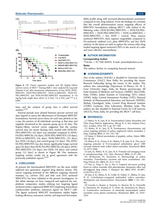
Antitumor activity of the different MWCNT formulations
was evaluated in tumor bearing Balb/c model after single iv
administration (5 mg/kg body weight). The DOX/ES-PEG-
MWCNT, DOX/FA-PEG-MWCNT, DOX/PEG-MWCNT,
DOX/ox-MWCNT, and free DOX formulations significantly
suppressed tumor growth compared to control group.
However, DOX/ES-PEG-MWCNT and DOX/FA-PEG-
MWCNT formulations showed higher tumor growth suppres-
sion than other formulations and free drug solution (Figure 9
A). The high antitumor activity of the FA- and ES-PEG-
conjugated MWCNTs can be attributed to its higher
accumulation in cancer cells through receptor-mediated uptake.
These antitumor activity results indicated that the PEGylation
of CNTs increases the overall tumor targeting efficiency of the
drug loaded ligand anchored MWCNT formulations.
The Kaplan−Meier survival curve analysis was used to
measure the fraction of the treated mice that survived over a
period of time. It is the simplest way of computing the survival
over time. The time starting from a defined point to the
occurrence of a given event, for example death, is called survival
Figure 8. Biodistribution patterns of free DOX, DOX/MWCNT, DOX/ox-MWCNT, DOX/PEG-MWCNT, DOX/ES-MWCNT, and DOX/FA-
PEG-MWCNT formulations in different tissues. Values represented as mean ± SD (n = 3).
Molecular Pharmaceutics Article
DOI: 10.1021/mp500720a
Mol. Pharmaceutics 2015, 12, 630−643
640
time, and the analysis of group data is called survival
analysis.2,4,42
Survival periods were plotted between percent survival and
days elapsed to assess the effectiveness of developed MWCNT
formulations. Survival curves show, for each time plotted on the
x-axis, the portion of all individuals surviving at that time and
regularly monitored in the separate group up to 45 days. The
Kaplan−Meier survival curves suggested that the median
survival time for tumor bearing mice treated with DOX/ES-
PEG-MWCNTs (43 days) was extended compared to DOX/
FA-PEG-MWCNs (42 days), DOX/PEG-MWCNTs (33 days),
DOX/ox-MWCNTs (24 days), DOX/MWCNTs (23 days),
free DOX (18 days), and control group (12 days). The DOX/
ES-PEG-MWCNTs has also shown significantly longer survival
span (43 days) than DOX/FA-PEG-MWCNs (42 days), DOX/
PEG-MWCNTs (33 days), free DOX (18 days), and control
group (12 days) as shown in Figure 9B. Our antitumor
targeting activity results are in good agreement with the
previously published reports.2,4,42
4. CONCLUSION
At present the functionalized MWCNTs are the most widely
explored new generation targeted drug delivery system. The
cancer targeting potential of the different targeting chemical
moieties, i.e., estrone (ES) and folic acid (FA) anchored
MWCNTs, has been validated in the present investigation. In
this debut attempt, we have compared and explored the one-
platform comparative studies utilizing two targeting ligand
anchored surface engineered MWCNTs employing doxorubicin
(anthracycline antibiotic; anticancer agent) on MCF-7 cells.
The ligand anchored MWCNT formulations exhibit higher
loading efficiency, anticancer activity, and improved biocompat-
ibility profile along with increased pharmacokinetic parameters
compared to free drug solution. From the findings we conclude
that the overall pharmaceutical cancer targeting efficacy of
MWCNTs formulations utilizing MCF-7 cells ranked in the
following order: DOX/ES-PEG-MWCNTs > DOX/FA-PEG-
MWCNTs > DOX/PEG-MWCNTs > DOX/ox-MWCNTs >
DOX/MWCNTs > free DOX > control. Thus, estrone
anchored MWCNTs show superior targetability compared to
FA-anchored, oxidized (ox), plain MWCNTs, and free DOX
solution. In the future we wish to explore the various other drug
loaded targeting ligand anchored CNTs in the search of a safer
and more effective nanomedicine.
■ AUTHOR INFORMATION
Corresponding Author
*Tel/fax: + 91-7582-265055. E-mail: jnarendr@yahoo.co.in.
Notes
The authors declare no competing financial interest.
■ ACKNOWLEDGMENTS
One of the authors (N.K.M.) is thankful to University Grants
Commission (UGC), New Delhi, for providing the Senior
Research Fellowship during the tenure of the studies. The
authors also acknowledge Department of Physics, Dr. H. S.
Gour University, Sagar, India, for Raman spectroscopy; All
India Institute of Medicine and Sciences (AIIMS), New Delhi,
India (TEM); Indian Institute of Technology (IIT), Indore
(AFM), India; Central Instruments Facilities (CIF), National
Institute of Pharmaceutical Education and Research (NIPER),
Mohali, Chandigarh, India; Central Drug Research Institute
(CDRI), Lucknow; Diya Laboratory, Mumbai, India. The
authors are also thankful to National Centre for Cell Sciences
(NCCS), Pune, India, for providing the MCF-7 cell lines.
■ REFERENCES
(1) Mehra, N. K.; Jain, N. K. Functionalized Carbon Nanotubes and
Their Drug Delivery Applications; Bhoop, B. S., Ed.; Studium Press,
LLC: London, 2014; Vol. 4, pp 327−369.
(2) Mehra, N. K.; Jain, N. K. Development, characterization and
cancer targeting potential of surface engineered carbon nanotubes. J.
Drug Targeting 2013, 21 (8), 745−758.
(3) Iijima, S. Helical microtubules of graphitic carbon. Nature 1991,
354, 56−58.
(4) Mehra, N. K.; Verma, A. K.; Mishra, P. R.; Jain, N. K. The cancer
targeting potential of D-α-tocopheryl polyethylene glycol 1000
succinate tethered multi walled carbon nanotubes. Biomaterials 2014,
35, 4573−4588.
(5) Jain, K.; Mehra, N. K.; Jain, N. K. Potential and emerging trends
in nanopharmacology. Curr. Opin. Pharmacol. 2014, 15, 97−106.
(6) Ali-Boucetta, H.; Kostarelos, K. Pharmacology of carbon
nanotubes: Toxicokinetics, excretion and tissue accumulation. Adv.
Drug Delivery Rev. 2013, 65, 2111−2119.
(7) Fan, J.; Zeng, F.; Xu, J.; Wu, S. Targeted anti-cancer prodrug
based on carbon nanotube with photodynamic therapeutic effect and
pH-triggered drug release. J. Nanopart. Res. 2013, DOI: 10.1007/
s11051-013-1911-z.
(8) Jain, A. K.; Mehra, N. K.; Lodhi, N.; Dubey, V.; Mishra, D.; Jain,
N. K. Carbohydrate-conjugated multiwalled carbon nanotubes:
development and characterization. Nanomedicine 2009, 5 (4), 432−
442.
(9) Lacerda, L.; Russier, J.; Pastorin, G.; Herrero, M. A.; Venturelli,
E.; Dumortier, H.; Al-Jamal, K. T.; Prato, M.; Kostarelos, K.; Bianco,
A. Translocation mechanisms of chemically functionalized carbon
nanotubes across plasma membranes. Biomaterials 2012, 33, 3334−
3343.
Figure 9. (A) Tumor regression analysis and (B) Kaplan−Meier
survival curves of MCF-7 bearing Balb/c mice analyzed by Log-rank
(Mantel−Cox) after intravenous administration of free DOX, DOX/
MWCNT, DOX/PEG-MWCNT, DOX/ox-MWCNT, DOX/FA-
PEG-MWCNT, and DOX/ES-PEG-MWCNT formulations at 5.0
mg/kg body weight dose, where *p ≤ 0.05, **p ≤ 0.01, and *** p ≤
0.001.
Molecular Pharmaceutics Article
DOI: 10.1021/mp500720a
Mol. Pharmaceutics 2015, 12, 630−643
641
(10) Niu, L.; Meng, L.; Lu, Q. Folate-conjugated PEG on single-
walled carbon nanotubes for targeting delivery of doxorubicin to
cancer cells. Macromol. Biosci. 2013, 13, 735−744.
(11) Fabbro, C.; Ali-Boucetta, H.; Ros, T. D.; Kostarelos, K.; Bianco,
A.; Prato, M. Targeting carbon nanotubes against cancer. Chem.
Coummun. 2012, 48, 3911−3926.
(12) Mehra, N. K.; Mishra, V.; Jain, N. K. A review of ligand tethered
surface engineered carbon nanotubes. Biomaterials 2014, 35 (4),
1267−1283.
(13) Liu, Z.; Sun, X.; Nakayama-Ratchford, N.; Dai, H. Supra-
molecular chemistry on water soluble carbon nanotubes for drug
loading and delivery. ACS Nano 2007, 1 (1), 50−56.
(14) Zhang, X.; Meng, L.; Lu, Q.; Fei, Z.; Dyson, P. J. Targeted
delivery and controlled release of doxorubicin to cancer cells using
modified single wall carbon nanotubes. Biomaterials 2009, 30, 6041−
6047.
(15) Huang, H.; Yuan, Q.; Shah, J. S.; Misra, R. D. K. A new family of
folate-decorated and carbon nanotubes-mediated drug delivery system:
synthesis and drug delivery response. Adv. Drug Delivery Rev. 2011, 63,
1332−1339.
(16) Liu, Z.; Chen, K.; Davis, C.; Sherlock, S.; Cao, Q.; Chen, X.;
Dai, H. Drug delivery with carbon nanotubes for in vivo cancer
treatment. Cancer Res. 2008, 68 (16), 6652−6660.
(17) Mody, N.; Tekade, R. K.; Mehra, N. K.; Chopdey, P.; Jain, N. K.
Dendrimer, liposomes, carbon nanotubes and PLGA nanoparticles:
one platform assessment of drug delivery potential. AAPS
PharmSciTechn 2014, 15 (2), 388−399.
(18) Singh, R.; Mehra, N. K.; Jain, V.; Jain, N. K. Folic acid
conjugated carbon nanotubes for gemcitabine HCL delivery. J. Drug
Targeting 2013, 21 (6), 581−592.
(19) Wu, W.; Wieckowski, S.; Pastorin, G.; Benincasa, M.; Klumpp,
C.; Briand, J. P. Targeted delivery of Amphotericin B to cells by using
functionalized carbon nanotubes. Angew. Chem., Int. Ed. 2005, 44,
6358−6362.
(20) Bhatnagar, I.; Venkatesan, J.; Kim, S.-K. Polymer functionalized
single walled carbon nanotubes mediated drug delivery of Gliotoxin in
cancer cells. J. Biomed. Nanotechnol. 2014, 10 (1), 120−130.
(21) Gupta, R.; Mehra, N. K.; Jain, N. K. Fucosylated multiwalled
carbon nanotubes for Kupffer cells targeting for the treatment of
cytokine-induced liver damage. Pharm. Res. 2013, 31 (2), 322−334.
(22) Wu, L.; Man, C.; Wang, H.; Lu, X.; Ma, Q.; Cai, Y.; Ma, W.
PEGylated multi-walled carbon nanotubes for encapsulation and
sustained release of oxaliplatin. Pharm. Res. 2013, 30, 412−423.
(23) Paliwal, S. R.; Paliwal, R.; Pal, H. C.; Saxena, A. K.; Sharma, P.
R.; Gupta, P. N.; Agrawal, G. P.; Vyas, S. P. Estrogen-anchored pH-
sensitive liposomes as nanomodule designed for site-specific delivery
of doxorubicin in breast cancer therapy. Mol. Pharmaceutics 2012, 9
(1), 176−186.
(24) Lodhi, N.; Mehra, N. K.; Jain, N. K. Development and
characterization of dexamethasone mesylate anchored on multi walled
carbon nanotubes. J. Drug Targeting 2013, 21 (1), 67−76.
(25) Lu, Y. J.; Wei, K. C.; Ma, C. C. M.; Yang, S. Y.; Chen, J. P. Dual
targeted delivery of doxorubicin to cancer cells using folate-conjugated
magnetic multi-walled carbon nanotubes. Colloids Surf., B 2012, 89, 1−
9.
(26) Mehra, N. K.; Mishra, V.; Jain, N. K. Receptor based therapeutic
targeting. Ther. Delivery 2013, 4 (3), 369−394.
(27) Das, M.; Singh, R. P.; Datir, S. R.; Jain, S. Intracellular drug
delivery and effective in vivo cancer therapy via estradiol-PEG-
appended multi walled carbon nanotubes. Mol. Pharmaceutics 2013, 10
(9), 3404−3416.
(28) Das, M.; Singh, R. P.; Datir, S. R.; Jain, S. Surface chemistry
dependent “switch” regulated the trafficking and therapeutic perform-
ance of drug-loaded carbon nanotubes. Bioconjugate Chem. 2013, 24,
626−639.
(29) Zalipsky, S.; Gilon, C.; Zalkha, A. Attachment of drugs to
polyethylene glycols. Eur. Polym. J. 1983, 19, 1177−1183.
(30) Rai, S.; Paliwal, R.; Vyas, S. P. Targeted delivery of doxorubicin
via estrone-appended liposomes. J. Drug Targeting 2008, 16 (6), 455−
463.
(31) Lee, R. J.; Low, P. S. Folate-mediated tumor cell targeting of
liposome entrapped doxorubicin in vitro. Biochim. Biophys. Acta 1995,
1233, 134−144.
(32) Kesharwani, P.; Tekade, R. K.; Jain, N. K. Formulation
development and in vitro−in vivo assessment of the fourth-generation
PPI dendrimer as a cancer-targeting vector. Nanomedicine 2014, 9
(15), 2291−2308 DOI: 10.2217/nnm.13.210.
(33) Lee, P. C.; Chiou, Y. C.; Wong, J. M.; Peng, C. L.; Shieh, M. J.
Targeting colorectal cancer cells with single-walled carbon nanotubes
conjugated to anticancer agent SN-38 and EGFR antibody.
Biomaterials 2013, 34, 8756−8765.
(34) Nima, Z. A.; Mahmood, M. W.; Karmakar, A.; Mustafa, T.;
Bourdo, S.; Xu, Y.; Biris, A. S. Single-walled carbon nanotubes as
specific targeting and Raman spectroscopic agents for detection and
discrimination of single human breast cancer cells. J. Biomed. Opt.
2013, 18 (5), 55003.
(35) Sacchetti, C.; Rapini, N.; Magrini, A.; Cirelli, E.; Bellucci, S.;
Matteri, M.; Rosato, N.; Bottini, N.; Bottini, M. In vivo targeting of
intracellular regulatory T cell using PEG-modified single-walled carbon
nanotubes. Bioconjugate Chem. 2013, 24, 852−858.
(36) Delogu, L. G.; Venturelli, E.; Manetti, R.; Pinna, G. A.; Carru,
C.; Madeddu, R.; Murgia, L.; Sgarrella, F.; Dumortier, H.; Bianco, A.
Ex vivo impact of functionalized carbon nanotubes on human immune
cells. Nanomedicine 2012, 7 (2), 231−243.
(37) Chen, J.; Li, S.; Shen, Q. Folic acid and cell-penetrating peptide
conjugated PLGA-PEG bifunctional nanoparticles for vincristine
sulfate delivery. Eur. J. Pharm. Sci. 2012, 47, 430−443.
(38) Han, Y.; Xu, J.; Li, Z.; Ren, G.; Yang, Z. In vitro toxicity of
multi-walled carbon nanotubes in C6 rat glioma cells. Neurotoxicology
2012, 33, 1128−1134.
(39) Kundu, P.; Mohanty, C.; Sahoo, S. K. Anti-glioma activity of
curcumin-loaded lipid nanoparticles and its enhanced bioavailability in
brain tissue for effective glioblastoma therapy. Acta Biomater. 2012, 8,
2670−2687.
(40) Mulvey, J. J.; Villa, C. H.; MCDevitt, M. R.; Escorcia, F. E.;
Casey, E.; Scheinberg, D. A. Self-assembly of carbon nanotubes and
antibodies on tumours for targeted amplified delivery. Nat. Nano-
technol. 2013, 8, 763−771.
(41) Lin, Z.; Xi, Z.; Huang, J. Biodistribution of single-walled carbon
nanotubes in rats. Toxicol. Res. 2014, DOI: 10.1039/C3TX50059D.
(42) Ren, J.; Shen, S.; Wang, D.; Xi, Z.; Guo, L.; Pang, Z.; Qian, Y.;
Sun, X.; Jiang, X. The targeted delivery of anticancer drugs to brain
glioma by PEGylated oxidized multi-walled carbon nanotubes
modified with angiopep-2. Biomaterials 2012, 33, 3324−3333.
(43) Ji, Z.; Lin, G.; Lu, Q.; Meng, L.; Shen, X.; Dong, L.; Fu, C.;
Zhang, X. Targeted therapy of SMMC-7721 liver cancer in vitro and in
vivo with carbon nanotubes based drug delivery systems. J. Colloid
Interface Sci. 2012, 365, 143−149.
(44) Muscella, A.; Vertugno, C.; Migoni, D.; Biagioni, F.; Fanizzi, F.
P.; Fornai, F.; De Pascali, S. A.; Marsigliante, S. Antitumor activity of
[Pt (O,O′-acac) (γ-acac) (DMS) in mouse xenograft model of breast
cancer. Cell Death Dis. 2014, 5, e1014.
(45) Cherukuri, P.; Gannon, C. J.; Leeuw, T. K.; Schmidt, H. K.;
Smalley, R. E.; Curley, S. A.; Weisman, R. B. Mammlian
pharmacokinetics of carbon nanotubes using intrinsic near-infrared
fluorescence. Proc. Natl. Acad. Sci. U.S.A. 2006, 103 (50), 18882−
18886.
(46) Al-Jamal, K. T.; Nunes, A.; Methven, L.; Ali-Boucetta, H.; Li, S.;
Toma, F. M.; Herrero, M. A.; Al-Jamal, W. T.; Eikelder, H. M. M.;
Foster, J.; Mather, S.; Prato, M.; Bianco, A.; Kostarelos, K. Degree of
chemical functionalization of carbon nanotubes determines tissue
distribution and excretion profile. Angew. Chem., Int. Ed. 2012, 51,
6389−6393.
(47) Bhirde, A.; Patel, V.; Gavard, J.; Zhang, G.; Sousa, A. A.;
Masedunskas, A.; Leapman, R. D.; Weigert, R.; Gutkind, J. S.; Rusling,
J. F. Targeted killing of cancer cells in vivo and in vitro with EGF-
Molecular Pharmaceutics Article
DOI: 10.1021/mp500720a
Mol. Pharmaceutics 2015, 12, 630−643
642
directed carbon nanotubes-based drug delivery. ACS Nano 2009, 3
(2), 307−316.
(48) Campagnolo, L.; Massimiani, M.; Palmieri, G.; Bernardini, R.;
Sacchetti, C.; Bergamaschi, A. Biodistribution and toxicity of pegylated
single wall carbon nanotubes in pregnant mice. Part. Fibre Toxicol.
2013, DOI: 10.1186/1743-8977-10-21.
(49) Jain, S.; Thakare, V. S.; Das, M.; Godugu, C.; Jain, A. K.;
Mathur, R.; Chuttani, K.; Mishra, A. K. Toxicity of multi walled carbon
nanotubes with end defects critically depends on their functionaliza-
tion density. Chem. Res. Toxicol. 2011, 24 (11), 2028−2040.
(50) Nunez, R. DNA measurement and cell cycle analysis by flow
cytometry. Curr. Issues Mol. Biol. 2001, 3 (3), 67−70.
(51) Dinan, N. M.; Atyabi, F.; Rouini, M. R.; Amini, M.; Golbchifar,
A. K.; Dinarvand, R. Doxorubicin loaded folate-targeted carbon
nanotubes: preparation, cellular internalization, in vitro cytotoxicity
and disposition kinetic study in the isolated perfused rat liver. Mater.
Sci. Eng. C 2014, 39, 47−55.
Molecular Pharmaceutics Article
DOI: 10.1021/mp500720a
Mol. Pharmaceutics 2015, 12, 630−643
643

More Related Content

What's hot

Drug target & delivery system
Drug target & delivery systemDrug target & delivery system
Drug target & delivery system
KING GEORGE MEDICAL UNIVERSITY
 
Drug targeting
Drug targetingDrug targeting
Drug targeting
Anvita Bharati
 
Polymers controlled delivery of therapeutic nucleic acid
Polymers controlled delivery of therapeutic nucleic acidPolymers controlled delivery of therapeutic nucleic acid
Polymers controlled delivery of therapeutic nucleic acid
Sivasangari Shanmugam
 
NANOPARTICULATE DRUG DELIVERY SYSTEM
NANOPARTICULATE DRUG DELIVERY SYSTEMNANOPARTICULATE DRUG DELIVERY SYSTEM
NANOPARTICULATE DRUG DELIVERY SYSTEM
Sagar Savale
 
Nano drug delivery
Nano drug deliveryNano drug delivery
Nano drug delivery
tabirsir
 
Monoclonal Antibodies
Monoclonal Antibodies Monoclonal Antibodies
Monoclonal Antibodies
Sarvan Mani
 
Drug targeting (Pravin Patil)
Drug targeting (Pravin Patil)Drug targeting (Pravin Patil)
Drug targeting (Pravin Patil)
Pravin176
 
Nanoparticles
NanoparticlesNanoparticles
Nanoparticles
Deepali Pandey
 
Nanoparticle
NanoparticleNanoparticle
Nanoparticle
biniyapatel
 
Nanoparticles targetted drug delivery system
Nanoparticles targetted drug delivery systemNanoparticles targetted drug delivery system
Nanoparticles targetted drug delivery system
shashankc10
 
Nano chemistry presentation (1)
Nano chemistry presentation (1)Nano chemistry presentation (1)
Nano chemistry presentation (1)
SapanaSinha1
 
targeted dds for cancer
targeted dds for cancertargeted dds for cancer
targeted dds for cancer
Neha Singh
 
Nano Drug Delivery Approaches and Importance of Quality by Design (QbD)
Nano Drug Delivery Approaches and Importance of Quality by Design (QbD)Nano Drug Delivery Approaches and Importance of Quality by Design (QbD)
Nano Drug Delivery Approaches and Importance of Quality by Design (QbD)
SABYA SACHI DAS
 
final ppt nano with corrections
final ppt nano with correctionsfinal ppt nano with corrections
final ppt nano with corrections
Aniket Shishupal
 
Nanomaterial drug delivery updated 20202
Nanomaterial drug delivery   updated 20202Nanomaterial drug delivery   updated 20202
Nanomaterial drug delivery updated 20202
Tejas Pitale
 
Drug delivery nanocarriers
Drug delivery nanocarriersDrug delivery nanocarriers
Drug delivery nanocarriers
Mohamed Ali
 
Nano Particles for Drug Delivery
Nano Particles for Drug DeliveryNano Particles for Drug Delivery
Nano Particles for Drug Delivery
Lendl Monterola
 
NANO TECHNOLOGY IN DRUG DELIVERY SYSTEM
NANO TECHNOLOGY IN DRUG DELIVERY SYSTEMNANO TECHNOLOGY IN DRUG DELIVERY SYSTEM
NANO TECHNOLOGY IN DRUG DELIVERY SYSTEM
sathish sak
 
Application of nanoparticals in drug delivery system
Application of nanoparticals in drug delivery systemApplication of nanoparticals in drug delivery system
Application of nanoparticals in drug delivery system
Malay Jivani
 
Targteted Drug Delivery - Technologies and Applications
Targteted Drug Delivery - Technologies and ApplicationsTargteted Drug Delivery - Technologies and Applications
Targteted Drug Delivery - Technologies and Applications
Aiswariya Chidambaram
 

What's hot (20)

Drug target & delivery system
Drug target & delivery systemDrug target & delivery system
Drug target & delivery system
 
Drug targeting
Drug targetingDrug targeting
Drug targeting
 
Polymers controlled delivery of therapeutic nucleic acid
Polymers controlled delivery of therapeutic nucleic acidPolymers controlled delivery of therapeutic nucleic acid
Polymers controlled delivery of therapeutic nucleic acid
 
NANOPARTICULATE DRUG DELIVERY SYSTEM
NANOPARTICULATE DRUG DELIVERY SYSTEMNANOPARTICULATE DRUG DELIVERY SYSTEM
NANOPARTICULATE DRUG DELIVERY SYSTEM
 
Nano drug delivery
Nano drug deliveryNano drug delivery
Nano drug delivery
 
Monoclonal Antibodies
Monoclonal Antibodies Monoclonal Antibodies
Monoclonal Antibodies
 
Drug targeting (Pravin Patil)
Drug targeting (Pravin Patil)Drug targeting (Pravin Patil)
Drug targeting (Pravin Patil)
 
Nanoparticles
NanoparticlesNanoparticles
Nanoparticles
 
Nanoparticle
NanoparticleNanoparticle
Nanoparticle
 
Nanoparticles targetted drug delivery system
Nanoparticles targetted drug delivery systemNanoparticles targetted drug delivery system
Nanoparticles targetted drug delivery system
 
Nano chemistry presentation (1)
Nano chemistry presentation (1)Nano chemistry presentation (1)
Nano chemistry presentation (1)
 
targeted dds for cancer
targeted dds for cancertargeted dds for cancer
targeted dds for cancer
 
Nano Drug Delivery Approaches and Importance of Quality by Design (QbD)
Nano Drug Delivery Approaches and Importance of Quality by Design (QbD)Nano Drug Delivery Approaches and Importance of Quality by Design (QbD)
Nano Drug Delivery Approaches and Importance of Quality by Design (QbD)
 
final ppt nano with corrections
final ppt nano with correctionsfinal ppt nano with corrections
final ppt nano with corrections
 
Nanomaterial drug delivery updated 20202
Nanomaterial drug delivery   updated 20202Nanomaterial drug delivery   updated 20202
Nanomaterial drug delivery updated 20202
 
Drug delivery nanocarriers
Drug delivery nanocarriersDrug delivery nanocarriers
Drug delivery nanocarriers
 
Nano Particles for Drug Delivery
Nano Particles for Drug DeliveryNano Particles for Drug Delivery
Nano Particles for Drug Delivery
 
NANO TECHNOLOGY IN DRUG DELIVERY SYSTEM
NANO TECHNOLOGY IN DRUG DELIVERY SYSTEMNANO TECHNOLOGY IN DRUG DELIVERY SYSTEM
NANO TECHNOLOGY IN DRUG DELIVERY SYSTEM
 
Application of nanoparticals in drug delivery system
Application of nanoparticals in drug delivery systemApplication of nanoparticals in drug delivery system
Application of nanoparticals in drug delivery system
 
Targteted Drug Delivery - Technologies and Applications
Targteted Drug Delivery - Technologies and ApplicationsTargteted Drug Delivery - Technologies and Applications
Targteted Drug Delivery - Technologies and Applications
 

Similar to 20 Molecular Pharmaceutics

Synthesis and Evaluation of the Cytotoxic and Anti-Proliferative Properties o...
Synthesis and Evaluation of the Cytotoxic and Anti-Proliferative Properties o...Synthesis and Evaluation of the Cytotoxic and Anti-Proliferative Properties o...
Synthesis and Evaluation of the Cytotoxic and Anti-Proliferative Properties o...
IJEAB
 
44 D DT Mehra Palakurthi
44 D DT Mehra Palakurthi44 D DT Mehra Palakurthi
44 D DT Mehra Palakurthi
Dr. Neelesh Mehra
 
44 D DT Mehra Palakurthi
44 D DT Mehra Palakurthi44 D DT Mehra Palakurthi
44 D DT Mehra Palakurthi
Dr. Neelesh Mehra
 
Summer Scholars Research Symposium
Summer Scholars Research SymposiumSummer Scholars Research Symposium
Summer Scholars Research Symposium
Elim Na
 
Formulation and in vitro evaluation of quercetin loaded carbon nanotubes for ...
Formulation and in vitro evaluation of quercetin loaded carbon nanotubes for ...Formulation and in vitro evaluation of quercetin loaded carbon nanotubes for ...
Formulation and in vitro evaluation of quercetin loaded carbon nanotubes for ...
IRJET Journal
 
Experimental and theoretical solubility advantage screening of bi-component s...
Experimental and theoretical solubility advantage screening of bi-component s...Experimental and theoretical solubility advantage screening of bi-component s...
Experimental and theoretical solubility advantage screening of bi-component s...
Maciej Przybyłek
 
Development and characterization of anionic liposaccharides for enhanced oral
Development and characterization of anionic liposaccharides for enhanced oralDevelopment and characterization of anionic liposaccharides for enhanced oral
Development and characterization of anionic liposaccharides for enhanced oral
Adel Abdelrahim, PhD
 
Polymeric Micelles.pptx
Polymeric Micelles.pptxPolymeric Micelles.pptx
Polymeric Micelles.pptx
MdSufiyan10
 
Impact of carbon nanomaterials on the formation of multicellular spheroids by...
Impact of carbon nanomaterials on the formation of multicellular spheroids by...Impact of carbon nanomaterials on the formation of multicellular spheroids by...
Impact of carbon nanomaterials on the formation of multicellular spheroids by...
Татьяна Гергелюк
 
Impact of carbon nanomaterials on the formation of multicellular spheroids by...
Impact of carbon nanomaterials on the formation of multicellular spheroids by...Impact of carbon nanomaterials on the formation of multicellular spheroids by...
Impact of carbon nanomaterials on the formation of multicellular spheroids by...
Татьяна Гергелюк
 
IAPST 2010
IAPST 2010IAPST 2010
IAPST 2010
SAIKATGHOSH58
 
curcumin nanoparticles_1599661643.pptx
curcumin nanoparticles_1599661643.pptxcurcumin nanoparticles_1599661643.pptx
curcumin nanoparticles_1599661643.pptx
AbdulhussienAljebory1
 
824746
824746824746
Nanoparticles in lung cancer treatment and diagnosis.
Nanoparticles in lung cancer treatment and diagnosis.Nanoparticles in lung cancer treatment and diagnosis.
Nanoparticles in lung cancer treatment and diagnosis.
mohamedAhmed1628
 
Biomedical ppt
Biomedical pptBiomedical ppt
Biomedical ppt
preet yadav
 
biomedicines-04-00022
biomedicines-04-00022biomedicines-04-00022
biomedicines-04-00022
Christian Schmidt
 
Engineering cells
Engineering cellsEngineering cells
Engineering cells
Oscar1Miranda2
 
Engineering cells
Engineering cellsEngineering cells
Engineering cells
Oscar1Miranda2
 
Engineering cells
Engineering cellsEngineering cells
Engineering cells
Oscar1Miranda2
 
Engineering cells
Engineering cellsEngineering cells
Engineering cells
Oscar1Miranda2
 

Similar to 20 Molecular Pharmaceutics (20)

Synthesis and Evaluation of the Cytotoxic and Anti-Proliferative Properties o...
Synthesis and Evaluation of the Cytotoxic and Anti-Proliferative Properties o...Synthesis and Evaluation of the Cytotoxic and Anti-Proliferative Properties o...
Synthesis and Evaluation of the Cytotoxic and Anti-Proliferative Properties o...
 
44 D DT Mehra Palakurthi
44 D DT Mehra Palakurthi44 D DT Mehra Palakurthi
44 D DT Mehra Palakurthi
 
44 D DT Mehra Palakurthi
44 D DT Mehra Palakurthi44 D DT Mehra Palakurthi
44 D DT Mehra Palakurthi
 
Summer Scholars Research Symposium
Summer Scholars Research SymposiumSummer Scholars Research Symposium
Summer Scholars Research Symposium
 
Formulation and in vitro evaluation of quercetin loaded carbon nanotubes for ...
Formulation and in vitro evaluation of quercetin loaded carbon nanotubes for ...Formulation and in vitro evaluation of quercetin loaded carbon nanotubes for ...
Formulation and in vitro evaluation of quercetin loaded carbon nanotubes for ...
 
Experimental and theoretical solubility advantage screening of bi-component s...
Experimental and theoretical solubility advantage screening of bi-component s...Experimental and theoretical solubility advantage screening of bi-component s...
Experimental and theoretical solubility advantage screening of bi-component s...
 
Development and characterization of anionic liposaccharides for enhanced oral
Development and characterization of anionic liposaccharides for enhanced oralDevelopment and characterization of anionic liposaccharides for enhanced oral
Development and characterization of anionic liposaccharides for enhanced oral
 
Polymeric Micelles.pptx
Polymeric Micelles.pptxPolymeric Micelles.pptx
Polymeric Micelles.pptx
 
Impact of carbon nanomaterials on the formation of multicellular spheroids by...
Impact of carbon nanomaterials on the formation of multicellular spheroids by...Impact of carbon nanomaterials on the formation of multicellular spheroids by...
Impact of carbon nanomaterials on the formation of multicellular spheroids by...
 
Impact of carbon nanomaterials on the formation of multicellular spheroids by...
Impact of carbon nanomaterials on the formation of multicellular spheroids by...Impact of carbon nanomaterials on the formation of multicellular spheroids by...
Impact of carbon nanomaterials on the formation of multicellular spheroids by...
 
IAPST 2010
IAPST 2010IAPST 2010
IAPST 2010
 
curcumin nanoparticles_1599661643.pptx
curcumin nanoparticles_1599661643.pptxcurcumin nanoparticles_1599661643.pptx
curcumin nanoparticles_1599661643.pptx
 
824746
824746824746
824746
 
Nanoparticles in lung cancer treatment and diagnosis.
Nanoparticles in lung cancer treatment and diagnosis.Nanoparticles in lung cancer treatment and diagnosis.
Nanoparticles in lung cancer treatment and diagnosis.
 
Biomedical ppt
Biomedical pptBiomedical ppt
Biomedical ppt
 
biomedicines-04-00022
biomedicines-04-00022biomedicines-04-00022
biomedicines-04-00022
 
Engineering cells
Engineering cellsEngineering cells
Engineering cells
 
Engineering cells
Engineering cellsEngineering cells
Engineering cells
 
Engineering cells
Engineering cellsEngineering cells
Engineering cells
 
Engineering cells
Engineering cellsEngineering cells
Engineering cells
 

20 Molecular Pharmaceutics

  • 1. One Platform Comparison of Estrone and Folic Acid Anchored Surface Engineered MWCNTs for Doxorubicin Delivery Neelesh Kumar Mehra†,‡ and N. K. Jain*,†,‡ † Pharmaceutics Research Laboratory, Department of Pharmaceutical Sciences, Dr. H. S. Gour University, Sagar 470 003, India ‡ Pharmaceutical Nanotechnology Research Laboratory, ISF College of Pharmacy, Moga 142 001, India ABSTRACT: Our main aim in the present investigation was to assess and compare the in vitro and in vivo cancer targeting propensity of doxorubicin (DOX) loaded folic acid (FA) and estrone (ES) anchored PEGylated multiwalled carbon nanotubes (MWCNTs) employing tumor bearing Balb/c mice. The DOX was loaded into the developed functionalized MWCNTs after proper characterization using dialysis diffusion method. The in vitro, ex vivo, and in vivo studies were performed on the MCF-7 cell line for assessment of the cancer targeting propensity. Both qualitative and quantitative cell uptake studies indicated the preferential higher uptake of estrone anchored nanotube formulation compared to other formulations and free DOX owing to the overexpression of estrogen receptors (ERs) on human breast MCF-7 cells. Similarly, the pharmacokinetic and increased antitumor activities also confirmed the elevated cancer targeting propensity of the estrone and folic acid anchored MWCNT formulations. The DOX/ES-PEG-MWCNTs has also shown significantly longer survival span (43 days) than free DOX (18 days) and control group (12 days). Present outcomes from the ex vivo and in vivo studies are deemed to be of great scientific value and shall assist targeted drug delivery formulation scientists for selection of the targeting moieties in the treatment of human breast cancer. KEYWORDS: carbon nanotubes, drug targeting, doxorubicin, pharmacokinetic, breast cancer, estrone, folic acid, antitumor activity 1. INTRODUCTION On recalling the “Magic bullet concept” tremendous sophisti- cation has been visualized in targeting concepts and delivery of bioactives. In the present scenario, carbon nanotubes (CNTs) have attracted escalating attention after attachment of huge targeting moieties to offer sustained/controlled release behavior with cellular targeting and enhanced specificity.1,2 CNTs were first discovered and fully explored in 1991.3 CNTs are unique, three-dimensional sp2 hybridized carbon nanomaterials that have attracted tremendous attention as a valuable, promising, safe and effective alternative nanoarchitec- ture for biomedical applications due to their unique physicochemical properties such as biocompatibility, non- immunogenicity, high loading efficiency, high aspect ratio, structural flexibility, and noncytotoxic nature.2−9 Functionalized CNTs (f-CNTs) have the capacity to readily cross the plasma membrane via an energy-dependent, endosomally mediated and direct translocation, which is known as tiny nanoneedle mechanism.9,10 Due to the tiny nanoneedle tubular shape, they pass through cell membrane without causing cell death, thus allowing efficient tissue and tumor specific drug delivery with minimal or no toxic effects. Compared with other nanocarrier systems, which have low entrapment efficiency due to steric hindrance, the f-CNTs have greater entrapment due to the availability of high surface area.4,11,12 Despite excellent progress in using surface engineered CNTs as targeted drug delivery carriers, more research is needed to further optimize their ability to selectively accumulate in diseased/targeted tissues and to release their toxic payload in a controlled manner. To date, numerous research reports are available considering functionalized CNTs for drug delivery including doxorubicin hydrochloride,13−15 paclitaxel,16 docetax- el,17 gemcitabine,18 amphotericin B,19 gliotoxin,20 sulfasala- zine,21 oxaliplatin,22 etc. The anthracycline antibiotic, doxorubicin hydrochloride, chosen for the present investigation as an anticancer agent is widely used in the treatment of cancers of the breast, prostate, brain, and cervix and Hodgkin lymphoma. Short half-life and severe toxicity (acute cardiotoxicity) associated with doxor- ubicin restrict its clinical application. According to the Biopharmaceutical Classification System (BCS) doxorubicin hydrochloride belongs to class III, i.e., high solubility and low permeability (BCS III). BCS is a scientific framework for classifying substances according to their aqueous solubility and permeability.23−25 Very recently our laboratory explored the delivery of DOX employing D-α-tocopheryl polyethylene glycol 1000 succinate (TPGS) as targeting ligand with improved therapeutic outcomes and antitumor targeting efficacy on the MCF-7 cell line.4 The purpose of the present investigation was to compare the cancer-targeting propensity of the folic acid (FA) and estrone (ES) appended surface functionalized Received: October 27, 2014 Revised: December 13, 2014 Accepted: December 17, 2014 Published: December 17, 2014 Article pubs.acs.org/molecularpharmaceutics © 2014 American Chemical Society 630 DOI: 10.1021/mp500720a Mol. Pharmaceutics 2015, 12, 630−643
  • 2. MWCNTs employing breast cancer cells on Balb/c mice. The cancerous cells overexpress the folate receptors (FRs) and estrogen receptors (ERs) belonging to folate and nuclear hormone receptor superfamily, respectively.26 The MWCNTs tethering ES and FA probably deliver DOX more efficiently into the cancerous cells after binding selectively to their respective receptors through endocytosis or tiny nanoneedle mechanism. The ligand-driven MWCNTs are an established, well-known concept, and these targeting moieties can be easily attached to f-MWCNTs. The comparative cancer targeting propensity of targeting ligand conjugated f-MWCNTs studied on a single platform reported herein is expected to be of greater scientific interest in targeted drug delivery. We have extensively characterized the ligand (ES and FA) conjugated MWCNTs in vitro, ex vivo, and in vivo in terms of biodistribution and pharmacokinetics, Kaplan−Meier survival analysis, and tumor targeting efficacy on Balb/c mice. 2. MATERIALS AND METHODS 2.1. Materials. Multiwalled carbon nanotubes (MWCNTs) produced by chemical vapor deposition (CVD) with purity 99.3% were purchased from Sigma-Aldrich Pvt. Ltd. (St. Louis, Missouri, USA). Doxorubicin hydrochloride was received as a benevolent gift from M/s Sun Pharm Advanced Research Centre (SPARC), Vadodara, India. Folic acid (FA), dimethyl sulfoxide (DMSO), 1-ethyl-3-(3-dimethylaminopropyl)- carbodiimide (EDC), and dialysis membrane (MWCO, 5−6 kDa) were purchased from HiMedia Pvt. Ltd. Mumbai, India. Estrone (ES) was purchased from Sigma-Aldrich Pvt. Ltd. USA. All the reagents and solvents were used as received. 2.2. Functionalization of MWCNTs. The procured pristine MWCNTs were initially purified and treated with strong oxidizing acid following a previously reported method from our and other laboratories after minor modifica- tions.2,4,10,13,27,28 Briefly, MWCNTs (500 mg) were treated with concentrated nitric acid and sulfuric acid (HNO3:H2SO4::1:3) mixture with continuous magnetic stirring at 100 rpm (Remi, Mumbai, India) maintaining 100 ± 5 °C temperature for 24 h. Then dispersed MWCNTs were washed with deionized water, ultracentrifuged, vacuum-dried, and collected. Treated MWCNTs (300 mg) were further treated with ammonium hydroxide (NH4OH) and hydrogen peroxide (H2O2) in 50:50 ratio in a round-bottom flask (RBF) at 80 ± 5 °C for 24 h. The treated oxidized MWCNTs (ox-MWCNTs) were repeatedly washed to neutral pH, ultracentrifuged (Z36HK, HERMLE LaborTchnik GmbH, Germany) at 20,000 rpm for 15 min, and finally lyophilized (Heto dry Winner, Germany). 2.3. Conjugation of Estrone to Surface Functionalized MWCNTs (ES-PEG-MWCNTs). The conjugation of estrone (ES) with oxidized-MWCNTs was performed using standard 1- ethyl-3-(3-dimethylaminopropyl)carbodiimide (EDC) chemis- try in three steps following the previously reported methods after minor modifications.23,27,29 2.4. Activation of Estrone. Briefly, estrone (ES; 8 mM), succinic anhydride (SA; 10 mM), dimethylaminopyridine (DMAP; 8 mM), and triethylamine (TEA; 8 mM) were dissolved in dioxane and magnetically stirred overnight at 25 ± 2 °C. The dioxane was evaporated under vacuum, and the obtained residue was dissolved in dichloromethane (DCM) and filtered. The filtrate was further dried and precipitated using diethyl ether to obtain the carboxylic acid derivative of estrone (ES-COOH). The ES-COOH (0.5 mM) was dissolved in DCM (0.5 mM) in equimolar concentration, and hydrox- ybenzotriazole (HOBT; 0.1 mM), PEG-3000-amine, and DCC were added to it and continually magnetically stirred at 25 ± 2 °C for 24 h until precipitate was formed. The pellet of ES-PEG- NH2 was collected by centrifugation (Scheme 1), repeatedly washed with deionized water, and collected.23,30 The COOH-MWCNTs (33.36 mg) and EDC (6.41 mg) were dissolved in DMSO with continuous magnetic stirring at 100 rpm. After 6 h, ES-PEG-NH2 in DMSO (4.60 mg/mL) solution was added and kept magnetic stirred up to 5 days. Then the unreacted biproducts/materials were separated out, lyophilized (Heto dry Winner, Denmark, Germany), collected, and stored (Scheme 2).27 2.5. Synthesis of FA-PEG Conjugation to Function- alized MWCNTs. The tert-butoxycarbonyl (t-Boc)-protected folic acid (FA) for amine protection of FA was synthesized, and its further activation was performed following the previously reported well-known method to obtain t-Boc-FA-NHS.2,31 2.5.1. Conjugation of t-Boc-FA to Surface Functionalized MWCNTs. The t-Boc-FA-NHS (16.25 mg) was mixed with PEG-3k bis amine (Sigma-Aldrich, USA) (225.0 mg) in DMSO in the presence of triethylamine (TEA) and magnetically stirred for 24 h at 25 ± 2 °C. The obtained t-Boc-FA-PEG-NH2 as a pale yellow solid was collected and dried. The t-Boc-FA-PEG-NH2 was mixed with ox-MWCNTs in DMSO followed by addition of EDC and continually magnetically stirred for 5 days maintaining the dark condition at 25 ± 2 °C (Scheme 3). The resultant conjugate was collected, dried, and characterized by FTIR spectroscopy.2,10,32 The activation and conjugation of both ligands with MWCNTs were extensively characterized by UV/vis spectros- copy (Shimadzu 1601, UV−visible spectrophotometer, Shi- madzu, Japan), and Fourier transform infrared spectroscopy (FTIR) was performed using the KBr pellet method (PerkinElmer 783, Pyrogon 1000 spectrophotometer, USA) scanned in the range from 4000 to 500 cm−1 . 2.6. Characterization of the MWCNT Conjugates. The developed MWCNT conjugates were characterized for particle size by photon correlation spectroscopy in a Malvern Zetasizer nano ZS90 (Malvern Instruments, Ltd., Malvern, U.K.) at room Scheme 1. Conjugation of PEG to Activated Estrone Molecular Pharmaceutics Article DOI: 10.1021/mp500720a Mol. Pharmaceutics 2015, 12, 630−643 631
  • 3. temperature, atomic force microscope (AFM) using Digital Nanoscope IV Bioscope (Veeco Innova Instruments, Santa Barbara, CA, USA), FTIR spectroscopy using PerkinElmer FTIR spectrophotometer (PerkinElmer 783, Pyrogon 1000 Spectrophotometer, Shelton, Connecticut), 1 H nuclear mag- netic resonance (NMR) spectroscopy (Bruker, DRX, USA), and Raman spectroscopy RINSHAW, inVia Raman spectro- photometer (Renishaw, Gloucestershire, U.K.). 2.7. DOX Loading Studies. DOX was loaded into the MWCNT dispersion using modified equilibrium dialysis method as reported previously.10,15,25 Briefly, DOX was initially dissolved in acetone, and aqueous triethylamine (TEA) solution was added in a 2:1 molar ratio (DOX:TEA). The DOX was magnetically stirred (100 rpm; Remi, India) with the developed MWCNT conjugates and pristine MWCNTs in phosphate buffer solution (PBS: pH 7.4) using Teflon beads overnight at 37 ± 0.5 °C for 24 h to facilitate DOX loading. MWCNT conjugates were then ultracentrifuged until the nanotube dispersion became color free. The amount of unbound DOX was determined measuring absorbance at λmax 480.2 nm spectrophotometrically (Shimadzu 1601, UV−visible spectrophotometer, Shimadzu, Japan). The developed DOX loaded MWCNT (DOX/MWCNTs, DOX/ox-MWCNTs, DOX/ES-PEG-MWCNTs, and DOX/FA-PEG-MWCNTs) conjugates were collected, dried, lyophilized (Heto dry Winner, Denmark, Germany), and stored at 2−8 °C for further use. The DOX loading efficiency (%) was calculated using the following formula: Scheme 2. Development of ES-PEG-MWCNT Conjugate Scheme 3. Conjugation Scheme of FA-PEG and ES-PEG Conjugated MWCNTs Molecular Pharmaceutics Article DOI: 10.1021/mp500720a Mol. Pharmaceutics 2015, 12, 630−643 632
  • 4. = − × % loading efficiency wt of loaded DOX wt of free DOX wt of loaded DOX 100 2.8. In Vitro Release Studies. The dispersion of DOX/ MWCNT, DOX/ox-MWCNT, DOX/ES-PEG-MWCNT, and DOX/FA-PEG-MWCNT conjugates was studied in sodium acetate buffer saline (pH 5.3) and phosphate buffer saline (pH 7.4) as recipient media using a modified dissolution method with maintenance of 37 ± 0.5 °C physiological temperature. The MWCNT conjugates were filled in pretreated dialysis membrane (MWCO 5−6 kDa, HiMedia, Mumbai, India) separately and kept in the releasing media under constant magnetic stirring (100 rpm; Remi, Mumbai, India) at 37 ± 0.5 °C. At definite time points, the MWCNT samples were withdrawn, and after each sampling, the withdrawn medium was replenished with fresh sink solution maintaining strict sink condition. The drug concentration was determined after proper dilution in a UV/visible spectrophotometer at λmax 480.2 nm (UV/vis, Shimadzu 1601, Kyoto, Japan). 2.9. Ex Vivo (Cell Line) Studies. The ex vivo (cell line) studies were performed on Michigan Cancer Foundation human breast cancer cells (MCF-7) cell line procured from National Center for Cell Sciences (NCCS), Pune, India. The MCF-7 cells were maintained in Dulbecco’s modified Eagle’s medium (DMEM; HiMedia, Mumbai, India) containing 10% fetal bovine serum (FBS; Sigma, St. Louis, Missouri) supplemented with 1% antibiotic (penicillin−streptomycin) solution grown to confluence in humidified atmosphere containing 5% CO2 at 37 °C. Cells were seeded at a density of 4 × 10³ cells/well and incubated for 24 h prior to commencement of the experiments. The DMEM medium was changed as per requirement every alternate day, and cells with approximately 80% confluence were used for further studies. After that, medium was decanted and washed with fresh PBS (6 mL for 25 cm2 ) and cells were trypsinized using 0.25% trypsin solution. Trypsin was removed completely, and subsequently medium was added to split the cells.4,14 2.10. Cell Viability Assay. The cell viability assay was performed using 3-(4,5-dimethylthiazol-2-yl)-2,5-diphenyltetra- zolium bromide (MTT) dye reduction assay to a blue formazan derivative by living cells to assess the cytotoxic propensity of the nanotube formulations at different concentrations.27,28,33 Briefly, MCF-7 cells were placed onto 96-well flat-bottomed tissue culture plates (Sigma, Germany) and allowed to adhere for 24 h at 37 °C prior to assay. The cells in quadruplicate were treated with free DOX, DOX/MWCNTs, DOX/ox-MWCNTs, DOX/FA-PEG-MWCNTs, and DOX/ES-PEG-MWCNTs with increasing concentration (0.001−100 μM) of DOX simultaneously under controlled environment for 24 and 48 h at 37 ± 0.5 °C in a humidified atmosphere with 5% CO2. Thereafter, medium was decanted and 50 μL of methylthiazole tetrazolium (MTT; 1 mg/mL) in DMEM (10 μL; 5 mg/mL in Hanks balanced salt solution; without phenol red) was added to each well and incubated for 4 h at 37 °C. Formazan crystals were solubilized in 50 μL of isopropanol by shaking at rt for 10 min. The absorbance was taken using a microplate reader (Medispec Ins. Ltd., Mumbai, India) at 570 nm wavelength, blanked with DMSO solution, and (%) cell viability was calculated using the following formula: = ×cell viability (%) [A] [A] 100test control where [A]test is the absorbance of the test sample and [A]control is the absorbance of control samples. 2.11. Cell Uptake Study. Cellular uptake studies were carried out through flow cytometry analysis using FACS Calibur flow cytometer (Becton, Dickinson Systems, FACS canto, USA) on MCF-7 cell line to determine the intracellular uptake efficiency of the developed surface engineered MWCNT formulations.20,27,34−36 The cellular uptake of free DOX and DOX loaded MWCNT formulations using MCF-7 cell line was performed through flow cytometry. The cell suspension was incubated at 25 ± 2 °C for 4 h with intermittent mixing every 20 min. The 4 × 10³ cells per well were seeded in a 12-well plate and incubated for 24 h at 37 ± 0.5 °C with 5% CO2, and then the medium in each well was replaced with 2 mL of serum-free and antibiotic-free medium. Then various concen- trations of MWCNT formulations (DOX solution, DOX/ MWCNTs, DOX/ox-MWCNTs, DOX/FA-PEG-MWCNTs, and DOX/ES-PEG-MWCNTs) were incubated for 3 h. Three hours post in vitro incubation, cells were washed three times with ice-cold PBS, trypsinized (0.1%; w/v), pelletized via centrifugation (1000g) to remove the trypsin, and finally resuspended in PBS. Cells associated with DOX were measured quantitatively (FACS Calibur flow cytometer; Becton, Dick- inson Systems, FACS canto, USA) and qualitatively (fluo- rescence microscope, Leica, Germany). 2.12. Cell Cycle Distribution Measurement. The DNA content in the different cell cycle phases was determined by flow cytometry for 24 h after treatment with the developed MWCNT formulations.4,37−39 Briefly, 4 × 10³ cells were seeded and allowed to attach overnight at 37 ± 0.5 °C; the cultured cells were treated with developed optimized nanotube formulation (2 nM/mL concentration of drug) under CO2 atmosphere, and the incubated cells were harvested, washed, and fixed using 70% cold ethanol overnight. Subsequently the cells were trypsinized, washed again, fixed, and further resuspended in hypotonic propidium iodide solution (PI; 50 μg/mL) containing ribonuclease A (RNase free, 100 μg/mL) and incubated for 30 min at 37 °C in dark before measurement. The distribution of cell cycle was determined with FACS Calibur flow cytometer and analyzed using Cell Quest software (Becton, Dickinson Systems, FACS canto, USA). 2.13. In Vivo Studies. 2.13.1. Animals and Dosing. The Balb/c mice of either sex (20−25 g) were used for in vivo studies, as mice present a more sensitive model for in vivo evaluations. The experimental design was duly approved by the Committee for the Purpose of Control and Supervision of Experiments on Animals (CPCSEA) [Registration No. 379/ 01/ab/CPCSEA/02] of Dr. H. S. Gour Vishwavidyalaya, Sagar- 470003 India. The Balb/c mice of uniform weight (20−25 g) were housed in ventilated plastic cages with access to water ad libitum. The mice were acclimatized at 25 ± 2 °C by maintaining the relative humidity (RH) 55−60% under natural light/dark condition prior to studies, avoiding any kind of stress on Balb/c mice.4,16,40−43 2.13.2. Antitumor Targeting Efficacy Studies. The present antitumor targeting efficacy study was performed on tumor bearing Balb/c mice employing tumor growth inhibition study and Kaplan−Meier survival curve analysis. The tumor was developed on Balb/c mice using the well-known right flank method4 by injecting serum-free cultured MCF-7 cells in the Molecular Pharmaceutics Article DOI: 10.1021/mp500720a Mol. Pharmaceutics 2015, 12, 630−643 633
  • 5. right hind leg of the mice. The development of tumor was regularly monitored by palpating the injected area with index finger and thumb. When the tumor grew to approximately 100 mm3 in size, mice were subjected to in vivo studies and Balb/c mice were accommodated in a pathogen-free laboratory environment.20,42,44 The sterilized free DOX, DOX/ MWCNT, DOX/ox-MWCNT, DOX/PEG-MWCNT, DOX/ FA-PEG-MWCNT, and DOX/ES-PEG-MWCNT formulations (equivalent dose = 5.0 mg/kg body weight) were intravenously administered into the mice through the tail vein route. The size of the tumor was measured using an electronic digital Vernier caliper and calculated using the formula =1/2 × length × width2 . The median survival time was also recorded, and the study was terminated 45 days post treatment. 2.13.3. Pharmacokinetic and Biodistribution Studies after Intravenous Administration. The pharmacokinetic and bio- distribution studies were performed on Balb/c mice after intravenous administration of the free DOX and DOX loaded MWCNT formulations (DOX/MWCNTs, DOX/ox- MWCNTs, DOX/ES-PEG-MWCNTs, DOX/FA-PEG- MWCNTs) at equivalent dose 5.0 mg/kg body weight dose. The blood was collected in to the Hi-Anticlot blood collecting vials (HiMedia, Mumbai, India) through retro-orbital plexus of the animal’s eyes under mild anesthetic condition at the different time points. The collected blood was centrifuged after addition of 100 μL of trichloroacetic acid (TCA) in methanol (10% w/v) to separate RBCs. Supernatant was collected, vortexed, and ultracentrifuged (Z36HK, HERMLE LaborTch- nik GmbH, Germany), and drug concentration was determined using the high performance liquid chromatography (HPLC) method. The pharmacokinetic parameters such as peak plasma concentration (Cmax), the area under the curve (AUC0−t), area under the first moment curve (AUMC), mean residence time (MRT), half-life (t1/2), and half value duration (HVD) were also calculated.4,16,18,45 The in vivo biodistribution study was also performed on tumor bearing Balb/c mice after administration of the same sterilized formulations. At 1, 6, 12, and 24 h time points mice were carefully sacrificed by the decapitation method. The different organs were separated out, washed with Ringer’s solution for removal of any adhered debris, dried with tissue paper, weighed, and stored frozen until used. Weighed tissues samples were immediately homogenized (York Scientific Instrument, New Delhi, India), vortexed, and ultracentrifuged at 3000 rpm for 15 min (Z36HK, HERMLE LaborTchnik GmbH, Germany). The obtained clear supernatant was collected in HPLC vials and loaded onto the HPLC system to determine the concentration of DOX (Shimadzu, C18, Japan) using a mobile phase consisting of buffer pH 4.0/ acetonitrile/methanol (60:24:16; v/v/v) with 1.2 mL/min flow rate at 102/101 bar.41,46−49 2.14. Statistical Analysis. The obtained data from the studies were presented as mean ± standard deviation (n = 3), and statistical analysis was performed using GraphPad InStat software (Version 6.00, Graph Pad Software, San Diego, California, USA) by one-way analysis of variance (ANOVA) followed by Tukey−Kramer multiple comparisons test. The comparison of the Kaplan−Meier survival analysis curve was performed using the Log-rank (Mantel−Cox) test (conserva- tive).The pharmacokinetic data analysis of the plasma concentration time profile was conducted using the Kinetica 5.0 software (Thermo Scientific, USA), followed by non- compartmental analysis. A probability p ≤ 0.05 was considered significant while p ≤ 0.001 was considered as extremely significant. 3. RESULTS AND DISCUSSION The surface engineered smart, multifunctional carbon nano- tubes appear to be useful in cancer chemotherapy as drug delivery systems, owing to their unique physicochemical properties and being continually explored in controlled and targeted drug delivery. As-synthesized pristine MWCNTs were procured and purified using different purification approaches rendering them suitable for further studies. The presence of impurities in pristine CNTs restricts their clinical application; thus emerges functionalization that renders them more biocompatible, safe, and effective for drug delivery. Function- alization is a well-known approach for surface alteration of CNTs. The f-MWCNTs used in the present investigation were prepared after proper functionalization and conjugation of targeting moieties, devoid of fluorescent dye, for cellular trafficking because doxorubicin has red-autofluorescence. The FTIR spectrum of procured unmodified (pristine) MWCNTs depicts absorption peaks at 1626 and 2400.24 cm−1 . The characteristic peak at 1626 cm−1 suggests the presence of carbon residue on the CNT surface, while a clear single characteristic peak at 2400.24 cm−1 is ascribed to the stretching of the carbon nanotube backbone. The oxidized MWCNTs shows characteristic peaks approximately at 1637.4 and 3425.6 cm−1 attributable to asymmetrical stretching of CO stretching vibration mode and O−H stretching vibration, respectively.4 The FTIR spectrum of the FA-PEG-MWCNTs showed characteristic peaks at 1025.0, 3438.6, 2920.0, 1650.0, 1435.2, and 1320.5 cm−1 . The peaks at 1650.0 and 1435.2 cm−1 of NH2 stretching and O−H deformation of phenyl skeleton and a strong clear sharp characteristic peak observed at 1025.0 cm−1 of C−O stretching of ether linkage suggest the attachment of FA-PEG to MWCNTs (Figure 1 A). The estradiol conjugated with surface engineered MWCNTs shows the characteristic peaks in the range of 1500−1655 cm−1 of −CO anhydride and amide bonding of estradiol conjugated MWCNTs. A strong and sharp characteristic peak Figure 1. FTIR spectra of (A) FA-PEG-MWCNTs and (B) ES-PEG- MWCNTs. Molecular Pharmaceutics Article DOI: 10.1021/mp500720a Mol. Pharmaceutics 2015, 12, 630−643 634
  • 6. was found at 1020.04 cm−1 of C−O stretch of ether linkage due to the presence of polyether backbone of PEG. The characteristic peak at 1650.94 cm−1 of C−O stretching of amide bond formation clearly indicates confirmation of conjugation of estrone to MWCNTs (Figure 1 B). The 1 H NMR spectra of the FA-PEG-MWCNTs and ES- PEG-MWCNTs are shown in Figures 2A and 2B. The characteristic signal of the pteridine ring proton and aromatic protons of the p-aminobenzoic acid (PABA) of FA was clearly present in the case of FA-PEG-MWCNTs (Figure 2 A). Similarly, ES-PEG-MWCNTs represent the protons character- istic of the phenyl ring of the steroidal moiety showing chemical shifts as presented in the NMR spectrum (Figure 2 B). Raman spectroscopy used for further characterization of the developed MWCNT conjugates provides information on hybridization state and defect site chemistry. It also gives information on changes in electronic structure upon anchoring of different chemical functional moieties. The MWCNTs show the disorder related to D mode (approximately at 1330−1360 cm−1 ) and high-energy mode known as tangential G mode (approximately at 1500−1600 cm−1 ).4 Raman spectra of ox-MWCNTs, FA-PEG-MWCNTs, and ES-PEG-MWCNTs are shown in Figures 3A, 3B, and 3C. The Raman spectrum of ox-MWCNTs shows the Raman shifts at 1584.73 and 1352.0 cm−1 , which correspond to the G band (graphite-like mode) and D band (disorder-induced band), respectively (Figure 3 A). The Raman spectrum of the FA- PEG-MWCNTs shows the G band around 1565 cm−1 and D band around 1310 cm−1 (Figure 3 B). The Raman spectrum of the ES-PEG-MWCNTs shows the G band around 1580 cm−1 and D band around 1320 cm−1 (Figure 3 C). The shifting of G and D band intensity toward higher intensity in the case of FA- PEG-MWCNTs and ES-PEG-MWCNTs is mainly due to increase in the extent of conjugation of FA/ES-PEG- with functionalized MWCNTs, which would increase the single bond characteristics in the functionalized systems. Figure 2. NMR spectra of (A) FA-PEG-MWCNTs and (B) ES-PEG-MWCNTs. Molecular Pharmaceutics Article DOI: 10.1021/mp500720a Mol. Pharmaceutics 2015, 12, 630−643 635
  • 7. The transmission electron microscopy (TEM) clearly revealed that the developed formulations were in nanometric size range, even after conjugation. The particle size and polydispersity index (PI) of the DOX/ MWCNTs, DOX/ox-MWCNTs, DOX/FA-PEG-MWCNTs, and DOX/ES-PEG-MWCNTs were found to be 350.02 ± 5.24, 230.12 ± 6.56, 242.28 ± 5.85, and 240.26 ± 4.50 and 0.22 ± 0.012, 0.32 ± 0.023, 0.55 ± 0.024, and 0.85 ± 0.034, respectively. The loading efficiency of DOX/MWCNTs, DOX/ ox-MWCNTs, DOX/FA-PEG-MWCNTs, and DOX/ES-PEG- MWCNTs was calculated to be 92.50 ± 2.62, 93.25 ± 3.52, 96.50 ± 2.52, and 96.80 ± 3.44, respectively, and determined by absorption peak at 480.2 nm. A significant improvement in the loading efficiency was observed in the case of ligand conjugated MWCNTs through various interaction mechanisms (such as π−π stacking, hydrogen bonding, and hydrophobic interactions) owing to the aromatic structure of the doxorubicin. Also large π-conjugated structures of the nano- tubes can form π−π stacking interaction with the quinine part of the DOX. One more possible reason is that cationically charged DOX molecule could easily be adsorbed at lower potential of ES-/FA-PEG-MWCNTs than pristine and ox- MWCNTs via electrostatic interaction as well as π−π stacking interaction that plays an important role with respect to DOX loading.2,4,15 Huang et al. reported approximately 91% of DOX loading efficiency in surface engineered SWCNTs.15 Similarly, the high surface area and conjugation chemistry of MWCNTs have been suggested to provide strong π−π stacking interaction between CNTs and drug moieties.25 According to Niu et al. DOX tends to strongly interact with side-walls of SWCNTs through π−π stacking and hydrophobic interactions owing to their aromatic nature.10 The DOX released from known amounts of DOX/FA-PEG- MWCNTs, DOX/ES-PEG-MWCNTs, DOX/PEG-MWCNTs, DOX/ox-MWCNTs, and DOX/MWCNTs was determined separately in PBS (pH 7.4) and sodium acetate buffer solution (pH 5.3) up to 200 h. The obtained release pattern exhibited a nonlinear profile characterized by relatively faster initial release followed by sustained and slower release at later periods in both DOX/FA-PEG-MWCNT and DOX/ES-PEG-MWCNT for- Figure 3. Raman spectra of (A) ox-MWCNTs, (B) ES-PEG-MWCNTs, and (C) FA-PEG-MWCNTs. Molecular Pharmaceutics Article DOI: 10.1021/mp500720a Mol. Pharmaceutics 2015, 12, 630−643 636
  • 8. mulations (Figure 4). Moreover DOX/ox-MWCNTs and DOX/MWCNTs show faster release of DOX in both media. The release of DOX from the MWCNT formulation mainly depends on the solubility and pH of the media as well as the interaction and cleavage of bonding; an appreciable DOX release could be observed in an acidic pH solution, suggesting pH-dependent release. It is clearly observed that pH-dependent DOX release is beneficial in targeted drug delivery to cancerous cells. However, the DOX molecules stacked on the surface engineered MWCNT formulations are very stable in a neutral solution (pH 7.4) at 37 °C. This pH-triggered DOX release may be caused by a larger degree of protonation of the carboxyl (−COOH) and amino (−NH2) groups on DOX at lower pH, which weakens the interaction between DOX and MWCNTs. At low pH, the hydrophilicity of DOX increases due to the protonation of the NH2 groups native to its structure. The increased hydrophilicity aids in overcoming the interaction (π−π) among the DOX and the f-MWCNTs while facilitating its detachment from the nanotubes4,14,15,51 Thus, the order of release from MWCNT formulations at all pH ranges is as follows: Zhang and co-workers reported pH-triggered drug release response from the modified nanotubes under normal physiological conditions and release at reduced pH, typical of microenvironments of intracellular lysosomes or endosomes and cancerous tissue.14 Our in vitro release data of DOX are in line with the previous reports.4,13−15,28 The MTT assay was performed to determine the cancer targeting propensity of the DOX loaded nanotube formulations at 0.001, 0.01, 1, 10, and 100 μM concentration for 24 and 48 h treatment. The higher anticancer activity was achieved with targeting ligand anchored MWCNTs most probably due to the interaction of activated overexpressed estrone and folate receptors on the cell surface hence higher targeting ligand− receptor interaction affinity leading to apoptosis by intercalat- ing DOX (anthracycline antibiotic) with DNA (Figure 5). Figure 4. In vitro cumulative release behavior of DOX from developed carbon nanotube formulations at different pH conditions. Values represented as mean ± SD (n = 3). Figure 5. Percent cell viability of MCF-7 cells after treatment with free DOX, DOX/MWCNTs, DOX/ox-MWCNTs, DOX/PEG-MWCNTs, DOX/FA-PEG-MWCNTs, and DOX/ES-PEG-MWCNTs at (A) 24 and (B) 48 h (n = 3). Molecular Pharmaceutics Article DOI: 10.1021/mp500720a Mol. Pharmaceutics 2015, 12, 630−643 637
  • 9. However, the nontargeted (DOX/MWCNTs and DOX/ox- MWCNTs) and PEGylated MWCNTs (DOX/PEG- MWCNTs) showed lesser cytotoxicity owing to low internal- ization in cancerous cells. The obtained results of cytotoxicity (MTT) assay are in good agreement with the previous published reports.14,27,33 Flow cytometric analysis was used to determine the cellular uptake of the DOX loaded MWCNT formulations by MCF-7 cells. The cellular uptake efficiency was performed on MCF-7 cells after 3 h treatment with the different MWCNT formulations (free DOX, DOX/MWCNTs, DOX/ox- MWCNTs, DOX/PEG-MWCNTs, DOX/FA-PEG-MWCNTs, and DOX/ES-PEG-MWCNTs) and is shown in Figures 6 and 7. The fluorescence intensity of DOX/ES-PEG-MWCNTs, DOX/FA-PEG-MWCNTs, DOX/PEG-MWCNTs, DOX/ox- MWCNTs, DOX/MWCNTs, and free DOX was found to be 78.65, 78.12, 66.22, 62.46, 60.25, and 58.15%, respectively. The higher red fluorescence intensity was achieved in the case of targeting ligand-anchored MWCNT formulations compared to other formulations and control group. It is well reported that the DOX has red-autofluorescence intensity. In line with the cytotoxicity assay, cell uptake study also showed higher cellular uptake of the targeting ligand anchored nanotube formulations possibly due to the targeting ligand−receptor interactions following receptor-mediated endocytosis and tiny nanoneedle specific mechanism and higher drug release. In spite of the high chemotherapeutic efficacy of DOX, clinical use is restricted due to its dose-limiting cardiotoxicity and renal and hepatic toxicity. The overall high cellular uptake efficiency was ranked in the following order: DOX/ES-PEG-MWCNTs > DOX/FA-PEG- MWCNTs > DOX/PEG-MWCNTs > DOX/ox-MWCNTs > DOX/MWCNTs > free DOX > control. The DNA cell cycle distribution study was performed to investigate the phases in which the developed nanotube formulations arrest the cancerous cells. The cell cycle analysis could be recognized by the four distinct phases in a proliferating cell population: G1, S, G2, and M phase.50 The DOX/ES-PEG-MWCNTs showed 78.64%, 13.52%, and 11.22% cell arrest in G1, G2, and S phase, respectively. However, DOX/FA-PEG-MWCNTs showed 76.97%, 12.17%, and 10.86% cell arrest in G1, G2, and S phase, respectively. Thus, we conclude that the doxorubicin loaded MWCNT formulations were more efficient at attacking and killing the cancerous cells in the G1 phase, i.e., DNA synthesis by accumulation into nucleus. As it is well reported that the doxorubicin interacts with DNA by intercalation, thus the functions of DNA are affected and it is believed that the Figure 6. Fluorescent images of breast cancer MCF-7 cells of (A) control and after treatment with (B) free DOX, (C) DOX/MWCNTs, (D)DOX/ PEG-MWCNTs, (E) DOX/ox-MWCNTs, (F) DOX/FA-PEG-MWCNTs, and (G) DOX/ES-PEG-MWCNTs (20 μM concentration). Molecular Pharmaceutics Article DOI: 10.1021/mp500720a Mol. Pharmaceutics 2015, 12, 630−643 638
  • 10. scission of DNA is mediated by binding of DOX to DNA and topoisomerase II enzyme.4 The pharmacokinetic parameters of DOX, DOX/ox- MWCNTs, DOX/MWCNTs, DOX/PEG-MWCNTs, DOX/ Figure 7. Quantitative cell uptake in breast cancer MCF-7 cells of (A) control and after treatment with (B) free DOX, (C) DOX/MWCNTs, (D)DOX/PEG-MWCNTs, (E) DOX/ox-MWCNTs, (F) DOX/FA-PEG-MWCNTs, and (G) DOX/ES-PEG-MWCNTs (20 μM concentration). Table 1. Pharmacokinetic Parameters of Free DOX, DOX/MWCNT, DOX/ox-MWCNT, DOX/PEG-MWCNT, DOX/FA-PEG- MWCNT, and DOX/ES-PEG-MWCNT Formulationsa pharmacokinetic parameters HVD (h) AUC0−t (μg/mL· h) AUC0−∞ (μg/ mL·h) AUMC0−t (μg/ mL·h2 ) AUMC0−∞ (μg/ mL·h2 ) t1/2 (h) MRT (h) free DOX 0.3564 ± 0.04 9.3066 ± 0.08 9.3292 ± 0.22 21.6860 ± 0.26 22.0079 ± 0.22 1.5648 ± 0.01 2.3590 ± 0.01 DOX/MWCNTs 0.8329 ± 0.03 22.3127 ± 1.20 22.9233 ± 0.42 131.0810 ± 4.66 149.8770 ± 3.86 4.7022 ± 0.02 6.5381 ± 0.02 DOX/ox- MWCNTs 1.5208 ± 0.02 26.2936 ± 1.60 26.4129 ± 0.62 182.6100 ± 3.65 189.7500 ± 5.40 8.2596 ± 0.20 7.18397 ± 0.04 DOX/PEG- MWCNTs 0.9319 ± 0.02 27.3680 ± 6.68 28.5475 ± 7.88 265.1670 ± 8.26 295.996 ± 6.64. 8.5706 ± 0.24 10.3685 ± 0.02 DOX/FA-PEG- MWCNTs 0.9318 ± 0.01 28.3430 ± 0.98 30.4799 ± 1.36 305.4730 ± 4.82 446.0940 ± 6.50 12.3431 ± 0.12 14.6357 ± 0.12 DOX/ES-PEG- MWCNTs 1.05418 ± 0.03 38.6518 ± 0.68 42.2180 ± 2.02 453.7930 ± 5.65 704.4600 ± 5.24 15.4495 ± 0.34 16.6863 ± 0.11 a Values represented as mean ± SD (n = 3). Abbreviations: Cmax = peak plasma concentration; Tmax = time taken to reach Cmax; t1/2 = elimination half-life; MRT = mean residence time; AUC0−∞ = area under plasma drug concentration over time curve; HVD = half value duration. Molecular Pharmaceutics Article DOI: 10.1021/mp500720a Mol. Pharmaceutics 2015, 12, 630−643 639
  • 11. FA-PEG-MWCNTs, and DOX/ES-PEG-MWCNTs were in- vestigated in the blood samples using the HPLC technique (Shimadzu, C18, Japan) after single iv administration in Balb/c mice (Table 1). The AUC0−∞ of DOX/ES-PEG-MWCNTs and DOX/FA-PEG-MWCNTs was 4.5-, 3.0-, and 1.8-, 1.2-fold higher compared to free DOX and DOX/MWCNTs, respectively. The AUMC0−∞ of DOX/ES-PEG-MWCNTs was 32-fold while MRT was found to be 7.07-fold higher compared to free DOX. The average half-life (t1/2) of DOX/ES-PEG-MWCNTs (15.4495 h) was 1.25, 1.80, 1.87, 3.28, and 9.87 times compared to DOX/FA-PEG-MWCNTs, DOX/PEG-MWCNTs, DOX/ ox-MWCNTs, DOX/MWCNTs, and free DOX, respectively. However, MRT values of DOX/ES-PEG-MWCNTs were 1.14, 1.60, 2.32, 2.55, and 7.07 times compared to DOX/FA-PEG- MWCNTs, DOX/PEG-MWCNTs, DOX/ox-MWCNTs, DOX/MWCNTs, and free DOX, respectively (Table 1). The pharmacokinetic analysis results ascribed to the improved biocompatibility and increased pharmacokinetic of highly functionalized MWCNTs allow longer residence time inside the animal body and impart the stealth characteristic. Liu et al. reported long-term fate of PEG functionalized SWCNTs after intravenous administration in animals and found longest blood circulation up to 1 day and nearly complete clearance of SWCNTs from the main organs approximately in 2 months. The intrinsic stability and structural flexibility of surface engineered CNTs may enhance the circulation time as well as the bioavailability of drug molecules.13,16 In vivo biodistribution studies were performed on tumor bearing Balb/c mice after iv administration of the developed nanotube formulations, and highest DOX level was found in tumor, liver, and kidney in 24 h in the case of DOX/ES-PEG- MWCNTs. Both DOX/FA-PEG-MWCNT and DOX/ES- PEG-MWCNT formulations showed prolonged systemic circulation in blood after single iv administration, and level of DOX recovered per organ is shown in Figure 8. The amount of DOX contents was found to be higher and accumulated significantly more in tumor tissue for ES-PEG and FA-PEG- MWCNTs compared to that of free DOX at all time points owing to the presence of targeting ligand and PEGylation. The acute cardiotoxicity is the main hurdle associated with doxorubicin chemotherapy. Thus, the concentration of DOX of the ligand anchored MWCNTs in the heart tissue was significantly lower than other nanotube formulations and efficiently delivered DOX at the target site through receptor- mediated endocytosis or tiny nanoneedle mechanisms. Higher amount of drug was delivered and accumulated at target sites by developed DOX/ES-PEG-MWCNTs formulations enhanc- ing the therapeutic index with minimal toxicity to healthy/ normal tissues like heart. Antitumor activity of the different MWCNT formulations was evaluated in tumor bearing Balb/c model after single iv administration (5 mg/kg body weight). The DOX/ES-PEG- MWCNT, DOX/FA-PEG-MWCNT, DOX/PEG-MWCNT, DOX/ox-MWCNT, and free DOX formulations significantly suppressed tumor growth compared to control group. However, DOX/ES-PEG-MWCNT and DOX/FA-PEG- MWCNT formulations showed higher tumor growth suppres- sion than other formulations and free drug solution (Figure 9 A). The high antitumor activity of the FA- and ES-PEG- conjugated MWCNTs can be attributed to its higher accumulation in cancer cells through receptor-mediated uptake. These antitumor activity results indicated that the PEGylation of CNTs increases the overall tumor targeting efficiency of the drug loaded ligand anchored MWCNT formulations. The Kaplan−Meier survival curve analysis was used to measure the fraction of the treated mice that survived over a period of time. It is the simplest way of computing the survival over time. The time starting from a defined point to the occurrence of a given event, for example death, is called survival Figure 8. Biodistribution patterns of free DOX, DOX/MWCNT, DOX/ox-MWCNT, DOX/PEG-MWCNT, DOX/ES-MWCNT, and DOX/FA- PEG-MWCNT formulations in different tissues. Values represented as mean ± SD (n = 3). Molecular Pharmaceutics Article DOI: 10.1021/mp500720a Mol. Pharmaceutics 2015, 12, 630−643 640
  • 12. time, and the analysis of group data is called survival analysis.2,4,42 Survival periods were plotted between percent survival and days elapsed to assess the effectiveness of developed MWCNT formulations. Survival curves show, for each time plotted on the x-axis, the portion of all individuals surviving at that time and regularly monitored in the separate group up to 45 days. The Kaplan−Meier survival curves suggested that the median survival time for tumor bearing mice treated with DOX/ES- PEG-MWCNTs (43 days) was extended compared to DOX/ FA-PEG-MWCNs (42 days), DOX/PEG-MWCNTs (33 days), DOX/ox-MWCNTs (24 days), DOX/MWCNTs (23 days), free DOX (18 days), and control group (12 days). The DOX/ ES-PEG-MWCNTs has also shown significantly longer survival span (43 days) than DOX/FA-PEG-MWCNs (42 days), DOX/ PEG-MWCNTs (33 days), free DOX (18 days), and control group (12 days) as shown in Figure 9B. Our antitumor targeting activity results are in good agreement with the previously published reports.2,4,42 4. CONCLUSION At present the functionalized MWCNTs are the most widely explored new generation targeted drug delivery system. The cancer targeting potential of the different targeting chemical moieties, i.e., estrone (ES) and folic acid (FA) anchored MWCNTs, has been validated in the present investigation. In this debut attempt, we have compared and explored the one- platform comparative studies utilizing two targeting ligand anchored surface engineered MWCNTs employing doxorubicin (anthracycline antibiotic; anticancer agent) on MCF-7 cells. The ligand anchored MWCNT formulations exhibit higher loading efficiency, anticancer activity, and improved biocompat- ibility profile along with increased pharmacokinetic parameters compared to free drug solution. From the findings we conclude that the overall pharmaceutical cancer targeting efficacy of MWCNTs formulations utilizing MCF-7 cells ranked in the following order: DOX/ES-PEG-MWCNTs > DOX/FA-PEG- MWCNTs > DOX/PEG-MWCNTs > DOX/ox-MWCNTs > DOX/MWCNTs > free DOX > control. Thus, estrone anchored MWCNTs show superior targetability compared to FA-anchored, oxidized (ox), plain MWCNTs, and free DOX solution. In the future we wish to explore the various other drug loaded targeting ligand anchored CNTs in the search of a safer and more effective nanomedicine. ■ AUTHOR INFORMATION Corresponding Author *Tel/fax: + 91-7582-265055. E-mail: jnarendr@yahoo.co.in. Notes The authors declare no competing financial interest. ■ ACKNOWLEDGMENTS One of the authors (N.K.M.) is thankful to University Grants Commission (UGC), New Delhi, for providing the Senior Research Fellowship during the tenure of the studies. The authors also acknowledge Department of Physics, Dr. H. S. Gour University, Sagar, India, for Raman spectroscopy; All India Institute of Medicine and Sciences (AIIMS), New Delhi, India (TEM); Indian Institute of Technology (IIT), Indore (AFM), India; Central Instruments Facilities (CIF), National Institute of Pharmaceutical Education and Research (NIPER), Mohali, Chandigarh, India; Central Drug Research Institute (CDRI), Lucknow; Diya Laboratory, Mumbai, India. The authors are also thankful to National Centre for Cell Sciences (NCCS), Pune, India, for providing the MCF-7 cell lines. ■ REFERENCES (1) Mehra, N. K.; Jain, N. K. Functionalized Carbon Nanotubes and Their Drug Delivery Applications; Bhoop, B. S., Ed.; Studium Press, LLC: London, 2014; Vol. 4, pp 327−369. (2) Mehra, N. K.; Jain, N. K. Development, characterization and cancer targeting potential of surface engineered carbon nanotubes. J. Drug Targeting 2013, 21 (8), 745−758. (3) Iijima, S. Helical microtubules of graphitic carbon. Nature 1991, 354, 56−58. (4) Mehra, N. K.; Verma, A. K.; Mishra, P. R.; Jain, N. K. The cancer targeting potential of D-α-tocopheryl polyethylene glycol 1000 succinate tethered multi walled carbon nanotubes. Biomaterials 2014, 35, 4573−4588. (5) Jain, K.; Mehra, N. K.; Jain, N. K. Potential and emerging trends in nanopharmacology. Curr. Opin. Pharmacol. 2014, 15, 97−106. (6) Ali-Boucetta, H.; Kostarelos, K. Pharmacology of carbon nanotubes: Toxicokinetics, excretion and tissue accumulation. Adv. Drug Delivery Rev. 2013, 65, 2111−2119. (7) Fan, J.; Zeng, F.; Xu, J.; Wu, S. Targeted anti-cancer prodrug based on carbon nanotube with photodynamic therapeutic effect and pH-triggered drug release. J. Nanopart. Res. 2013, DOI: 10.1007/ s11051-013-1911-z. (8) Jain, A. K.; Mehra, N. K.; Lodhi, N.; Dubey, V.; Mishra, D.; Jain, N. K. Carbohydrate-conjugated multiwalled carbon nanotubes: development and characterization. Nanomedicine 2009, 5 (4), 432− 442. (9) Lacerda, L.; Russier, J.; Pastorin, G.; Herrero, M. A.; Venturelli, E.; Dumortier, H.; Al-Jamal, K. T.; Prato, M.; Kostarelos, K.; Bianco, A. Translocation mechanisms of chemically functionalized carbon nanotubes across plasma membranes. Biomaterials 2012, 33, 3334− 3343. Figure 9. (A) Tumor regression analysis and (B) Kaplan−Meier survival curves of MCF-7 bearing Balb/c mice analyzed by Log-rank (Mantel−Cox) after intravenous administration of free DOX, DOX/ MWCNT, DOX/PEG-MWCNT, DOX/ox-MWCNT, DOX/FA- PEG-MWCNT, and DOX/ES-PEG-MWCNT formulations at 5.0 mg/kg body weight dose, where *p ≤ 0.05, **p ≤ 0.01, and *** p ≤ 0.001. Molecular Pharmaceutics Article DOI: 10.1021/mp500720a Mol. Pharmaceutics 2015, 12, 630−643 641
  • 13. (10) Niu, L.; Meng, L.; Lu, Q. Folate-conjugated PEG on single- walled carbon nanotubes for targeting delivery of doxorubicin to cancer cells. Macromol. Biosci. 2013, 13, 735−744. (11) Fabbro, C.; Ali-Boucetta, H.; Ros, T. D.; Kostarelos, K.; Bianco, A.; Prato, M. Targeting carbon nanotubes against cancer. Chem. Coummun. 2012, 48, 3911−3926. (12) Mehra, N. K.; Mishra, V.; Jain, N. K. A review of ligand tethered surface engineered carbon nanotubes. Biomaterials 2014, 35 (4), 1267−1283. (13) Liu, Z.; Sun, X.; Nakayama-Ratchford, N.; Dai, H. Supra- molecular chemistry on water soluble carbon nanotubes for drug loading and delivery. ACS Nano 2007, 1 (1), 50−56. (14) Zhang, X.; Meng, L.; Lu, Q.; Fei, Z.; Dyson, P. J. Targeted delivery and controlled release of doxorubicin to cancer cells using modified single wall carbon nanotubes. Biomaterials 2009, 30, 6041− 6047. (15) Huang, H.; Yuan, Q.; Shah, J. S.; Misra, R. D. K. A new family of folate-decorated and carbon nanotubes-mediated drug delivery system: synthesis and drug delivery response. Adv. Drug Delivery Rev. 2011, 63, 1332−1339. (16) Liu, Z.; Chen, K.; Davis, C.; Sherlock, S.; Cao, Q.; Chen, X.; Dai, H. Drug delivery with carbon nanotubes for in vivo cancer treatment. Cancer Res. 2008, 68 (16), 6652−6660. (17) Mody, N.; Tekade, R. K.; Mehra, N. K.; Chopdey, P.; Jain, N. K. Dendrimer, liposomes, carbon nanotubes and PLGA nanoparticles: one platform assessment of drug delivery potential. AAPS PharmSciTechn 2014, 15 (2), 388−399. (18) Singh, R.; Mehra, N. K.; Jain, V.; Jain, N. K. Folic acid conjugated carbon nanotubes for gemcitabine HCL delivery. J. Drug Targeting 2013, 21 (6), 581−592. (19) Wu, W.; Wieckowski, S.; Pastorin, G.; Benincasa, M.; Klumpp, C.; Briand, J. P. Targeted delivery of Amphotericin B to cells by using functionalized carbon nanotubes. Angew. Chem., Int. Ed. 2005, 44, 6358−6362. (20) Bhatnagar, I.; Venkatesan, J.; Kim, S.-K. Polymer functionalized single walled carbon nanotubes mediated drug delivery of Gliotoxin in cancer cells. J. Biomed. Nanotechnol. 2014, 10 (1), 120−130. (21) Gupta, R.; Mehra, N. K.; Jain, N. K. Fucosylated multiwalled carbon nanotubes for Kupffer cells targeting for the treatment of cytokine-induced liver damage. Pharm. Res. 2013, 31 (2), 322−334. (22) Wu, L.; Man, C.; Wang, H.; Lu, X.; Ma, Q.; Cai, Y.; Ma, W. PEGylated multi-walled carbon nanotubes for encapsulation and sustained release of oxaliplatin. Pharm. Res. 2013, 30, 412−423. (23) Paliwal, S. R.; Paliwal, R.; Pal, H. C.; Saxena, A. K.; Sharma, P. R.; Gupta, P. N.; Agrawal, G. P.; Vyas, S. P. Estrogen-anchored pH- sensitive liposomes as nanomodule designed for site-specific delivery of doxorubicin in breast cancer therapy. Mol. Pharmaceutics 2012, 9 (1), 176−186. (24) Lodhi, N.; Mehra, N. K.; Jain, N. K. Development and characterization of dexamethasone mesylate anchored on multi walled carbon nanotubes. J. Drug Targeting 2013, 21 (1), 67−76. (25) Lu, Y. J.; Wei, K. C.; Ma, C. C. M.; Yang, S. Y.; Chen, J. P. Dual targeted delivery of doxorubicin to cancer cells using folate-conjugated magnetic multi-walled carbon nanotubes. Colloids Surf., B 2012, 89, 1− 9. (26) Mehra, N. K.; Mishra, V.; Jain, N. K. Receptor based therapeutic targeting. Ther. Delivery 2013, 4 (3), 369−394. (27) Das, M.; Singh, R. P.; Datir, S. R.; Jain, S. Intracellular drug delivery and effective in vivo cancer therapy via estradiol-PEG- appended multi walled carbon nanotubes. Mol. Pharmaceutics 2013, 10 (9), 3404−3416. (28) Das, M.; Singh, R. P.; Datir, S. R.; Jain, S. Surface chemistry dependent “switch” regulated the trafficking and therapeutic perform- ance of drug-loaded carbon nanotubes. Bioconjugate Chem. 2013, 24, 626−639. (29) Zalipsky, S.; Gilon, C.; Zalkha, A. Attachment of drugs to polyethylene glycols. Eur. Polym. J. 1983, 19, 1177−1183. (30) Rai, S.; Paliwal, R.; Vyas, S. P. Targeted delivery of doxorubicin via estrone-appended liposomes. J. Drug Targeting 2008, 16 (6), 455− 463. (31) Lee, R. J.; Low, P. S. Folate-mediated tumor cell targeting of liposome entrapped doxorubicin in vitro. Biochim. Biophys. Acta 1995, 1233, 134−144. (32) Kesharwani, P.; Tekade, R. K.; Jain, N. K. Formulation development and in vitro−in vivo assessment of the fourth-generation PPI dendrimer as a cancer-targeting vector. Nanomedicine 2014, 9 (15), 2291−2308 DOI: 10.2217/nnm.13.210. (33) Lee, P. C.; Chiou, Y. C.; Wong, J. M.; Peng, C. L.; Shieh, M. J. Targeting colorectal cancer cells with single-walled carbon nanotubes conjugated to anticancer agent SN-38 and EGFR antibody. Biomaterials 2013, 34, 8756−8765. (34) Nima, Z. A.; Mahmood, M. W.; Karmakar, A.; Mustafa, T.; Bourdo, S.; Xu, Y.; Biris, A. S. Single-walled carbon nanotubes as specific targeting and Raman spectroscopic agents for detection and discrimination of single human breast cancer cells. J. Biomed. Opt. 2013, 18 (5), 55003. (35) Sacchetti, C.; Rapini, N.; Magrini, A.; Cirelli, E.; Bellucci, S.; Matteri, M.; Rosato, N.; Bottini, N.; Bottini, M. In vivo targeting of intracellular regulatory T cell using PEG-modified single-walled carbon nanotubes. Bioconjugate Chem. 2013, 24, 852−858. (36) Delogu, L. G.; Venturelli, E.; Manetti, R.; Pinna, G. A.; Carru, C.; Madeddu, R.; Murgia, L.; Sgarrella, F.; Dumortier, H.; Bianco, A. Ex vivo impact of functionalized carbon nanotubes on human immune cells. Nanomedicine 2012, 7 (2), 231−243. (37) Chen, J.; Li, S.; Shen, Q. Folic acid and cell-penetrating peptide conjugated PLGA-PEG bifunctional nanoparticles for vincristine sulfate delivery. Eur. J. Pharm. Sci. 2012, 47, 430−443. (38) Han, Y.; Xu, J.; Li, Z.; Ren, G.; Yang, Z. In vitro toxicity of multi-walled carbon nanotubes in C6 rat glioma cells. Neurotoxicology 2012, 33, 1128−1134. (39) Kundu, P.; Mohanty, C.; Sahoo, S. K. Anti-glioma activity of curcumin-loaded lipid nanoparticles and its enhanced bioavailability in brain tissue for effective glioblastoma therapy. Acta Biomater. 2012, 8, 2670−2687. (40) Mulvey, J. J.; Villa, C. H.; MCDevitt, M. R.; Escorcia, F. E.; Casey, E.; Scheinberg, D. A. Self-assembly of carbon nanotubes and antibodies on tumours for targeted amplified delivery. Nat. Nano- technol. 2013, 8, 763−771. (41) Lin, Z.; Xi, Z.; Huang, J. Biodistribution of single-walled carbon nanotubes in rats. Toxicol. Res. 2014, DOI: 10.1039/C3TX50059D. (42) Ren, J.; Shen, S.; Wang, D.; Xi, Z.; Guo, L.; Pang, Z.; Qian, Y.; Sun, X.; Jiang, X. The targeted delivery of anticancer drugs to brain glioma by PEGylated oxidized multi-walled carbon nanotubes modified with angiopep-2. Biomaterials 2012, 33, 3324−3333. (43) Ji, Z.; Lin, G.; Lu, Q.; Meng, L.; Shen, X.; Dong, L.; Fu, C.; Zhang, X. Targeted therapy of SMMC-7721 liver cancer in vitro and in vivo with carbon nanotubes based drug delivery systems. J. Colloid Interface Sci. 2012, 365, 143−149. (44) Muscella, A.; Vertugno, C.; Migoni, D.; Biagioni, F.; Fanizzi, F. P.; Fornai, F.; De Pascali, S. A.; Marsigliante, S. Antitumor activity of [Pt (O,O′-acac) (γ-acac) (DMS) in mouse xenograft model of breast cancer. Cell Death Dis. 2014, 5, e1014. (45) Cherukuri, P.; Gannon, C. J.; Leeuw, T. K.; Schmidt, H. K.; Smalley, R. E.; Curley, S. A.; Weisman, R. B. Mammlian pharmacokinetics of carbon nanotubes using intrinsic near-infrared fluorescence. Proc. Natl. Acad. Sci. U.S.A. 2006, 103 (50), 18882− 18886. (46) Al-Jamal, K. T.; Nunes, A.; Methven, L.; Ali-Boucetta, H.; Li, S.; Toma, F. M.; Herrero, M. A.; Al-Jamal, W. T.; Eikelder, H. M. M.; Foster, J.; Mather, S.; Prato, M.; Bianco, A.; Kostarelos, K. Degree of chemical functionalization of carbon nanotubes determines tissue distribution and excretion profile. Angew. Chem., Int. Ed. 2012, 51, 6389−6393. (47) Bhirde, A.; Patel, V.; Gavard, J.; Zhang, G.; Sousa, A. A.; Masedunskas, A.; Leapman, R. D.; Weigert, R.; Gutkind, J. S.; Rusling, J. F. Targeted killing of cancer cells in vivo and in vitro with EGF- Molecular Pharmaceutics Article DOI: 10.1021/mp500720a Mol. Pharmaceutics 2015, 12, 630−643 642
  • 14. directed carbon nanotubes-based drug delivery. ACS Nano 2009, 3 (2), 307−316. (48) Campagnolo, L.; Massimiani, M.; Palmieri, G.; Bernardini, R.; Sacchetti, C.; Bergamaschi, A. Biodistribution and toxicity of pegylated single wall carbon nanotubes in pregnant mice. Part. Fibre Toxicol. 2013, DOI: 10.1186/1743-8977-10-21. (49) Jain, S.; Thakare, V. S.; Das, M.; Godugu, C.; Jain, A. K.; Mathur, R.; Chuttani, K.; Mishra, A. K. Toxicity of multi walled carbon nanotubes with end defects critically depends on their functionaliza- tion density. Chem. Res. Toxicol. 2011, 24 (11), 2028−2040. (50) Nunez, R. DNA measurement and cell cycle analysis by flow cytometry. Curr. Issues Mol. Biol. 2001, 3 (3), 67−70. (51) Dinan, N. M.; Atyabi, F.; Rouini, M. R.; Amini, M.; Golbchifar, A. K.; Dinarvand, R. Doxorubicin loaded folate-targeted carbon nanotubes: preparation, cellular internalization, in vitro cytotoxicity and disposition kinetic study in the isolated perfused rat liver. Mater. Sci. Eng. C 2014, 39, 47−55. Molecular Pharmaceutics Article DOI: 10.1021/mp500720a Mol. Pharmaceutics 2015, 12, 630−643 643