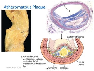
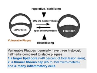
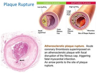
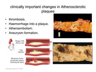
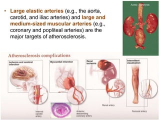
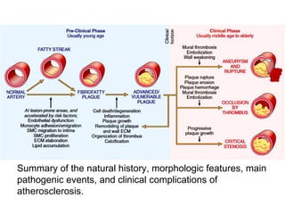
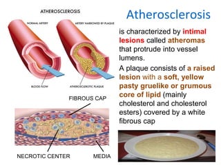
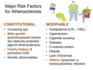
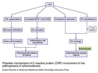
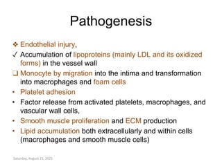
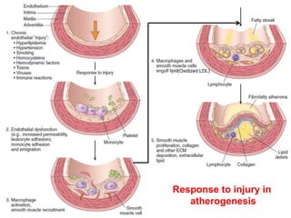
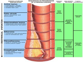
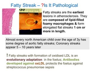
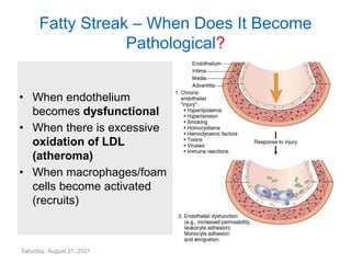
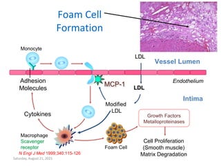
Pathology of
Atherosclerosis
• PA 27.1: Describe the pathogenesis of
atherosclerosis
• PA 27.5 Describe the epidemiology,risk
factors of Ischemic Heart Disease
Saturday, August 21, 2021
SPECIFIC LEARNING OBJECTIVES
• Define atherosclerosis, enumerate risk factors of and outline
the steps of the proposed pathogenesis
• Describe the morphology of atherosclerosis with special
reference to the morphologic changes in a complicated
plaques
• Discuss the systemic effects ( organ specific) and the clinical
significance (complications) of
atherosclerosis
Saturday, August 21, 2021
Atherosclerosis
is characterized by intimal
lesions called atheromas
that protrude into vessel
lumens.
A plaque consists of a raised
lesion with a soft, yellow
pasty gruelike or grumous
core of lipid (mainly
cholesterol and cholesterol
esters) covered by a white
fibrous cap
FIBROUS CAP
NECROTIC CENTER MEDIA
Major Risk Factors
for Atherosclerosis
CONSTITUTIONAL
• Increasing age
• Male gender
(premenopausal women
are relatively protected
against atherosclerosis)
• Family history of
premature CAD
• Genetic abnormalities
MODIFIABLE
• Dyslipidemia (LDL; HDL)
• Hypertension
• Cigarette smoking
• Diabetes
• C-reactive protein
• Obesity
• Lack of Exercise
• Others: lipoprotein a;
homocystinemia; infection
Age &
Gender
• clinically manifests in middle
age or later. Between ages
40 and 60 the incidence of MI
increases fivefold.
• premenopausal women are
relatively protected against
atherosclerosis .
• After menopause, the
incidence increases and at
older ages actually exceeds
that of men.
• Family history is the most
significant independent risk
factor for atherosclerosis.
• Familial predisposition to
atherosclerosis and IHD is
usually multifactorial.
• Certain mendelian disorders
(e.g., familial
hypercholesterolemia)are
associated with small % of
atherosclerosis.
Hyperlipidemia
• hypercholesterolemia—is a major risk factor for
atherosclerosis;
• The major component associated with increased
risk is
• Low-density lipoprotein (LDL) cholesterol (“bad
cholesterol”);
• higher levels of HDL correlate with reduced risk.
• High dietary intake of cholesterol, saturated fats
(present in egg yolks, animal fats, and butter, for
example), trans-unsaturated fats Raises plasma
cholesterol levels.
• Conversely, diets with higher ratios of
polyunsaturated fats & Omega-3 fatty acids
Lower cholesterol
Saturday, August 21, 2021
Ratio of total Cholesterol to HDL Ratio of LDL to HDL
Risk Men women Men Women
Very low
(1/2 average)
<3.4 <3.3 1 1.5
low risk 4.0 3.8
Average risk 5.0 4.5 3.6 3.2
Moderate
(2x average)
9.5 7.0 6.3 5.0
High risk (3x risk) >23 >11 8 6.1
Risk LDL Total C Triglycerides HDL
Optimal <100 Undesirable <40
Near optimal 100-129 Desirable >60
Desirable <200 < 130
(All in mg/dL)
Normal < 150
Borderline 130-159 200-239 150-199
High 160-189 >240 200-499
Very High >190 >= 500
Hypertension
• both systolic & diastolic
levels are important.
• On its own, hypertension
increases the risk of IHD by
approximately 60%
• Hypertension is the most
important cause of left
ventricular hypertrophy
Cigarette smoking
• Uncombustable ROS, Nic, Actn
of endothelium
• well-established risk factor.
• Prolonged (years) smoking of
one pack of cigarettes or more
daily doubles the death rate
from IHD.
• Cessation of Smoking reduces
the risk.
Diabetes mellitus
Induces
• Hypercholesterolemia &
glycation end prdts
• increases the risk of
atherosclerosis.
• the incidence of MI is twice
as high
• increased risk of stroke
• 100-fold increased risk of
atherosclerosis-induced
gangrene of the lower
extremities.
Others
• Inflammation: is present during
all stages of atherogenesis. CRP
levels are used in risk
stratification algorithms
• Hyperhomocystinemia.
Degrades fibers
• The metabolic syndrome:
abnormalities associated with
insulin resistance and adipose
tissue signaling
• Lipoprotein (a) (altered LDL that
contains apolipoprotein B – 100
portion of LDL linked with APO
LP-A levels are associated with
coronary and cerebrovascular
disease risk.
thrombosis
Pathogenesis
❖ Endothelial injury,
✓ Accumulation of lipoproteins (mainly LDL and its oxidized
forms) in the vessel wall
❑ Monocyte by migration into the intima and transformation
into macrophages and foam cells
• Platelet adhesion
• Factor release from activated platelets, macrophages, and
vascular wall cells,
• Smooth muscle proliferation and ECM production
• Lipid accumulation both extracellularly and within cells
(macrophages and smooth muscle cells)
Saturday, August 21, 2021
(Oxidized LDL)
Response to injury in
atherogenesis
Saturday, August 21, 2021
MORPHOLOGY
Saturday, August 21, 2021
Fatty Streak – ?Is It Pathological
! Fatty streaks with formation of oxidised LDL is an
evolutionary adaptation in the foetus; Antibodies
developed against oxLDL protects the foetus against
streptococcus pneumoniae sepsis
Almost every north American child over the age of 3y has
some degree of aortic fatty streaks; Coronary streaks
appear 5 – 10 years later
Fatty streaks are the earliest
lesions in atherosclerosis. They
are composed of lipid-filled
foamy macrophages & form
elongated flat streaks 1 cm or
more in length.
Saturday, August 21, 2021
Fatty Streak – When Does It Become
Pathological?
• When endothelium
becomes dysfunctional
• When there is excessive
oxidation of LDL
(atheroma)
• When macrophages/foam
cells become activated
(recruits)
Saturday, August 21, 2021
Foam Cell
Formation
N Engl J Med 1999;340:115-126
LDL
LDL
Endothelium
Vessel Lumen
Monocyte
Macrophage
Scavenger
receptor
Adhesion
Molecules
Foam Cell
Intima
Modified
LDL
Cytokines
Cell Proliferation
(Smooth muscle)
Matrix Degradation
Growth Factors
Metalloproteinases
MCP-1
Saturday, August 21, 2021
Role of HDL (also Apo A1 – Milano)
LDL
LDL
Endothelium
Vessel Lumen
Monocyte
Modified LDL
Macrophage
MCP-1
Adhesion
Molecules
Cytokines
Intima
HDL Inhibit
Oxidation
of LDL
HDL Inhibit Adhesion Molecule Expression
Foam
Cell
HDL Promote Cholesterol Efflux
“Inflammation impairs reverse cholesterol transport in vivo”
Cockerill GW et al. Arterioscler Thromb Vasc Biol 1995;15:1987-1994.
Circulation. 2009 Mar 3;119(8):1135-45. Epub 2009 Feb 16.
http://my.clevelandclinic.org/heart/news/hot/hdlapoa1.aspx
Saturday, August 21, 2021
Homocysteine
Homocysteine degrades and inhibits the formation of the three main structural
components of arteries: collagen, elastin and proteoglycans. In proteins, homocysteine
permanently degrades cysteine disulfide bridges and lysine amino acid
residues,[7] affecting structure and function.
Saturday, August 21, 2021
Atheromatous Plaque
Vulnerable Plaques generally have three histologic
hallmarks compared to stable plaques
1.a larger lipid core (>40 percent of total lesion area),
2. a thinner fibrous cap (65 to 150 micro-meters),
and 3. many inflammatory cells
Vulnerable Plaque
Atherosclerotic plaque rupture. Acute
coronary thrombosis superimposed on
an atherosclerotic plaque with focal
disruption of the fibrous cap, triggering
fatal myocardial infarction.
An arrow points to the site of plaque
rupture.
Plaque Rupture
clinically important changes in Atherosclerotic
plaques
• thrombosis.
• Haemorrhage into a plaque.
• Atheroembolism.
• Aneurysm formation.
Saturday, August 21, 2021
• Large elastic arteries (e.g., the aorta,
carotid, and iliac arteries) and large and
medium-sized muscular arteries (e.g.,
coronary and popliteal arteries) are the
major targets of atherosclerosis.
Summary of the natural history, morphologic features, main
pathogenic events, and clinical complications of
atherosclerosis.

17970_27.1Atherosclerosis.pptx

  • 1.
  • 2.
    • PA 27.1:Describe the pathogenesis of atherosclerosis • PA 27.5 Describe the epidemiology,risk factors of Ischemic Heart Disease Saturday, August 21, 2021
  • 3.
    SPECIFIC LEARNING OBJECTIVES •Define atherosclerosis, enumerate risk factors of and outline the steps of the proposed pathogenesis • Describe the morphology of atherosclerosis with special reference to the morphologic changes in a complicated plaques • Discuss the systemic effects ( organ specific) and the clinical significance (complications) of atherosclerosis Saturday, August 21, 2021
  • 4.
    Atherosclerosis is characterized byintimal lesions called atheromas that protrude into vessel lumens. A plaque consists of a raised lesion with a soft, yellow pasty gruelike or grumous core of lipid (mainly cholesterol and cholesterol esters) covered by a white fibrous cap FIBROUS CAP NECROTIC CENTER MEDIA
  • 5.
    Major Risk Factors forAtherosclerosis CONSTITUTIONAL • Increasing age • Male gender (premenopausal women are relatively protected against atherosclerosis) • Family history of premature CAD • Genetic abnormalities MODIFIABLE • Dyslipidemia (LDL; HDL) • Hypertension • Cigarette smoking • Diabetes • C-reactive protein • Obesity • Lack of Exercise • Others: lipoprotein a; homocystinemia; infection
  • 6.
    Age & Gender • clinicallymanifests in middle age or later. Between ages 40 and 60 the incidence of MI increases fivefold. • premenopausal women are relatively protected against atherosclerosis . • After menopause, the incidence increases and at older ages actually exceeds that of men. • Family history is the most significant independent risk factor for atherosclerosis. • Familial predisposition to atherosclerosis and IHD is usually multifactorial. • Certain mendelian disorders (e.g., familial hypercholesterolemia)are associated with small % of atherosclerosis.
  • 7.
    Hyperlipidemia • hypercholesterolemia—is amajor risk factor for atherosclerosis; • The major component associated with increased risk is • Low-density lipoprotein (LDL) cholesterol (“bad cholesterol”); • higher levels of HDL correlate with reduced risk. • High dietary intake of cholesterol, saturated fats (present in egg yolks, animal fats, and butter, for example), trans-unsaturated fats Raises plasma cholesterol levels. • Conversely, diets with higher ratios of polyunsaturated fats & Omega-3 fatty acids Lower cholesterol
  • 8.
    Saturday, August 21,2021 Ratio of total Cholesterol to HDL Ratio of LDL to HDL Risk Men women Men Women Very low (1/2 average) <3.4 <3.3 1 1.5 low risk 4.0 3.8 Average risk 5.0 4.5 3.6 3.2 Moderate (2x average) 9.5 7.0 6.3 5.0 High risk (3x risk) >23 >11 8 6.1 Risk LDL Total C Triglycerides HDL Optimal <100 Undesirable <40 Near optimal 100-129 Desirable >60 Desirable <200 < 130 (All in mg/dL) Normal < 150 Borderline 130-159 200-239 150-199 High 160-189 >240 200-499 Very High >190 >= 500
  • 9.
    Hypertension • both systolic& diastolic levels are important. • On its own, hypertension increases the risk of IHD by approximately 60% • Hypertension is the most important cause of left ventricular hypertrophy Cigarette smoking • Uncombustable ROS, Nic, Actn of endothelium • well-established risk factor. • Prolonged (years) smoking of one pack of cigarettes or more daily doubles the death rate from IHD. • Cessation of Smoking reduces the risk.
  • 10.
    Diabetes mellitus Induces • Hypercholesterolemia& glycation end prdts • increases the risk of atherosclerosis. • the incidence of MI is twice as high • increased risk of stroke • 100-fold increased risk of atherosclerosis-induced gangrene of the lower extremities. Others • Inflammation: is present during all stages of atherogenesis. CRP levels are used in risk stratification algorithms • Hyperhomocystinemia. Degrades fibers • The metabolic syndrome: abnormalities associated with insulin resistance and adipose tissue signaling • Lipoprotein (a) (altered LDL that contains apolipoprotein B – 100 portion of LDL linked with APO LP-A levels are associated with coronary and cerebrovascular disease risk.
  • 11.
  • 12.
    Pathogenesis ❖ Endothelial injury, ✓Accumulation of lipoproteins (mainly LDL and its oxidized forms) in the vessel wall ❑ Monocyte by migration into the intima and transformation into macrophages and foam cells • Platelet adhesion • Factor release from activated platelets, macrophages, and vascular wall cells, • Smooth muscle proliferation and ECM production • Lipid accumulation both extracellularly and within cells (macrophages and smooth muscle cells) Saturday, August 21, 2021
  • 13.
    (Oxidized LDL) Response toinjury in atherogenesis
  • 14.
    Saturday, August 21,2021 MORPHOLOGY
  • 15.
  • 16.
    Fatty Streak –?Is It Pathological ! Fatty streaks with formation of oxidised LDL is an evolutionary adaptation in the foetus; Antibodies developed against oxLDL protects the foetus against streptococcus pneumoniae sepsis Almost every north American child over the age of 3y has some degree of aortic fatty streaks; Coronary streaks appear 5 – 10 years later Fatty streaks are the earliest lesions in atherosclerosis. They are composed of lipid-filled foamy macrophages & form elongated flat streaks 1 cm or more in length.
  • 17.
    Saturday, August 21,2021 Fatty Streak – When Does It Become Pathological? • When endothelium becomes dysfunctional • When there is excessive oxidation of LDL (atheroma) • When macrophages/foam cells become activated (recruits)
  • 18.
    Saturday, August 21,2021 Foam Cell Formation N Engl J Med 1999;340:115-126 LDL LDL Endothelium Vessel Lumen Monocyte Macrophage Scavenger receptor Adhesion Molecules Foam Cell Intima Modified LDL Cytokines Cell Proliferation (Smooth muscle) Matrix Degradation Growth Factors Metalloproteinases MCP-1
  • 19.
    Saturday, August 21,2021 Role of HDL (also Apo A1 – Milano) LDL LDL Endothelium Vessel Lumen Monocyte Modified LDL Macrophage MCP-1 Adhesion Molecules Cytokines Intima HDL Inhibit Oxidation of LDL HDL Inhibit Adhesion Molecule Expression Foam Cell HDL Promote Cholesterol Efflux “Inflammation impairs reverse cholesterol transport in vivo” Cockerill GW et al. Arterioscler Thromb Vasc Biol 1995;15:1987-1994. Circulation. 2009 Mar 3;119(8):1135-45. Epub 2009 Feb 16. http://my.clevelandclinic.org/heart/news/hot/hdlapoa1.aspx
  • 20.
    Saturday, August 21,2021 Homocysteine Homocysteine degrades and inhibits the formation of the three main structural components of arteries: collagen, elastin and proteoglycans. In proteins, homocysteine permanently degrades cysteine disulfide bridges and lysine amino acid residues,[7] affecting structure and function.
  • 21.
    Saturday, August 21,2021 Atheromatous Plaque
  • 22.
    Vulnerable Plaques generallyhave three histologic hallmarks compared to stable plaques 1.a larger lipid core (>40 percent of total lesion area), 2. a thinner fibrous cap (65 to 150 micro-meters), and 3. many inflammatory cells Vulnerable Plaque
  • 23.
    Atherosclerotic plaque rupture.Acute coronary thrombosis superimposed on an atherosclerotic plaque with focal disruption of the fibrous cap, triggering fatal myocardial infarction. An arrow points to the site of plaque rupture. Plaque Rupture
  • 24.
    clinically important changesin Atherosclerotic plaques • thrombosis. • Haemorrhage into a plaque. • Atheroembolism. • Aneurysm formation. Saturday, August 21, 2021
  • 25.
    • Large elasticarteries (e.g., the aorta, carotid, and iliac arteries) and large and medium-sized muscular arteries (e.g., coronary and popliteal arteries) are the major targets of atherosclerosis.
  • 26.
    Summary of thenatural history, morphologic features, main pathogenic events, and clinical complications of atherosclerosis.