2. Case History 1
A 60-year old women was referred to a
hospital. She was noted to have hypertension.
The plasma cholesterol level was 390 mg/dL.
An angiogram of the right carotid artery
demonstrated a narrowed lumen and the
concentration of LDL was elevated.
Questions:
1. What is the probable diagnosis?
2. What is the normal plasma cholesterol
level?
3. Explain the clinical condition &
pathogenesis of narrowed lumen
3. S.No Cholesterol /
Lipoprotein
Normal
Range
(mg / dl)
1 Total cholesterol 150 - 200
2 LDL -Cholesterol < 130
3 VLDL -Cholesterol < 40
4 HDL -Cholesterol > 40
5 Triglyceride 50 - 150
Normal level of various types of Cholesterol in Blood /
Plasma
4. Cardiovascular Disease (CVD)
• Main type of CVD is Atherosclerosis (AS)
• Endothelial dysfunction is one of earliest
changes in AS.
• Mechanical, chemical, inflammatory
mediators can trigger endothelial
dysfunction:
– High blood pressure
– Smoking (free radicals that oxidatively
damage endothelium)
– Inflammatory stimuli
– Hyperlipidemia
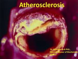
5. Atherosclerosis
Atherosclerosis thickening / hardening of
blood vessels due to the deposition of lipids.
Atherosclerosis – Greek word
“Athere” - accumulation of lipid
“Sclerosis” - thickening / hardening
6. Atherosclerosis
• Occurs in large and medium size arteries:
• Characterized by:
-endothelial dysfunction,
-vascular inflammation,
-deposition of cholesterol,
-calcium and cellular debris
within the intima of the arteries –
known as atheromatous plague.
7.
8. Atheromatous plaque
• It consists of a raised lesion with a
soft,yellow, core of lipid
(cholesterol & cholesterol ester)
covered by a firm, white fibrous cap
• It narrows the lumen of the vessels and
obstructs the blood flow (ischemia).
• It weakens the underlying media, ruptures
and causes thrombosis
10. Atherosclerosis & LDL
Stage I: Formation of foam cells
Increased levels of cholesterol for prolonged
periods
Free radical induced oxidative damage of LDL
Oxidised LDL particles are deposited in the walls
of the arteries.
Macrophages become overloaded with cholesterol
Foam cells
11. • Monocytes in the blood adhere to endothelial
cells at sites of injury/inflammation, & then pass
into the subendothelial space where they
differentiate into macrophages.
• Lipoproteins (e.g., LDL) leak across the
endothelium & accumulate in the subendothelial
space.
• Over time, exposure to oxygen radicals results in
oxidation of LDL & modification of the
apolipoprotein.
12. Atherosclerosis & LDL
Stage II : Progression of Atherosclerosis
Macrophages or Smooth muscle cells containing Lipid
droplets
Can be reversible if plasma lipid levels are lowered
When lipid accumulates the lesion progresses
Irreversible arterial changes
13. • Macrophages take up oxidized lipoproteins
becoming "foam cells" that have many
cytoplasmic lipid droplets.
• Although in humans foam cells mainly develop
from macrophages, smooth muscle cells may also
migrate into the subendothelial space & become
foam cells.
blood vessel lumen
smooth muscle cells
endothelial
cells
foam cell
LDL
14. Atherosclerosis & LDL
Stage III : Fibrous proliferation
Due to liberation of Growth factors by
Macrophages & platelets
Accumulation of Lipoproteins, Glycosaminoglycans
& collagen
Proliferative changes
Inflammatory changes in atherosclerosis
15. Atherosclerosis & LDL
Stage IV : Advancing Fibrous plaque
Narrowing of vessel wall
Clot formation
Myocardial Infarction
23. Prevention of cardiovascular
diseases
• No smoking
• Physical activities / Exercise
• Restrict saturated fat /animal fat
• Include: PUFA – rich in vegetable oil
omega 3 fatty acids - rich in fish
• Avoid trans fatty acids (hydrogenation of
vegetable oils)
• Dietary fiber
• Hypolipidemic Drugs (Simvostatin, Cholestyramine, Aspirin)
• Effective Treatment:
Hypercholesterolrmia, Hypertension, DM, Obesity
24. • Watch this video on pathogenesis of
atherosclerosis
• https://youtu.be/N33JsBeziEY
25. 1. A 52 year old male is brought to the casualty
with chest pain of 6 hours duration. He has
been having chest discomfort for the past 3
months and increased with unaccustomed
exertion. ECG revealed ST segment
elevation.
(a) What are the cardiac markers which can
be tested in this scenario? (2)
(b) Briefly explain their importance. (3)
26. Cardiac Biomarkers
• Cardiac Biomarkers are protein molecules
released into the blood stream from damaged
heart muscle
• Since ECG………Inconclusive……biomarkers!!!!!!??
Myocardial injury
• These markers have a characteristic rise & fall
pattern…..
27. Definition
Biomarker is defined as “a characteristic that is
objectively measured & evaluated to aid in
understanding on
prediction of disease,
its cause
the diagnosis
response to intervention”
28. CHARACTERISTIC OF AN IDEAL
MARKER
• High cardiac specificity
• Easy diagnosis
• Marker should play a designed role in the
treatment and management of clinical subject
29. Markers of Myocardial Injury
• Markers of Myocardial Necrosis
- Cardiac Troponins
- CKMB
- Myoglobin
• Markers of Myocardial Ischemia
- Ischemia Modified Albumin
- Heart-type fatty acid binding protein
(H-FAPP)
30. Ideal Cardiac markers
• High concentration in myocardium
• Absence from non-myocadrial tissue (high
specificity)
• Rapid release into plasma following MI
• Correlation between blood level & extent of
MI for prognosis
• Detectable by low concentration in blood.
31. Cardiac Markers
After the loss of integrity of
cardiac myocyte membranes,
intracellular macromolecules
diffuse into the interstitium ,
Lymphatics, microvasculature.
33. • Two types of Troponins:
• Troponin T : are found in the cardiac and
skeletal muscle.
• Troponin I are found only in cardiac muscle,
more specific.
• Early rise after Myocardial infarction.
Cardiac Markers
34. Troponin Onset Peak Duration
Troponin I (TnI) Within 4-6 hrs 12-24 hrs 7-10 days
Troponin T
(TnT)
Within 6 hr 24 hr 10-14 days
Ref.level in adults for TnT: 0 – 0.1 ng/mL.
Ref.level in adults for for TnI: 0-0.4 ng/mL
36. Characteristic feature of hs Cardiac Troponin
• Can detect Troponin at concentrations
10 to 100 fold lower than coventional
assays
• Reported as nanogram per litre
• High precision at lower concentration
37. CPK / CK: isoenzyme
• CPK / CK is dimeric enzyme made of two subunits of
different types:
• B submit (Brain) &
• M submit (sk.Muslce)
Normal Range for Total CPK in serum:
15 – 100 U/L for males
10 – 80 U/L for females
38. CPK / CK: isoenzyme..
Isoenzyme Subunit
composition
Principal
tissue/organ
Percent
of
normal
serum
Diagnostic
significance
CK1 BB Brain 1%
CK2 MB Heart 5-10% ↑ MI
CK3 MM Sk.Mucle 90%
CK-MB predominates in cardiac muscle
39.
40.
41. CK-MB relative index
• Relative Index = CK-MB X 100
Total CK
>5 indicates MI
< 3 skeletal muscle source
Total CK- Muscular dystrophies
CK-MB isoenzme - MI
42. Why CKMB is recommended?????
• Used for early confirmation of diagnosis
• High sensitivity
• To rule out skeletal muscle disorder,
when total CK is elevated
• High specificity
44. Obsolete markers…..
• 1. Myoglobin
- It is not specific
- Released as early as 1-2 hrs after onset (4)
- Normal range: 30-90ng/ml
• 2.Lactate Dehydrogenase (LDH)
- Increased only after 6-12 hrs after onset
- Highly non specific
• 3. Aspartate Transaminase (AST)
• Increased only after 24 hrs after onset
52. LIPOPROTEINS = Lipids + Proteins
• Spherical macromolecular complexes which
help in the transport of TGL and cholesterol
through blood stream between various
tissues.
• The lipoproteins consist of a core of
hydrophobic lipids (cholesterol ester & TGL)
surrounded by a shell of amphipathic
lipids(PL & free cholesterol) along with
proteins (apolipoproteins).
53.
54. Lipoproteins
Lipoproteins are classified into five
different types according to size and
density:
Apoprotein is the protein part of the
lipoprotein
• Chylomicrons: apoprotein B-48
• VLDL : apo B-100 (Pre-beta)
• IDL : apo B-100 (broad beta)
• LDL : apo B-100 (Beta )
• HDL : apo - A
55. Functions of Lipoproteins
Lipoprotein Site of
synthesis
Functions
Chylomicrons Intestine Transport of dietary lipids
from intestine to peripheral
tissues
VLDL Liver Transport endogenous
triacylglycerol from liver to
peripheral tissues
LDL Plasma
VLDL
Transport cholesterol from
liver to peripheral tissues
HDL Liver &
intestine
Transport free cholesterol
from peripheral tissues to the
liver where it can be
catabolized (Reverse
cholesterol transport)
56. A 10 year old girl was brought to the OPD
with fatty eruptions on the right elbow and
Achilles tendon. These were identified as
subcutaneous xanthomas. Her mother
informed that the child’s father had similar
eruptions and had died of heart attack at
the age of 32. Examination showed
bilateral corneal arcus.
61. Classes of Primary Hyperlipidemias
Class Cause Abnormalities Features
I. Familial
Hyperchylomicronemia
LPL deficiency
CM
No risk for CAD.
Eruptive xanthoma
Hepatomegaly,
Lipemia retinalis
IIa.Familial
Hypercholesterolemia
LDL receptor
defect
LDL; TC
Atherosclerosis &
CAD
Tuberous
xanthoma
IIb. Familial
Combined
Hyperlipidemia
Similar to type
IIa, in VLDL
VLDL, LDL Corneal arcus
III. Familial broad-
beta-lipoproteinemia
Defect in
remnant
clearance.
Defective
Apo E. Lack E3
&E4, have E2.
IDL,
LDL
Xanthomas &
atherosclerosis
Palmar xanthoma,
High incidence of
vascular disease
IV. Familial
Hypertriglyceridemia
Overproduction
of VLDL with
glucose
intolerance &
hyperinsulinemia
.
TG, associated
with CAD, type IIDM,
obesity
V. Familial mixed
Hypertriglyceridemia
Abnormal VLDL
& CM metabolism
secondary to other
causes
64. Summary
• Hyperlipoproteinemias – Frederickson’s
classification
• Type IIA – LDL receptor defect
• Lipid profile – Total cholesterol, TGL, LDL-c,
HDL-c
• Cardiac markers are CK-MB, Troponins I & T
and Myoglobin
65. 1. Elevated levels of all of the following are
considered as risk factors for cardiac
disease EXCEPT
(A) Total cholesterol
(B) LDL-cholesterol
(C) HDL-Cholesterol
(D) Triglycerides
66. 2. Deficiency of LPL and apo C-II causes
(A) Type I hyperlipoproteinemia
(B) Type III hyperlipoproteinemia
(C) Type V hyperlipoproteinemia
(D) Tangier disease
3. Earliest change in atherosclerosis is
(A) Formation of clot
(B) Endothelial dysfunction
(C) Foam cell formation
(D) Migration of smooth muscle cells
67. 4. The cardiac marker to be elevated first in
myocardial infarction is
(A) Troponin I
(B) Creatine kinase-MB
(C) Aspartate transaminase
(D) Myoglobin