Download to read offline
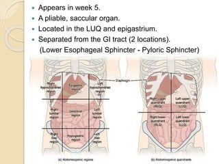
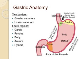
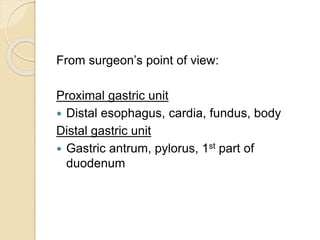
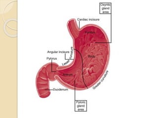
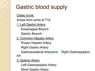
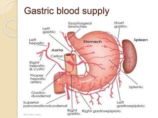
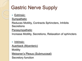
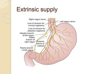
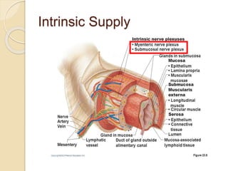
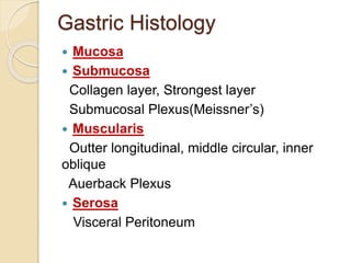
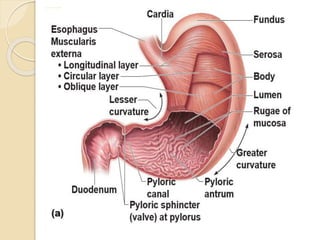
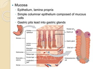
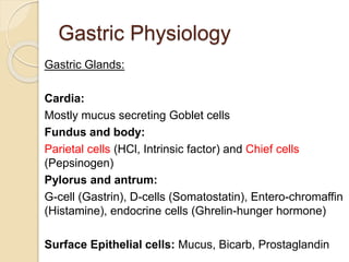
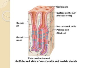
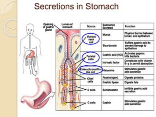
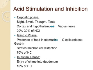


The document discusses the anatomy, blood supply, histology, and physiology of the stomach and duodenum, describing their locations, structures like the gastric glands and layers, innervation by the sympathetic and parasympathetic nervous systems, and secretions including hydrochloric acid and intrinsic factor. Key aspects covered include the divisions of the stomach, its blood supply from the celiac trunk, and how gastric acid secretion is stimulated by foods in the stomach and inhibited by foods entering the duodenum.


![Stomach by kp [autosaved]](https://cdn.slidesharecdn.com/ss_thumbnails/stomachbykpautosaved-140422141057-phpapp02-thumbnail.jpg?width=640&height=640&fit=bounds)


![SURGICAL ANATOMY OF STOMACH [Autosaved].pptx](https://cdn.slidesharecdn.com/ss_thumbnails/surgicalanatomyofstomachautosaved-230608035808-b982958c-thumbnail.jpg?width=640&height=640&fit=bounds)


