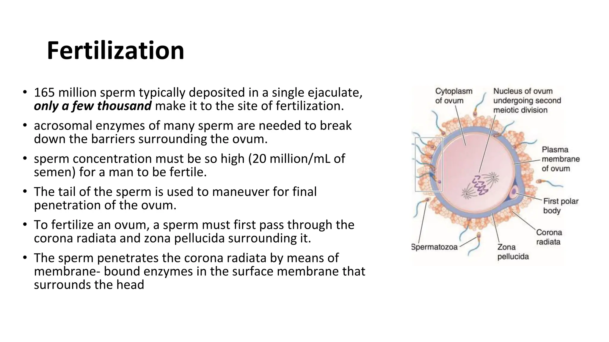
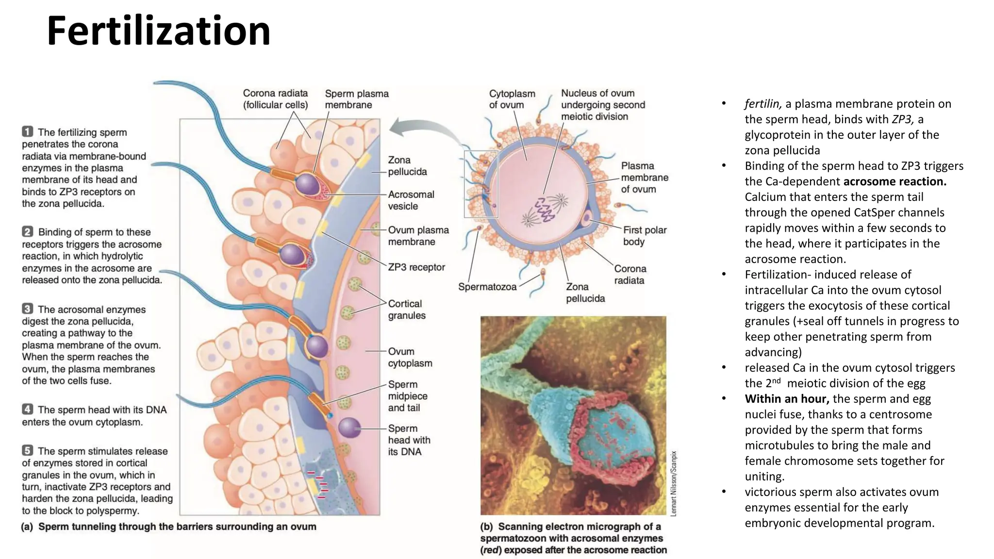
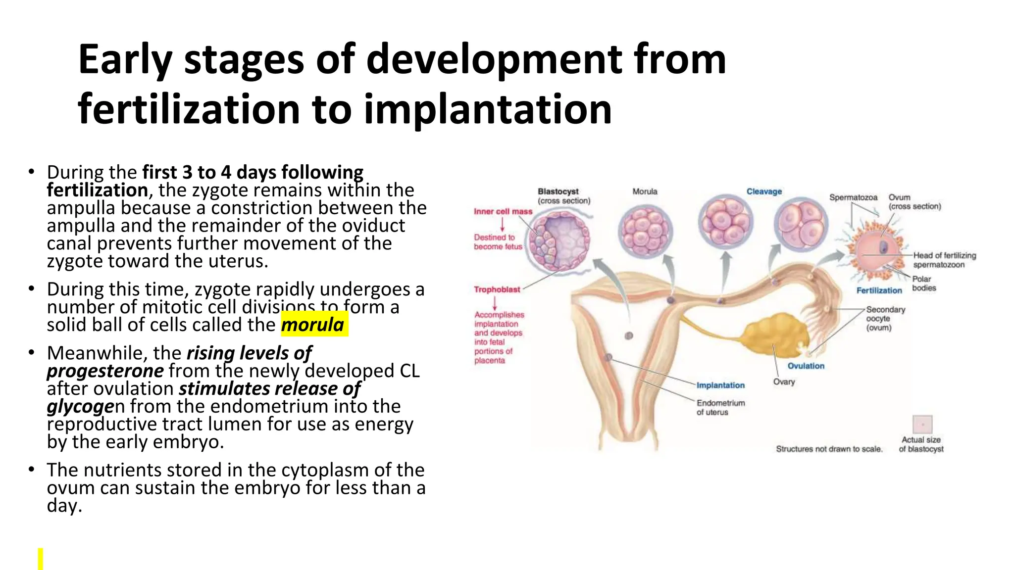
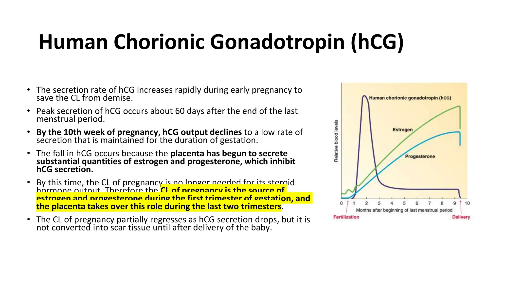
The document summarizes key stages of human fertilization and early pregnancy development. It describes how the ovum is transported to the oviduct where fertilization occurs with sperm. It then discusses early embryo development through the morula and blastocyst stages. It explains implantation of the blastocyst in the uterine lining and the subsequent development of the placenta and amniotic sac to support the growing embryo and fetus. The roles of hormones like hCG in supporting early pregnancy are also outlined.
This document summarizes the key stages in human reproduction from ovulation through implantation. It describes how ovulation is triggered by hormones, followed by fertilization if sperm penetrate the egg's barriers. The zygote then undergoes cell division as it develops into a blastocyst, which implants in the uterine wall. Successful implantation depends on cellular interactions between the trophoblast and endometrium.
The document provides information about the process of pregnancy from fertilization through birth in 3 sentences:
Fertilization occurs when the genetic material from a sperm and egg merge in the fallopian tube, forming a zygote which undergoes cell division and implants in the uterus, developing into an embryo and fetus over 38 weeks until birth. The placenta forms and secretes hormones like progesterone, estrogen, hCG, and others to support the pregnancy and development of the fetus, while the fetus develops all its major organ systems, tripling in size by the third trimester before birth. Maternal changes include support of the corpus luteum in early pregnancy and taking over hormone production by the placenta, enlarg
1. Fertilization occurs when a sperm and egg fuse, forming a single diploid cell called a zygote containing genetic material from both parents.
2. The journey of sperm is difficult, with millions being overcome by acidity or blocked, and thousands destroyed before reaching the egg.
3. During its journey, sperm undergo capacitation to prepare for fertilization by improving motility and membrane changes.
4. Fertilization must occur in the uterine tube, where the egg is swept after ovulation. Hundreds of sperm help make a path for one to fuse with the egg's membrane.
First week of development: Ovulation to Implantation Jwan AlSofi
The document summarizes key aspects of ovulation, fertilization, and early embryonic development. It describes the ovarian and menstrual cycles controlled by hormones like FSH and LH. Ovulation occurs mid-cycle due to an LH surge, releasing an egg. Sperm travel through the reproductive tract while undergoing capacitation. Fertilization typically occurs in the fallopian tubes, involving penetration of the egg's layers and fusion of gametes. This activates the egg and forms pronuclei, leading to cell division and pregnancy if implantation occurs. Otherwise, the corpus luteum regresses and menstruation begins.
1) Fertilization involves several sperm penetrating the corona radiata and zona pellucida to fuse with the egg nucleus to form a zygote, preventing additional sperm from entering.
2) The fertilized egg travels through the fallopian tubes to the uterus within 3-5 days, propelled by cilia and fluid currents.
3) The blastocyst implants in the uterine endometrium through enzymes secreted by trophoblast cells, forming the placenta which facilitates nutrient/gas exchange between mother and fetus and produces important hormones.
3 GEN EMBRYOLOGY third Week 3 germ layer .pptxAkhilaV16
third wk there is formation of three germ layers. primitive streek and prochordal plate forms the intra embyyonic mesoderm. the epiblast cells replace all the 3 germ layers. iem is converted to paraxial, intermediate and lateral plate mesoderms
Anatomy of male and female reproductive system, Functions of male and female
reproductive system, sex hormones, physiology of menstruation, fertilization,
spermatogenesis, oogenesis, pregnancy and parturition
The document summarizes key events in oogenesis and ovulation in cattle. It describes the LH and FSH hormone surge which stimulates meiosis and follicle development. It also discusses capacitation of sperm in the oviducts allowing fertilization to occur, and the prevention of polyspermy through the zona reaction and vitelline block. Early embryonic development is also summarized, including cleavage, formation of the morula and blastocyst, hatching from the zona pellucida, and initial implantation in the uterus.
The ovarian cycle is controlled by hormones from the hypothalamus and pituitary gland. During each cycle, follicles in the ovaries mature under the influence of FSH and LH, with usually one follicle ovulating and releasing an egg. If the egg is fertilized, the corpus luteum forms and secretes hormones to support early pregnancy. Fertilization occurs in the fallopian tubes, where a sperm penetrates the egg's corona and zona pellucida. The sperm and egg membranes then fuse, restoring the diploid number of chromosomes and initiating cell division of the new embryo.
The document summarizes key stages in human fertilization and early embryonic development. It describes:
1) How sperm capacitation and the acrosome reaction allow sperm to penetrate the zona pellucida and fuse with the egg.
2) The formation of pronuclei and how the zygote undergoes cell division to become a morula and then a blastocyst.
3) The process of blastocyst implantation in the uterine lining and how trophoblast cells mediate attachment to the endometrium.
Fertilization is the union of a sperm and an egg to form a zygote. There are two types: external fertilization, where gametes are released into the environment, and internal fertilization, which occurs in nearly all terrestrial animals. For internal fertilization to occur, sperm must undergo a process of transport and capacitation to prepare for fertilizing the egg. Upon fertilization, a series of cellular events take place to prevent polyspermy, including cortical reaction, acrosomal reaction, and nuclear reaction, leading to the formation of a diploid zygote.
Fertilization, implantaion and embryologyobgymgmcri
1. The document summarizes key stages in human development from fertilization through embryonic and fetal development. It describes fertilization, cleavage, implantation, and the formation of the three germ layers and extraembryonic membranes.
2. Implantation of the blastocyst in the endometrium occurs around 7 days after fertilization. The trophoblast erodes into the endometrium and connections are made between embryonic and maternal blood vessels in the placenta.
3. The embryonic phase lasts until 8 weeks when major organ systems have begun to form and it is then called a fetus. By the end of the third month the placenta is functioning to exchange gases, nutrients, and waste
The female reproductive system produces eggs and provides an environment for fertilization and embryo development. The ovaries contain primordial follicles which mature through various stages in response to hormones. Most follicles undergo atresia, while one becomes dominant and ovulates monthly. The released egg is transported by the uterine tubes to the uterus. If fertilized, it implants and the placenta develops to support fetal growth. The endometrium thickens monthly under hormonal control but is shed as menstruation if implantation does not occur.
The placenta is a temporary organ that facilitates nutrient and waste exchange between the fetus and mother. It is composed of fetal and maternal tissues. Parturition begins with the fetus triggering hormonal changes that relax the cervix and stimulate contractions. It occurs in three stages: 1) initiation of contractions through decreasing progesterone and increasing prostaglandins and estrogen, 2) expulsion of the fetus aided by hormones like cortisol and relaxin, 3) expulsion of fetal membranes due to villus detachment from maternal tissues. Placentas are classified by villus distribution and layers separating fetal and maternal blood.
This presentation was prepared by Isaac Monyrun Meen Ngeny, medical student in university of Juba. South Sudan.
It's meant for online readers who might be searching for any information about embryology
Ovulation, fertilization, implantation (1 st weekMarami Mustapa
The document discusses the processes of ovulation, fertilization, and implantation. It explains that ovulation is triggered by hormones like LH and FSH, leading to the release of an egg. If fertilization by sperm occurs, the fertilized egg undergoes cell division and develops into a blastocyst over a week. The blastocyst then implants in the uterus by attaching to the endometrial lining with the help of hormonal changes brought on by the corpus luteum.
1) Fertilization occurs when a sperm fuses with an egg in the fallopian tube. This initiates cell division and the development of an embryo.
2) By day 5, the embryo has developed into a blastocyst stage and begins to implant in the uterine wall. Implantation is completed by day 12.
3) For successful implantation, the blastocyst must attach to the endometrium and the trophoblast cells must invade the uterine tissues to establish blood flow from the mother. This allows the embryo to receive nutrients for growth and development of a pregnancy.
1. Capacitation is the process by which sperm gain the ability to fertilize an egg after leaving the male reproductive tract. It involves changes to the sperm membrane and motility that occur as sperm travel through the female reproductive tract.
2. For fertilization to occur, sperm must navigate barriers like cervical mucus, penetrate the zona pellucida surrounding the egg, and fuse with the egg membrane. Binding of sperm to zona pellucida protein ZP3 triggers the acrosome reaction which releases enzymes to penetrate the zona.
3. Fusion of sperm with the egg membrane activates the egg through release of calcium ions and cortical granules, allowing the male and female pronu
USMLE GENERAL EMBRYOLOGY 008 First week of development A embryo .pdfAHMED ASHOUR
The initial week of embryonic development is a vital period commencing with fertilization, leading to the creation of the zygote and early cell divisions. It's noteworthy that, throughout this week, the developing embryo remains in the pre-implantation stage, journeying from the fallopian tube toward the uterus. Key events such as fertilization, cleavage, and the formation of the blastocyst are crucial for the embryo's early development.
These events lay the foundation for subsequent processes in the following weeks. The successful implantation of the blastocyst into the uterus marks the transition from the first week to the second week of embryonic development.
Fertilization is the process where an egg is fertilized by a sperm. It begins with the maturation and release of an egg (ovum) from the ovaries. In the fallopian tubes, millions of sperm travel and undergo changes to be able to fertilize the egg, involving enzymes that allow penetration of the egg's outer layers. If a sperm successfully fertilizes the egg within 24 hours, its nucleus fuses with the egg nucleus to form a single-celled zygote, marking the beginning of a new human life. Rapid cell division of the zygote then occurs as it travels down the fallopian tube.
Fertilization occurs when a sperm fertilizes an egg to form a zygote. In Islam, legal fertilization is the union of a husband's sperm with his wife's egg in her fallopian tube. Hundreds of millions of sperm are deposited but only a few hundred reach the fallopian tube where the egg is located. The sperm must undergo capacitation and the acrosome reaction to penetrate the egg's barriers. Fusion of the sperm and egg nuclei forms the zygote with a full set of chromosomes, starting the process of cell division and human development.
The female reproductive system produces eggs and sex hormones, transports eggs to the uterus, sustains a pregnancy, and nourishes a fetus. It includes ovaries, fallopian tubes, uterus, vagina, and external genitals. The ovaries contain eggs at various stages of development and release an egg monthly during ovulation. The fallopian tubes transport eggs from the ovaries to the uterus. The uterus provides support and nutrients for a developing fetus. The male reproductive system produces and transports sperm. It includes testes, epididymis, vas deferens, seminal vesicles, prostate, and penis. The testes produce sperm and hormones. Mature sperm are stored in the epididymis and vas deferens until ejaculation
Mammalian development begins with fertilization inside the female's body. The zygote then undergoes cleavage divisions that are slower than in other animals. Around the 8-cell stage, the cells compact together and later form two distinct cell types - the inner cell mass and trophoblast cells. The trophoblast cells go on to form the blastocyst, with an inner cell mass surrounded by trophoblast cells. The blastocyst hatches from the zona pellucida and implants in the uterus, where the trophoblast cells invade the uterine tissue and the inner cell mass forms the embryo.
- A zygote is formed when a sperm fertilizes an ovum in the fallopian tube. It then undergoes cleavage, dividing rapidly through mitosis as it moves towards the uterus.
- By day 4 it has developed into a morula, a solid ball of 16 cells. It then forms a blastocyst with an inner cell mass and outer trophoblast layer.
- The blastocyst implants in the uterine wall around day 6, and the zona pellucida disintegrates allowing it to hatch out of its protective covering.
The document describes the female reproductive system, including the ovaries, fallopian tubes, and uterus. It discusses the follicular growth and development in the ovaries, from primordial follicles to mature Graafian follicles. It describes the layers of the fallopian tube walls and the regions of the fallopian tubes. It also outlines the layers of the uterine wall, including the perimetrium, myometrium, and endometrium.
The document summarizes key stages in human embryonic development from fertilization through the end of the embryonic period at 8 weeks. It describes fertilization and implantation, then outlines development by week. The first week includes cleavage and blastocyst formation. The second week brings germ layer formation and implantation. The third week involves organogenesis and formation of the heart and blood vessels. Weeks 4-5 see further organ development and limb buds. Weeks 6-8 are a period of rapid growth and tissue maturation as the embryonic period concludes.
Similar to 10. Fertilization , Pregnancy, Parturition.pptx (20)
The document discusses different aspects of neurophysiology related to synapses. It defines a synapse as the junction between neurons where communication occurs. There are two main types of synapses - chemical synapses, where a neurotransmitter is released to activate receptors, and electrical synapses, where neurons are directly connected through gap junctions allowing faster transmission. The processes of chemical synapses involve the presynaptic neuron releasing neurotransmitters into the synaptic cleft upon an action potential, which then bind to and activate receptors on the postsynaptic neuron. There are multiple ways neurons can connect at synapses, including axon to dendrite, axon to soma, and axon to axon terminals.
The endocrine system regulates physiological processes through glands and hormones. Key components include glands that produce hormones, hormones that act as chemical messengers, and target organs that hormones affect. Feedback mechanisms maintain hormonal balance, such as negative feedback that reduces hormone production when levels rise. Imaging modalities used to study the endocrine system include ultrasound, CT, MRI, and radionuclide imaging using radiotracers. Specific scans include thyroid scans, parathyroid scans, and adrenal scans. Positron emission tomography (PET) also provides functional information about endocrine glands and is used in research and to detect cancers.
The document summarizes information about bubonic plague, the most common form of the Black Death pandemic that killed around 50 million people in the 14th century. It describes how bubonic plague is caused by the Yersinia pestis bacteria typically found in small animals like rats. The bacteria is transmitted to humans via the bite of infected fleas. Key symptoms of bubonic plague include swollen and painful lymph nodes, called buboes, as well as high fever, chills, and muscle cramps. Prompt treatment with appropriate antibiotics can reduce the mortality rate from over 50% to just 1-15%.
This document provides an overview of the process of metastasis. It discusses the major steps cancer cells undergo during metastasis including invasion, intravasation, transport, extravasation, and metastatic colonization. It describes the tools cancer cells use for invasion like cell adhesion molecules and proteases. It also covers dormancy of micrometastases, pre-metastatic niche formation, angiogenesis, and potential therapeutic strategies like MMP inhibitors, anti-angiogenic therapy, and targeting multiple steps of metastasis.
This document discusses organizational culture and its importance in healthcare organizations. It defines organizational culture as the shared norms, values and behaviors of an organization. Culture is shaped by both internal and external forces and can be interpreted by what employees see and hear. Subcultures within different departments may exist within an overall organizational culture. A strong, consistent culture can benefit an organization, but subcultures allow flexibility for different needs. Leaders must balance developing the overall culture while allowing for variation in subcultures.
Healthy Eating Habits:
Understanding Nutrition Labels: Teaches how to read and interpret food labels, focusing on serving sizes, calorie intake, and nutrients to limit or include.
Tips for Healthy Eating: Offers practical advice such as incorporating a variety of foods, practicing moderation, staying hydrated, and eating mindfully.
Benefits of Regular Exercise:
Physical Benefits: Discusses how exercise aids in weight management, muscle and bone health, cardiovascular health, and flexibility.
Mental Benefits: Explains the psychological advantages, including stress reduction, improved mood, and better sleep.
Tips for Staying Active:
Encourages consistency, variety in exercises, setting realistic goals, and finding enjoyable activities to maintain motivation.
Maintaining a Balanced Lifestyle:
Integrating Nutrition and Exercise: Suggests meal planning and incorporating physical activity into daily routines.
Monitoring Progress: Recommends tracking food intake and exercise, regular health check-ups, and provides tips for achieving balance, such as getting sufficient sleep, managing stress, and staying socially active.
About this webinar: This talk will introduce what cancer rehabilitation is, where it fits into the cancer trajectory, and who can benefit from it. In addition, the current landscape of cancer rehabilitation in Canada will be discussed and the need for advocacy to increase access to this essential component of cancer care.
Chandrima Spa Ajman is one of the leading Massage Center in Ajman, which is open 24 hours exclusively for men. Being one of the most affordable Spa in Ajman, we offer Body to Body massage, Kerala Massage, Malayali Massage, Indian Massage, Pakistani Massage Russian massage, Thai massage, Swedish massage, Hot Stone Massage, Deep Tissue Massage, and many more. Indulge in the ultimate massage experience and book your appointment today. We are confident that you will leave our Massage spa feeling refreshed, rejuvenated, and ready to take on the world.
Visit : https://massagespaajman.com/
Call : 052 987 1315
At Malayali Kerala Spa Ajman, Full Service includes individualized care for every client. We specifically design each massage session for the individual needs of the client. Our therapists are always willing to adjust the treatments based on the client's instruction and feedback. This guarantees that every client receives the treatment they expect.
By offering a variety of massage services, our Ajman Spa Massage Center can tackle physical, mental, and emotional illnesses. In addition, efficient identification of specific health conditions and designing treatment plans accordingly can significantly enhance the quality of massaging.
At Malayali Kerala Spa Ajman, we firmly believe that everyone should have the option to experience top-quality massage services regularly. To achieve that goal we offer cheap massage services in Ajman.
If you are interested in experiencing transformative massage treatment at Malayali Kerala Spa Ajman, you can use our Ajman Massage Center WhatsApp Number to schedule your next massage session.
Contact @ +971 529818279
Visit @ https://malayalikeralaspaajman.com/
Comprehensive Rainy Season Advisory: Safety and Preparedness Tips.pdfDr Rachana Gujar
The "Comprehensive Rainy Season Advisory: Safety and Preparedness Tips" offers essential guidance for navigating rainy weather conditions. It covers strategies for staying safe during storms, flood prevention measures, and advice on preparing for inclement weather. This advisory aims to ensure individuals are equipped with the knowledge and resources to handle the challenges of the rainy season effectively, emphasizing safety, preparedness, and resilience.
The best massage spa Ajman is Chandrima Spa Ajman, which was founded in 2023 and is exclusively for men 24 hours a day. As of right now, our parent firm has been providing massage services to over 50,000+ clients in Ajman for the past 10 years. It has about 8+ branches. This demonstrates that Chandrima Spa Ajman is among the most reasonably priced spas in Ajman and the ideal place to unwind and rejuvenate. We provide a wide range of Spa massage treatments, including Indian, Pakistani, Kerala, Malayali, and body-to-body massages. Numerous massage techniques are available, including deep tissue, Swedish, Thai, Russian, and hot stone massages. Our massage therapists produce genuinely unique treatments that generate a revitalized sense of inner serenely by fusing modern techniques, the cleanest natural substances, and traditional holistic therapists.
TEST BANK FOR Health Assessment in Nursing 7th Edition by Weber Chapters 1 - ...rightmanforbloodline
TEST BANK FOR Health Assessment in Nursing 7th Edition by Weber Chapters 1 - 34.
At Apollo Hospital, Lucknow, U.P., we provide specialized care for children experiencing dehydration and other symptoms. We also offer NICU & PICU Ambulance Facility Services. Consult our expert today for the best pediatric emergency care.
For More Details:
Map: https://cutt.ly/BwCeflYo
Name: Apollo Hospital
Address: Singar Nagar, LDA Colony, Lucknow, Uttar Pradesh 226012
Phone: 08429021957
Opening Hours: 24X7
MBC Support Group for Black Women – Insights in Genetic Testing.pdfbkling
Christina Spears, breast cancer genetic counselor at the Ohio State University Comprehensive Cancer Center, joined us for the MBC Support Group for Black Women to discuss the importance of genetic testing in communities of color and answer pressing questions.
R3 Stem Cell Therapy: A New Hope for Women with Ovarian FailureR3 Stem Cell
Discover the groundbreaking advancements in stem cell therapy by R3 Stem Cell, offering new hope for women with ovarian failure. This innovative treatment aims to restore ovarian function, improve fertility, and enhance overall well-being, revolutionizing reproductive health for women worldwide.
2. Fertilization
• Fertilization, the union of male and female
gametes, normally occurs in the ampulla, the
upper third of the oviduct.
• Thus, both the ovum and the sperm must be
to the ampulla.
3. Ovum Transport to the Oviduct
• the ovaries are not in direct contact with the
reproductive tract. The ovum is released into the
abdominal cavity at ovulation. Normally, however,
the oviduct quickly picks up the egg.
• The dilated end of the oviduct cups around the
ovary and contains fimbriae, fingerlike projections
that contract in a sweeping motion to guide the
released ovum into the oviduct. Furthermore, the
fimbriae are lined by cilia that beat in waves toward
the interior of the oviduct— further assuring the
ovum’s passage into the oviduct
• Within the oviduct, the ovum is rapidly propelled by
peristaltic contractions and ciliary action to the
ampulla.
4. Ovum Transport to the Oviduct
• Conception can take place during a limited time span in each cycle (the fertile period).
• If not fertilized, the ovum begins to disintegrate within 12 to 24 hours and is subsequently
phagocytized by cells that line the reproductive tract.
• Fertilization must therefore occur within 24 hours after ovulation, when the ovum is still viable.
• Sperm typically survive about 48 hours but can survive up to 5 days in the female reproductive
tract, so sperm deposited from 5 days before ovulation to 24 hours after ovulation may be able
to fertilize the released ovum, although these times vary considerably.
• Occasionally, an ovum fails to be transported into the oviduct and remains instead in the
abdominal cavity. Rarely, such an ovum gets fertilized, resulting in an ectopic abdominal
pregnancy, in which the fertilized egg implants in the rich vascular supply to the digestive organs
rather than in its usual site in the uterus. An abdominal pregnancy often leads to life-threatening
hemorrhage because the digestive organ blood supply is not primed to respond appropriately to
implantation as the endometrium is. If this unusual pregnancy proceeds to term, probability of
maternal complications at birth is greatly increased because the digestive vasculature is not
designed to “seal itself off ” after birth as the endometrium does.
5. Sperm Transport to the Oviduct
• After sperm are deposited in the vagina, they must travel through the cervical canal, through the uterus, and then up to the egg in the upper
third of the oviduct.
• The first sperm arrive in the oviduct within half an hour after ejaculation. Even though sperm are mobile by means of whiplike contractions of
their tails, 30 minutes is too soon for a sperm’s mobility to transport it to the site of fertilization. To make this journey, sperm need the help of
the female reproductive tract.
• The first difficulty is passage through the cervical canal. Throughout most of the cycle, the cervical mucus is too thick to permit sperm
penetration. The cervical mucus becomes thin and watery enough to permit sperm to penetrate only when estrogen levels are high. The canal
remains penetrable for only 2 or 3 days during each cycle, around the time of ovulation.
• Once sperm have entered the uterus, contractions of the myometrium churn them around. This action quickly disperses sperm throughout the
uterine cavity. When sperm reach the oviduct, they are propelled to the fertilization site by upward contractions of the oviduct smooth muscle.
• These myometrial and oviduct contractions that facilitate sperm transport are induced by the high estrogen level just before ovulation,
aided by seminal prostaglandins.
• There are several chemoattractants released by follicular cells (corona radiata) that further attract sperm for fertilization, including
progesterone, which binds with fast-responding nongenomic surface membrane receptors on the sperm.
• On binding, progesterone opens Ca-permeable cation channels called CatSper channels found exclusively in the plasma membrane of a sperm
tail. The resultant Ca entry is crucial for the following fertilization- related events in sperm: (1) capacitation -enhancement of sperm’s capacity
to fertilize in the male and female reproductive tracts. (2) hyperactivated motility, and (3) the acrosome reaction. Thus CatSper activation is
essential for male fertility.
• When Ca floods into the cell on progesterone-induced opening of CatSper channels, sperm switch from their usual smooth swimming motion
to a highly asymmetric, frantic beating of the tail called hyperactivated motility. This more powerful type of motility generates the extra
thrust needed for sperm to penetrate the corona radiata and zona pellucida to enter the egg.
6. Fertilization
• 165 million sperm typically deposited in a single ejaculate,
only a few thousand make it to the site of fertilization.
• acrosomal enzymes of many sperm are needed to break
down the barriers surrounding the ovum.
• sperm concentration must be so high (20 million/mL of
semen) for a man to be fertile.
• The tail of the sperm is used to maneuver for final
penetration of the ovum.
• To fertilize an ovum, a sperm must first pass through the
corona radiata and zona pellucida surrounding it.
• The sperm penetrates the corona radiata by means of
membrane- bound enzymes in the surface membrane that
surrounds the head
7. Fertilization
• fertilin, a plasma membrane protein on
the sperm head, binds with ZP3, a
glycoprotein in the outer layer of the
zona pellucida
• Binding of the sperm head to ZP3 triggers
the Ca-dependent acrosome reaction.
Calcium that enters the sperm tail
through the opened CatSper channels
rapidly moves within a few seconds to
the head, where it participates in the
acrosome reaction.
• Fertilization- induced release of
intracellular Ca into the ovum cytosol
triggers the exocytosis of these cortical
granules (+seal off tunnels in progress to
keep other penetrating sperm from
advancing)
• released Ca in the ovum cytosol triggers
the 2nd meiotic division of the egg
• Within an hour, the sperm and egg
nuclei fuse, thanks to a centrosome
provided by the sperm that forms
microtubules to bring the male and
female chromosome sets together for
uniting.
• victorious sperm also activates ovum
enzymes essential for the early
embryonic developmental program.
8. Early stages of development from
fertilization to implantation
• During the first 3 to 4 days following
fertilization, the zygote remains within the
ampulla because a constriction between the
ampulla and the remainder of the oviduct
canal prevents further movement of the
zygote toward the uterus.
• During this time, zygote rapidly undergoes a
number of mitotic cell divisions to form a
solid ball of cells called the morula
• Meanwhile, the rising levels of
progesterone from the newly developed CL
after ovulation stimulates release of
glycogen from the endometrium into the
reproductive tract lumen for use as energy
by the early embryo.
• The nutrients stored in the cytoplasm of the
ovum can sustain the embryo for less than a
day.
9. Descent of the Morula to the Uterus
• About 3 to 4 days after ovulation, progesterone is being produced in sufficient quantities to relax
the oviduct constriction, thus permitting the morula to be rapidly propelled into the uterus by
oviductal peristaltic contractions and ciliary activity.
• The temporary delay before the developing embryo passes into the uterus lets enough nutrients
accumulate in the uterine lumen to support the embryo until implantation can take place. If the
morula arrives prematurely, it dies.
• When the morula descends to the uterus, it floats freely within the uterine cavity for another 3
to 4 days, living on endometrial secretions and continuing to divide.
• During the first 6 to 7 days after ovulation, while the developing embryo is in transit in the
oviduct and floating in the uterine lumen, the uterine lining is simultaneously being prepared for
implantation under the influence of luteal-phase progesterone. During this time, the uterus is in
its secretory, or progestational phase, storing up glycogen and becoming richly vascularized.
• Occasionally, the morula fails to descend into the uterus and continues to develop and implant in
the lining of the oviduct. This leads to an ectopic tubal pregnancy, which must be terminated.
10. Implantation of the Blastocyst in the
Prepared Endometrium
• By the time the endometrium is suitable for
implantation (about a week after ovulation), the
morula has descended to the uterus and
continued to proliferate and differentiate into a
blastocyst capable of implantation.
• A blastocyst is a single-layer hollow ball of about
50 cells encircling a fluid-filled cavity, with a
dense mass of cells known as the inner cell
mass grouped together at one side and thin
outermost layer, the trophoblast.
11. Implantation of the Blastocyst
in the Prepared Endometrium
• When the blastocyst is ready to implant, its surface becomes sticky.
• Endometrium too has become more adhesive through increased formation of cell adhesion
molecules (CAMs).
• The blastocyst adheres to the uterine lining on the side of its inner cell mass.
• Implantation begins when, on contact with the endometrium, the trophoblastic cells overlying
the inner cell mass release protein-digesting enzymes, which digest pathways between the
endometrial cells, permitting fingerlike cords of trophoblastic cells to penetrate into the depths
of the endometrium, where they continue to digest uterine cells (step 2 ).
• trophoblast performs the dual functions of accomplishing implantation and making metabolic
fuel and raw materials available for the developing embryo as the advancing trophoblastic
projections break down the nutrient-rich endometrial tissue.
• The plasma membranes of the advancing trophoblastic cells degenerate, forming a
multinucleated syncytium that eventually becomes the fetal portion of the placenta.
• endometrial tissue at the contact site undergoes dramatic changes that enhance its ability to
support the implanting embryo. underlying endometrial cells secrete prostaglandins, which
locally increase vascularization, produce edema, and enhance nutrient storage. The
endometrial tissue so modified at the implantation site is called the decidua. After the
blastocyst burrows into the decidua, a layer of endometrial cells covers over the surface of the
hole, completely burying the blastocyst within the uterine lining (step 3 ).
• The trophoblastic layer continues to digest the surrounding decidual cells, providing energy for
the embryo until the placenta develops.
12. Placenta • The glycogen stores in the endometrium are
sufficient to nourish the embryo only during its first
few weeks. Thereafter, placenta takes over.
• The placenta is derived from both trophoblastic and
decidual tissue. It is an unusual organ because it is
composed of tissues of two organisms: the embryo–
fetus and the mother.
• By day 12, the embryo is completely embedded in
the decidua. By this time, the trophoblastic layer is
two cell layers thick and is called the chorion.
• As the chorion continues to release enzymes and
expand, it forms an extensive network of cavities
within the decidua. As the expanding chorion erodes
decidual capillary walls, maternal blood leaks from
the capillaries and fills these cavities.
• The blood is kept from clotting by an anticoagulant
produced by the chorion.
• Fingerlike projections of chorionic tissue extend into
the pools of maternal blood. Soon the developing
embryo sends out capillaries into these chorionic
projections to form placental villi. Some villi extend
completely across the blood-filled spaces to anchor
the fetal portion of the placenta to the endometrial
tissue, but most simply project into the pool of
maternal blood.
13. Placenta
• Each placental villus contains embryonic (later fetal)
capillaries surrounded by a thin layer of chorionic tissue,
which separates the embryonic–fetal blood from the pools of
maternal blood in the intervillous spaces.
• Maternal and fetal blood do not actually mingle, but the
barrier between them is extremely thin.
• All exchanges between these two bloodstreams take place
across this extremely thin barrier.
• This entire system of interlocking maternal (decidual) and
fetal (chorionic) structures makes up the placenta.
• Even though not fully developed, the placenta is well
established and operational by 5 weeks after implantation. By
this time, the heart of the developing embryo is pumping
blood into the placental villi and to the embryonic tissues.
• Throughout gestation, fetal blood continuously traverses
between the placental villi and the circulatory system of the
fetus by means of two umbilical arteries and one umbilical
vein, which are wrapped within the umbilical cord, a lifeline
between the fetus and the placenta.
• The maternal blood within the placenta is continuously
replaced as fresh blood enters through uterine arterioles;
percolates through the intervillous spaces, where it exchanges
substances with fetal blood in the surrounding villi; and then
exits through uterine venules.
14. Amnion
• Meanwhile, during the time of implantation and early placental development, the inner cell
mass forms a fluid-filled amniotic cavity between the trophoblast–chorion and the portion
of the inner cell mass destined to become the fetus.
• The epithelial layer that encloses the amniotic cavity is called the amniotic sac, or amnion.
• As it continues to develop, the amniotic sac eventually fuses with the chorion, forming a
single combined membrane that surrounds the embryo–fetus.
• The fluid in the amniotic cavity, the amniotic fluid, which is similar in composition to
normal ECF, surrounds and cushions the fetus throughout gestation
15. Functions of the Placenta
• Nutrients and O2 move from the maternal blood across the thin
placental barrier into the fetal blood, whereas CO2 and other
metabolic wastes simultaneously move from the fetal blood into the
maternal blood.
• Thus, the mother’s digestive tract, respira- tory system, and kidneys
serve the fetus’s needs and her own.
• The means by which materials move across the placenta depends on
the substance: some cross by simple diffusion, others through special
transporters or endocytosis
• Placenta also becomes a temporary endocrine organ during
pregnancy
16. Placental Hormones
• The fetally derived portion of the placenta has the remarkable capacity to secrete a number of hormones essential for maintaining
pregnancy.
• Placenta is unique among endocrine tissues in two regards:
1. it is a transient tissue
2. secretion of its hormones is not subject to extrinsic control. Instead, the type and rate of placental hormone secretion depend
primarily on the stage of pregnancy.
17. Human Chorionic Gonadotropin (hCG)
• secreted by the developing chorion
• peptide hormone that prolongs the life span of the corpus luteum (binds to the
same receptor as LH, stimulates and maintains the CL so that it does not
degenerate. LH itself is suppressed through feedback inhibition by the high levels
of progesterone).
• corpus luteum of pregnancy, as a result, grows even larger and produces
increasingly greater amounts of estrogen and progesterone for an additional 10
weeks until the placenta takes over secretion of these steroid hormones
• implanted blastocyst saves itself from being flushed out in menstrual flow by
producing hCG: because of the persistence of estrogen and progesterone, the
thick, pulpy endometrial tissue is maintained instead of sloughing. Accordingly,
menstruation ceases during pregnancy.
• In a male fetus, hCG also stimulates the precursor Leydig cells in the fetal testes
to secrete testosterone, which masculinizes the developing reproductive tract.
18. Human Chorionic Gonadotropin (hCG)
• The secretion rate of hCG increases rapidly during early pregnancy to
save the CL from demise.
• Peak secretion of hCG occurs about 60 days after the end of the last
menstrual period.
• By the 10th week of pregnancy, hCG output declines to a low rate of
secretion that is maintained for the duration of gestation.
• The fall in hCG occurs because the placenta has begun to secrete
substantial quantities of estrogen and progesterone, which inhibit
hCG secretion.
• By this time, the CL of pregnancy is no longer needed for its steroid
hormone output, Therefore the CL of pregnancy is the source of
estrogen and progesterone during the first trimester of gestation, and
the placenta takes over this role during the last two trimesters.
• The CL of pregnancy partially regresses as hCG secretion drops, but it is
not converted into scar tissue until after delivery of the baby.
19. Human Chorionic Gonadotropin (hCG)
• hCG is eliminated from the body in the urine.
• Pregnancy tests can detect hCG in urine as early as the first month
of pregnancy, about 2 weeks after the first missed menstrual
period.
• A frequent early clinical sign of pregnancy is morning sickness - daily
bout of nausea and vomiting that often occurs in the morning but can
take place at any time of day.
• Because this condition usually appears shortly after implantation and
coincides with the time of peak hCG production, this can be caused
by hCG acting on the chemoreceptor trigger zone next to the
vomiting center.
20. Estrogen and Progesterone
• In the case of estrogen, the placenta does not have all the enzymes needed for
complete synthesis of this hormone. Estrogen synthesis requires a complex
interaction between the placenta and the fetus.
• The placenta converts the DHEA produced by the fetal adrenal cortex into
estrogen.
• The placenta cannot produce estrogen until the fetus has developed to the
point that its adrenal cortex is secreting DHEA into the blood.
• The placenta extracts DHEA from the fetal blood and converts it into estrogen,
which it then secretes into the maternal blood.
• The primary estrogen synthesized by the placenta is estriol, in contrast to the
main estrogen product of the ovaries, estradiol.
• In the case of progesterone, the placenta can synthesize it soon after
implantation. Even though the early placenta has the enzymes necessary to
convert cholesterol extracted from the maternal blood into progesterone, it
does not produce much of it because the amount of progesterone produced is
proportional to placental weight.. The notable increase in circulating
progesterone in the last 7 months of gestation reflects placental growth during
this period.
21. Roles of Estrogen and Progesterone During
Pregnancy
ESTROGEN
• Stimulates growth of myometrium (increases size)
🡪 stronger uterine musculature needed to expel
fetus during labour
• Promotes development of ducts within mammary
glands, through which milk is ejected during
lactation
PROGESTERONE
• Suppresses contractions of uterine myoetrium
(prevents miscarriage)
• Promotes formation of a thick mucus plug in
cervical canal 🡪 prevention of vaginal
contaminants reaching uterus
• Stimulates development of milk glands in
breats in preparation of lactation
22. Changes during late gestation preparing for
parturition
• Parturition (labor, delivery, or birth) requires:
(1) dilation of the cervical canal to accommodate passage of the fetus
(2) contractions of the uterine myometrium that are sufficiently strong to expel the fetus.
• Changes take place during late gestation in preparation for the onset of parturition.
• During the first two trimesters of gestation, the uterus remains relatively quiet because
of the inhibitory effect of the high levels of progesterone on the uterine muscle. During
the last trimester, however, the uterus becomes progressively more excitable, so mild
contractions (Braxton–Hicks contractions) are experienced.
• Throughout gestation, the exit of the uterus remains sealed by tightly closed cervix. As
parturition approaches, the cervix begins to soften (or “ripen”) as a result of the
dissociation of its tough connective tissue (collagen) fibers. This is caused largely by
relaxin, a peptide hormone produced by the CL of pregnancy and by the placenta.
• Relaxin also “relaxes” the birth canal by loosening the connective tissue between pelvic
bones.
23. Parturition: Role of Estrogen
1. high levels of estrogen promote synthesis of connexons within the
uterine smooth muscle cells. These myometrial cells are not functionally
linked to any extent throughout most of gestation. The newly manufactured
connexons are inserted in the myometrial plasma membranes to form gap
junctions that electrically link together the uterine smooth muscle cells so
that they become able to contract as a coordinated unit
2. increase the concentration of myometrial receptors for oxytocin 🡪
increased uterine responsiveness to oxytocin
3. increasing estrogen levels promote production of local
prostaglandins that contribute to cervical ripening by stimulating cervical
enzymes that degrade local collagen fibers. These prostaglandins also
increase uterine responsiveness to oxytocin.
24. Parturition: Role of Oxytocin
• Oxytocin is a peptide hormone produced by the hypothalamus,
stored in the posterior pituitary, and released into the blood from the
posterior pituitary on nervous stimulation by the hypothalamus
• oxytocin plays the key role in the progression of labor.
• powerful uterine muscle stimulant
• circulating levels of oxytocin remain constant before the onset of
labor, however, labor begins when myometrial responsiveness to
oxytocin reaches a critical thresh- old that permits onset of strong,
coordinated contractions in response to ordinary levels of circulating
oxytocin.
25. Parturition: Role of CRH
CRH (secreted by the fetal portion of the placenta into both the maternal and the fetal circulations) drives the
manufacture of placental estrogen, ultimately dictating the timing of the onset of labor, promotes changes in the
fetal lungs needed for breathing air
• In the fetus, much of the CRH comes from the placenta rather than solely from the fetal hypo- thalamus. The
additional cortisol secretion summoned by the extra CRH promotes fetal lung maturation.
• bumped-up rate of DHEA secretion by the adrenal cor- tex in response to placental CRH leads to the rising levels
of placental estrogen secretion because the placenta converts DHEA from the fetal adrenal gland into estrogen,
which enters the maternal bloodstream
• When sufficiently high, this estrogen sets in motion the events that initiate labor.
• Thus, pregnancy duration and delivery timing are determined largely by the placenta’s rate of CRH production.
(“placental clock”)
• The ticking of the placental clock is measured by the rate of placental secretion of CRH. As the pregnancy
progresses, CRH levels in maternal plasma rise. data suggest that when a critical level of placental CRH is reached,
parturition is triggered.
• What controls placental secretion of CRH? Yet unkown
26. Parturition: Role of Inflammation
• new evidence suggests that inflammation plays a central role in the labor process.
• Key to this inflammatory response is activation of nuclear factor kB (NF-kB) in the
uterus.
• NF-kB boosts production of inflammatory cytokines such as IL-8 and prostaglandins that
increase the sensitivity of the uterus to contraction-inducing chemical messengers and
help soften the cervix.
• What activates NF-kB?
stretching of the uterine muscle and the presence of a specific pulmonary surfactant
protein SP-A (stimulated by the action of CRH on the fetal lungs) in the amniotic fluid from
the fetus. SP-A promotes migration of fetal macrophages (see p. 393) to the uterus. These
macrophages, in turn, produce the inflammatory cytokine interleukin 1b (IL-1b) that
activates NF-kB. In this way, fetal lung maturation contributes to the onset of labor.
• Bacterial infections and allergic reactions can lead to premature labor by activating NF-
kB. Also, multiple- fetus pregnancies are at risk for premature labor, likely because the
increased uterine stretching triggers earlier activation of NF-kB.
27. Parturition
• Once uterine responsiveness to oxytocin reaches a critical level and regular uterine contractions begin,
myometrial contractions progressively increase in frequency, strength, and duration throughout labor until
they expel the uterine contents.
• At the beginning of labor, contractions lasting 30 seconds or less occur about every 25 to 30 minutes; by the
end, they last 60 to 90 seconds and occur every 2 to 3 minutes.
• As labor progresses, a positive-feedback cycle involving oxytocin and prostaglandin ensues, increasing
myometrial contractions.
• Each uterine contraction begins at the top of the uterus and sweeps downward, forcing the fetus toward
the cervix. Pressure of the fetus against the cervix does two things:
1. fetal head pushing against the softened cervix wedges open the cervical canal.
2. stimulation of receptors in the cervix in response to fetal pressure sends a neural signal up the spinal
cord to the hypothalamus, which in turn triggers oxytocin release from the posterior pituitary 🡪 more
powerful uterine contractions. As a result, the fetus is pushed more forcefully against the cervix,
stimulating the release of even more oxytocin, and so on. This cycle is reinforced as oxytocin stimulates
prostaglandin production by the decidua. As a powerful myometrial stimulant, prostaglandin further
enhances uterine contractions.
Oxytocin secretion, prostaglandin production, and uterine contractions continue to increase in positive-
feedback fashion throughout labor until delivery relieves the pressure on the cervix.
29. Stages of Labor
• At the onset of labor or sometime during the first stage, the amniotic sac ruptures. As amniotic
fluid escapes out of the vagina, it helps lubricate the birth canal.
Stage 1: Cervical Dilation
• Longest stage, (several hours to as long as 24
hours in a first pregnancy)
• cervix forced to dilate to accommodate the
diameter of the baby’s head, (usually to a
max of 10 cm)
• head - largest diameter of the baby’s body. If
the baby approaches the birth canal feet first,
the feet may not dilate the cervix enough to
let the head pass
Stage 2: Delivery of Baby
• stretch receptors in the vagina activate a
neural reflex that triggers contractions of the
abdominal wall in synchrony with the uterine
contractions 🡪 greatly increasing force
pushing the baby through the birth canal.
• Can be done voluntarily too - in unison with
each uterine contraction ( “pushing” with each
“labor pain”).
• lasting 30 to 90 minutes.
Stage 3: Delivery of Placenta
• Shortly after delivery of the baby, a second
series of uterine contractions separates the
placenta from the myometrium and expels it
through the vagina.
• shortest stage, being completed within 15 to 30
minutes after the baby is born.
• After the placenta is expelled, continued
contractions of the myometrium constrict the
uterine blood vessels supplying the site of
placental attachment to prevent hemorrhage.
30. Uterine Involution
• After delivery, the uterus shrinks to its pregestational size, a process known as
involution, which takes 4 to 6 weeks to complete.
• During involution, the remaining endometrial tissue not expelled with the
placenta gradually disintegrates and sloughs off, producing a vaginal discharge
called lochia that continues for 3 to 6 weeks following parturition. After this
period, the endometrium is restored to its nonpregnant state.
• Involution occurs largely because of the precipitous fall in circulating estrogen
and progesterone when the placental source of these steroids is lost at delivery.
• The process is facilitated in mothers who breast-feed their infants because
oxytocin is released in response to suckling. this periodic nursing-induced release
of oxytocin promotes myometrial contractions that help maintain uterine muscle
tone, enhancing involution.
• Involution is usually complete in about 4 weeks in nursing mothers but takes
about 6 weeks in those who do not breast-feed.