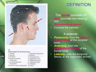
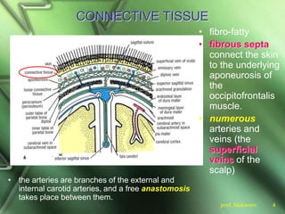
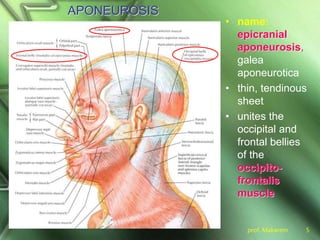
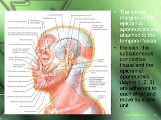
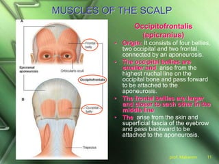
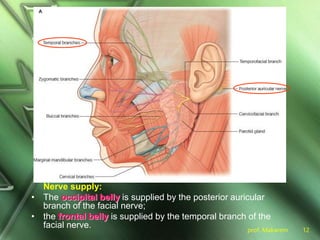
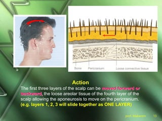
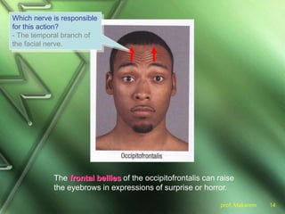
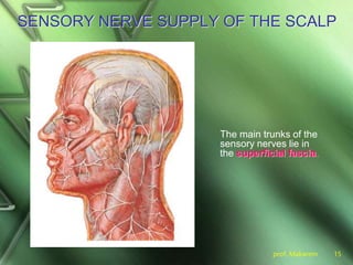
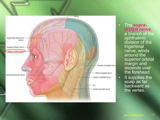
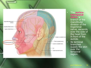
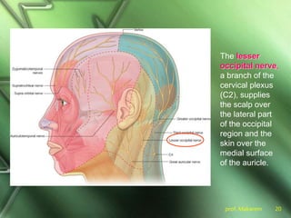
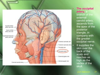
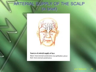
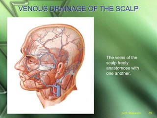
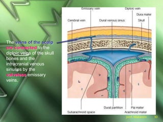
The scalp comprises skin, subcutaneous tissue, and five layers: skin, connective tissue, aponeurosis, loose areolar tissue, and pericranium, covering the calvaria. It is richly supplied with arteries and veins, including branches from the external and internal carotid arteries, and is innervated by various sensory nerves from the trigeminal nerve and cervical plexus. The scalp's structure allows for movement and expression, with significant lymphatic drainage into regional lymph nodes.


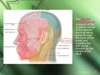
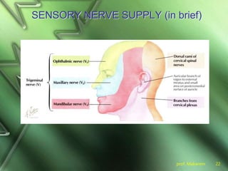
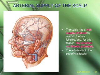

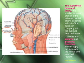


![Scalp[1]](https://cdn.slidesharecdn.com/ss_thumbnails/scalp1-170504174806-thumbnail.jpg?width=640&height=640&fit=bounds)


