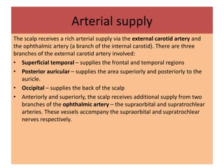
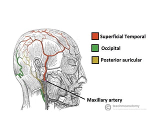
Anatomy
sir shoaib ali
Student Mujtaba H
Bs MLT
First Topic
Scalp
Define Scalp
• The scalp is composed of soft tissue layers
that cover the cranium. It is an anatomic
region bordered anteriorly by the human face,
and laterally and posteriorly by the neck. It
extends from the superior nuchal lines and
occipital turbulences to the supraorbital
foramen.
Layers of the scalp
• The scalp consists of five layers. The first three layers are tightly bound together and move as a
collective structure.
• The mnemonic ‘SCALP’ can be a useful way to remember the layers of the scalp: Skin, Dense
Connective Tissue, Epicranial Aponeurosis, Loose Areolar Connective Tissue and Periosteum.
• Skin– contains numerous hair follicles and sebaceous glands (thus a common site for
sebaceous cysts).
• Dense Connective tissue – connects the skin to the epicranial aponeurosis. It is
richly vascularised and innervated.
– The blood vessels within the layer are highly adherent to the connective tissue. This
renders them unable to constrict fully if lacerated – and so the scalp can be a site of
profuse bleeding.
• Epicranial Aponeurosis – a thin, tendon-like structure that connects the occipitalis
and frontalis muscles.
• Loose Areolar Connective Tissue – a thin connective tissue layer that separates
the periosteum of the skull from the epicranial aponeurosis.
– It contains numerous blood vessels, including emissary veins which connect the veins of
the scalp to the diploic veins and intracranial venous sinuses.
• Periosteum – the outer layer of the skull bones. It becomes continuous with the endosteum
at the suture lines.
Sturture of layers
Arterial supply
The scalp receives a rich arterial supply via the external carotid artery and
the ophthalmic artery (a branch of the internal carotid). There are three
branches of the external carotid artery involved:
• Superficial temporal – supplies the frontal and temporal regions
• Posterior auricular – supplies the area superiorly and posteriorly to the
auricle.
• Occipital – supplies the back of the scalp
• Anteriorly and superiorly, the scalp receives additional supply from two
branches of the ophthalmic artery – the supraorbital and supratrochlear
arteries. These vessels accompany the supraorbital and supratrochlear
nerves respectively.
Venous Drainage
• The venous drainage of the scalp can be divided into superficial and
deep components.
• The superficial drainage follows the arterial supply: superficial
temporal, occipital, posterior auricular, supraorbital and
supratrochlear veins.
• The deep (temporal) region of the skull is drained by the pterygoid
venous plexus. This is a large plexus of veins situated between the
temporalis and lateral pterygoid muscles,and drains into the
maxillary vein.
• Importantly, the veins of the scalp connect to the diploic veins of
the skull via valveless emissary veins. This establishes a connection
between the scalp and the dural venous sinuses.
• Innervation
• The scalp receives cutaneous innervation from branches of the trigeminal
nerve or the cervical nerve roots.
• Trigeminal Nerve
• Supratrochlear nerve – branch of the ophthalmic nerve which supplies the
anteromedial forehead.
• Supraorbital nerve – branch of the ophthalmic nerve which supplies a
large portion of the scalp between the anterolateral forehead and the
vertex.
• Zygomaticotemporal nerve – branch of the maxillary nerve, this supplies
the temple.
• Auriculotemporal nerve – branch of the mandibular nerve
which supplies skin anterosuperior to the auricle.
• Cervical Nerves
• Lesser occipital nerve – derived from the anterior ramus
(division) of C2 and supplies the skin posterior to the ear
• Greater occipital nerve – derived from the posterior ramus
(division) of C2 and supplies the skin of the occipital region.
• Great auricular nerve – derived from the anterior rami of C2
and C3 and supplies the skin posterior to the ear and over the
angle of the mandible.
• Third occipital nerve – derived from the posterior ramus of C3
and supplies the skin of the inferior occipital region.
•
• Thank you
• KMU IHS KURRAM Batch 1
• Email >hussainmujtaba91 @yahoo.com
• whatapp no :03038492224

Anatomy frist topic Scalp.pptx

  • 1.
    Anatomy sir shoaib ali StudentMujtaba H Bs MLT First Topic Scalp
  • 2.
    Define Scalp • Thescalp is composed of soft tissue layers that cover the cranium. It is an anatomic region bordered anteriorly by the human face, and laterally and posteriorly by the neck. It extends from the superior nuchal lines and occipital turbulences to the supraorbital foramen.
  • 3.
    Layers of thescalp • The scalp consists of five layers. The first three layers are tightly bound together and move as a collective structure. • The mnemonic ‘SCALP’ can be a useful way to remember the layers of the scalp: Skin, Dense Connective Tissue, Epicranial Aponeurosis, Loose Areolar Connective Tissue and Periosteum. • Skin– contains numerous hair follicles and sebaceous glands (thus a common site for sebaceous cysts). • Dense Connective tissue – connects the skin to the epicranial aponeurosis. It is richly vascularised and innervated. – The blood vessels within the layer are highly adherent to the connective tissue. This renders them unable to constrict fully if lacerated – and so the scalp can be a site of profuse bleeding. • Epicranial Aponeurosis – a thin, tendon-like structure that connects the occipitalis and frontalis muscles. • Loose Areolar Connective Tissue – a thin connective tissue layer that separates the periosteum of the skull from the epicranial aponeurosis. – It contains numerous blood vessels, including emissary veins which connect the veins of the scalp to the diploic veins and intracranial venous sinuses. • Periosteum – the outer layer of the skull bones. It becomes continuous with the endosteum at the suture lines.
  • 4.
  • 5.
    Arterial supply The scalpreceives a rich arterial supply via the external carotid artery and the ophthalmic artery (a branch of the internal carotid). There are three branches of the external carotid artery involved: • Superficial temporal – supplies the frontal and temporal regions • Posterior auricular – supplies the area superiorly and posteriorly to the auricle. • Occipital – supplies the back of the scalp • Anteriorly and superiorly, the scalp receives additional supply from two branches of the ophthalmic artery – the supraorbital and supratrochlear arteries. These vessels accompany the supraorbital and supratrochlear nerves respectively.
  • 7.
    Venous Drainage • Thevenous drainage of the scalp can be divided into superficial and deep components. • The superficial drainage follows the arterial supply: superficial temporal, occipital, posterior auricular, supraorbital and supratrochlear veins. • The deep (temporal) region of the skull is drained by the pterygoid venous plexus. This is a large plexus of veins situated between the temporalis and lateral pterygoid muscles,and drains into the maxillary vein. • Importantly, the veins of the scalp connect to the diploic veins of the skull via valveless emissary veins. This establishes a connection between the scalp and the dural venous sinuses.
  • 8.
    • Innervation • Thescalp receives cutaneous innervation from branches of the trigeminal nerve or the cervical nerve roots. • Trigeminal Nerve • Supratrochlear nerve – branch of the ophthalmic nerve which supplies the anteromedial forehead. • Supraorbital nerve – branch of the ophthalmic nerve which supplies a large portion of the scalp between the anterolateral forehead and the vertex. • Zygomaticotemporal nerve – branch of the maxillary nerve, this supplies the temple. • Auriculotemporal nerve – branch of the mandibular nerve which supplies skin anterosuperior to the auricle.
  • 9.
    • Cervical Nerves •Lesser occipital nerve – derived from the anterior ramus (division) of C2 and supplies the skin posterior to the ear • Greater occipital nerve – derived from the posterior ramus (division) of C2 and supplies the skin of the occipital region. • Great auricular nerve – derived from the anterior rami of C2 and C3 and supplies the skin posterior to the ear and over the angle of the mandible. • Third occipital nerve – derived from the posterior ramus of C3 and supplies the skin of the inferior occipital region.
  • 10.
    • • Thank you •KMU IHS KURRAM Batch 1 • Email >hussainmujtaba91 @yahoo.com • whatapp no :03038492224