SlideShare a Scribd company logo
1 of 1
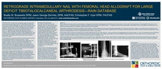
RETROGRADE INTRAMEDULLARY NAIL WITH FEMORAL HEAD ALLOGRAFT FOR LARGE DEFICIT TIBIOTALOCALCANEAL ARTHRODESIS—RAIN DATABASE Bradly W. Bussewitz DPM; Jason George DeVries, DPM, AACFAS; Christopher F. Hyer DPM, FACFAS ORTHOPEDIC FOOT & ANKLE CENTER | Columbus, OH  |   614-895-8747  |   www.orthofootankle.com INTRODUCTION: The lower limb is designed to adapt to terrain as the foot encounters ground reactive forces. To effectively accomplish this, the foot must be stable, plantigrade, and able to reach the ground. Ankle and subtalar joint (STJ) pain, arthritis, and deformity, both natural and iatrogenic, limits this ability. As one is challenged with severe hindfoot pathology a salvage procedure may involve tibiotalocalcaneal (TTC) fusion. TTC arthrodesis was originally described by Russotti and Johnson in 1988 to surgically correct these challenging scenarios (1). Correction is driven at achieving a painless, stable, plantigrade foot, with or without bracing. Additional complexity is encountered by the foot and ankle surgeon intraoperatively when a large bone void is presented during a TTC fusion. Loss of quality bone and/or loss of length in the lower extremity typically presents following avascular necrosis of the talus, Charcot neuroarthropathy of the ankle, longstanding gross malalignment, and iatrogenic changes, both acute and revisional. Regardless of the etiology, surgical correction must address the loss of height. One method of regaining bony height is by adding a structural graft at the time of fusion. There is considerable morbidity with taking large volume structural autograft. Large bone block frozen femoral heads are readily available and meet the volume and structural demands necessitated to restore bony defects at the level of the ankle.  Incorporating a large bone block allograft into a TTC arthrodesis in a single stage is a difficult task. Few reports exist to guide the foot and ankle surgeon regarding outcome expectations. The purpose of this study was to use the Retrograde Arthrodesis Intramedullary Nail (RAIN) Database to retrospectively review patients with TTC arthrodesis utilizing femoral head allograft and a retrograde intramedullary rod as a treatment for the morbidity of the pathologic hindfoot associated with substantial loss of bone. MATERIALS AND METHODS: This review was taken from the RAIN Database, which is a comprehensive chart and radiographic review constructed of all patients who have undergone extended hindfoot and ankle arthrodesis with the use of a retrograde arthrodesis nail at a single institution. Patients with femoral head allograft utilization were selected out of the entire group for evaluation. All conditions that were surgically treated with a retrograde intramedullary nail and femoral head allograft were evaluated.  All cases had failed conservative care, including some with joint sparing surgical options. The arthrodesis was performed to relieve pain and disability, including restoration and/or maintenance of limb length. Patients were all counseled on the risk of operative management.  Inclusion criteria in this specific study were all patients entered into the RAIN Database that were treated with a femoral head allograft, and surgery at least 1 year prior to data collection. Exclusion criteria included patients treated primarily with forms of fixation other than an intramedullary nail, and included plates, screws, and external fixation, and restoration of bony voids with options other than femoral head allograft.  RESULTS:  Eleven patients fit inclusion criteria and were evaluated.  Demographic data showed the average age of the patients was 54.6 years (range, 47 to 64), and included eight males. The average BMI of the group was 31.7 (range, 21.3 to 46.2).  Significant risk factors revealed five smokers and three patients with diabetes mellitus.  The etiology was Charcot neuroarthropathy in three patients, AVN in three patients, revisional fusion in two, failed total ankle replacement in two patients, and post-traumatic DJD in one patient. Surgical data revealed that the left side was affected in seven patients. All eleven patients had some type of orthobiologic augmentation to the fusion site at the time of surgery. Ten patients had bone stimulation at the time of the index procedure; four patients had implantable bone stimulation, and six used external combined magnetic field bone stimulation. Adjunctive procedures included a posterior muscle group lengthening in seven patients and complete talectomy in four. No patients had a tendon transfer or tarsal fusions. During followup after the surgery, the average time to protected weight bearing was 78.5 days (range, 42 – 204), and the time to full weightbearing was 124.8 days (range, 43 – 204). The average time to the final followup visit was 102.2 weeks (range, 28 to 180). Complications occurred in a total of six patients. This included postoperative incision and drainage including soft-tissue only in one patient and bone in one patient, partial hardware removal in one, stress fractures in two, and need for intravenous (IV) antibiotics in two. Overall, three patients went on to solid radiographic union of the ankle, and six radiographic unions of the STJ. Eight limbs were clinically stable with or without bracing. Seven patients required bracing. One patient was able to ambulate in shoes postoperatively and five walked unaided, two used a walker, and one other.  Based on our criteria for success or failure, eight patients were successful, while the three transtibial amputations were considered failures.  REFERENCES: Russotti GM, Johnson KA, Cass JR. Tibiotalocalcaneal arthrodesis for arthritis and deformity of the hind part of the foot. J Bone Joint Surg. 70A:1304-1307, 1988. CONCLUSION: The treatment of combined hindfoot and ankle pathology, regardless of etiology, is difficult to treat.  When faced with large bony deficits, the complexity increases dramatically.  The authors have reported on the results of intramedullary nail arthrodesis of the hindfoot and ankle with the use of a large volume, femoral head allograft.  This is a powerful, one-stage method of treating very complex pathology.  However, there is considerable risk as relatively small number of solid unions at the distal and proximal graft interfaces were observed.  Still, the authors feel this is a useful technique as the success rate was reasonable for this difficult population, and the results can be useful to any foot and ankle surgeon faced with this difficult clinical presentation.  Initial stabilization with external fixator Immediate post-op with internal fixation and femoral head 18 months post-op, with femoral head incorporated

More Related Content

What's hot

2009 ghassemi-comparsion of donor beckjoms
2009 ghassemi-comparsion of donor beckjoms2009 ghassemi-comparsion of donor beckjoms
2009 ghassemi-comparsion of donor beckjomsKlinikum Lippe GmbH
 
Radiological parameters in patients with patellofemoral pathology
Radiological parameters in patients with patellofemoral pathologyRadiological parameters in patients with patellofemoral pathology
Radiological parameters in patients with patellofemoral pathologyProfessor M. A. Imam
 
Displasia anca ISTA Dubai
Displasia anca ISTA DubaiDisplasia anca ISTA Dubai
Displasia anca ISTA Dubaibestmix
 
The Direct Anterior Hip Replacement
The Direct Anterior Hip ReplacementThe Direct Anterior Hip Replacement
The Direct Anterior Hip Replacementwashingtonortho
 
Crimson Publishers-Abdominal Pain Caused by Bilateral Acetabular Fractures Se...
Crimson Publishers-Abdominal Pain Caused by Bilateral Acetabular Fractures Se...Crimson Publishers-Abdominal Pain Caused by Bilateral Acetabular Fractures Se...
Crimson Publishers-Abdominal Pain Caused by Bilateral Acetabular Fractures Se...CrimsonPublishersOPROJ
 
Percutaneous fixation of bilateral anterior column acetabular fractures: A ca...
Percutaneous fixation of bilateral anterior column acetabular fractures: A ca...Percutaneous fixation of bilateral anterior column acetabular fractures: A ca...
Percutaneous fixation of bilateral anterior column acetabular fractures: A ca...Apollo Hospitals
 
A comparative study on the clinical and functional outcome of limb salvage su...
A comparative study on the clinical and functional outcome of limb salvage su...A comparative study on the clinical and functional outcome of limb salvage su...
A comparative study on the clinical and functional outcome of limb salvage su...NAAR Journal
 
Guidelines for DVT Prophylaxis
Guidelines for DVT ProphylaxisGuidelines for DVT Prophylaxis
Guidelines for DVT ProphylaxisArun Shanbhag
 
Two treatment methods for spiral fracture of the lower third of the tibia sha...
Two treatment methods for spiral fracture of the lower third of the tibia sha...Two treatment methods for spiral fracture of the lower third of the tibia sha...
Two treatment methods for spiral fracture of the lower third of the tibia sha...Clinical Surgery Research Communications
 
Anterolateral ligament in pediatric knees a radiographic study
Anterolateral ligament in pediatric knees a radiographic studyAnterolateral ligament in pediatric knees a radiographic study
Anterolateral ligament in pediatric knees a radiographic studyProf. Hesham N. Mustafa
 
Anatomical Glenoid Reconstruction for Recurrent Anterior Glenohumeral Instabi...
Anatomical Glenoid Reconstruction for Recurrent Anterior Glenohumeral Instabi...Anatomical Glenoid Reconstruction for Recurrent Anterior Glenohumeral Instabi...
Anatomical Glenoid Reconstruction for Recurrent Anterior Glenohumeral Instabi...Peter Millett MD
 

What's hot (20)

2009 ghassemi-comparsion of donor beckjoms
2009 ghassemi-comparsion of donor beckjoms2009 ghassemi-comparsion of donor beckjoms
2009 ghassemi-comparsion of donor beckjoms
 
Df w recon
Df w reconDf w recon
Df w recon
 
Radiological parameters in patients with patellofemoral pathology
Radiological parameters in patients with patellofemoral pathologyRadiological parameters in patients with patellofemoral pathology
Radiological parameters in patients with patellofemoral pathology
 
Rotationplasty
RotationplastyRotationplasty
Rotationplasty
 
Displasia anca ISTA Dubai
Displasia anca ISTA DubaiDisplasia anca ISTA Dubai
Displasia anca ISTA Dubai
 
The Direct Anterior Hip Replacement
The Direct Anterior Hip ReplacementThe Direct Anterior Hip Replacement
The Direct Anterior Hip Replacement
 
eidelman2016.pdf
eidelman2016.pdfeidelman2016.pdf
eidelman2016.pdf
 
Crimson Publishers-Abdominal Pain Caused by Bilateral Acetabular Fractures Se...
Crimson Publishers-Abdominal Pain Caused by Bilateral Acetabular Fractures Se...Crimson Publishers-Abdominal Pain Caused by Bilateral Acetabular Fractures Se...
Crimson Publishers-Abdominal Pain Caused by Bilateral Acetabular Fractures Se...
 
Percutaneous fixation of bilateral anterior column acetabular fractures: A ca...
Percutaneous fixation of bilateral anterior column acetabular fractures: A ca...Percutaneous fixation of bilateral anterior column acetabular fractures: A ca...
Percutaneous fixation of bilateral anterior column acetabular fractures: A ca...
 
A comparative study on the clinical and functional outcome of limb salvage su...
A comparative study on the clinical and functional outcome of limb salvage su...A comparative study on the clinical and functional outcome of limb salvage su...
A comparative study on the clinical and functional outcome of limb salvage su...
 
BMAC in cuff repairs
BMAC in cuff repairsBMAC in cuff repairs
BMAC in cuff repairs
 
Cementless TKA
Cementless TKACementless TKA
Cementless TKA
 
68th publication sjmcr - 1st name
68th publication  sjmcr - 1st name68th publication  sjmcr - 1st name
68th publication sjmcr - 1st name
 
Guidelines for DVT Prophylaxis
Guidelines for DVT ProphylaxisGuidelines for DVT Prophylaxis
Guidelines for DVT Prophylaxis
 
Two treatment methods for spiral fracture of the lower third of the tibia sha...
Two treatment methods for spiral fracture of the lower third of the tibia sha...Two treatment methods for spiral fracture of the lower third of the tibia sha...
Two treatment methods for spiral fracture of the lower third of the tibia sha...
 
GOODAY.ARTICLE.Final
GOODAY.ARTICLE.FinalGOODAY.ARTICLE.Final
GOODAY.ARTICLE.Final
 
Anterolateral ligament in pediatric knees a radiographic study
Anterolateral ligament in pediatric knees a radiographic studyAnterolateral ligament in pediatric knees a radiographic study
Anterolateral ligament in pediatric knees a radiographic study
 
Aao2016 recon
Aao2016 reconAao2016 recon
Aao2016 recon
 
Re-operation Rates After Combined ACL and Anterolateral Ligament Reconstruction
Re-operation Rates After Combined ACL and Anterolateral Ligament ReconstructionRe-operation Rates After Combined ACL and Anterolateral Ligament Reconstruction
Re-operation Rates After Combined ACL and Anterolateral Ligament Reconstruction
 
Anatomical Glenoid Reconstruction for Recurrent Anterior Glenohumeral Instabi...
Anatomical Glenoid Reconstruction for Recurrent Anterior Glenohumeral Instabi...Anatomical Glenoid Reconstruction for Recurrent Anterior Glenohumeral Instabi...
Anatomical Glenoid Reconstruction for Recurrent Anterior Glenohumeral Instabi...
 

Similar to Retrograde Intramedullary Nail with Femoral Head Allograft for Large Deficit Tibiotalocalcaneal Arthrodesis: RAIN Database

Revision Total Ankle Arthroplasty to Tibial Stemmed Implant: A Case Report
Revision Total Ankle Arthroplasty to Tibial Stemmed Implant: A Case ReportRevision Total Ankle Arthroplasty to Tibial Stemmed Implant: A Case Report
Revision Total Ankle Arthroplasty to Tibial Stemmed Implant: A Case Reportskisnfeet
 
Comparison Results between Patients with Developmental Hip Dysplasia Treated ...
Comparison Results between Patients with Developmental Hip Dysplasia Treated ...Comparison Results between Patients with Developmental Hip Dysplasia Treated ...
Comparison Results between Patients with Developmental Hip Dysplasia Treated ...CrimsonPublishersOPROJ
 
AUTOLOGUS CHONDROCYTE IMPLANTATION PRIYANK
AUTOLOGUS CHONDROCYTE IMPLANTATION PRIYANKAUTOLOGUS CHONDROCYTE IMPLANTATION PRIYANK
AUTOLOGUS CHONDROCYTE IMPLANTATION PRIYANKDr Khushbu
 
AUTOLOGUS CHONDROCYTE IMPLANTATION PRIYANK
AUTOLOGUS CHONDROCYTE IMPLANTATION PRIYANKAUTOLOGUS CHONDROCYTE IMPLANTATION PRIYANK
AUTOLOGUS CHONDROCYTE IMPLANTATION PRIYANKDr Khushbu
 
British Columbia Medical Journal - November 2010: Knee Arthroplasty
British Columbia Medical Journal - November 2010: Knee ArthroplastyBritish Columbia Medical Journal - November 2010: Knee Arthroplasty
British Columbia Medical Journal - November 2010: Knee ArthroplastyBritish Columbia Medical Journal
 
TKA for severe valgus
TKA for severe valgusTKA for severe valgus
TKA for severe valgusFernando Gf
 
In hospital complications after total joint arthroplasty
In hospital complications after total joint arthroplastyIn hospital complications after total joint arthroplasty
In hospital complications after total joint arthroplastyFUAD HAZIME
 
Megaprosthetic replacement of knee in a young boy of 14 years
Megaprosthetic replacement of knee in a young boy of 14 yearsMegaprosthetic replacement of knee in a young boy of 14 years
Megaprosthetic replacement of knee in a young boy of 14 yearsApollo Hospitals
 
Comparative study of functional outcome of lateral locking plate fixation an...
Comparative study of functional outcome of  lateral locking plate fixation an...Comparative study of functional outcome of  lateral locking plate fixation an...
Comparative study of functional outcome of lateral locking plate fixation an...Om Patil
 
Corrigendum to “Special surgical technique for knee arthroplasty”
Corrigendum to “Special surgical technique for knee arthroplasty”Corrigendum to “Special surgical technique for knee arthroplasty”
Corrigendum to “Special surgical technique for knee arthroplasty”Apollo Hospitals
 
Bone conserving hip arthroplasty
Bone conserving hip arthroplastyBone conserving hip arthroplasty
Bone conserving hip arthroplastyApollo Hospitals
 
Biocartilage to Treat Osteochondral Defects of the Talus: Case Report and Tec...
Biocartilage to Treat Osteochondral Defects of the Talus: Case Report and Tec...Biocartilage to Treat Osteochondral Defects of the Talus: Case Report and Tec...
Biocartilage to Treat Osteochondral Defects of the Talus: Case Report and Tec...Jennifer Gerres, DPM
 
Management of extensor mechanism deficit as a consequence of patellar tendon ...
Management of extensor mechanism deficit as a consequence of patellar tendon ...Management of extensor mechanism deficit as a consequence of patellar tendon ...
Management of extensor mechanism deficit as a consequence of patellar tendon ...FUAD HAZIME
 

Similar to Retrograde Intramedullary Nail with Femoral Head Allograft for Large Deficit Tibiotalocalcaneal Arthrodesis: RAIN Database (20)

Revision Total Ankle Arthroplasty to Tibial Stemmed Implant: A Case Report
Revision Total Ankle Arthroplasty to Tibial Stemmed Implant: A Case ReportRevision Total Ankle Arthroplasty to Tibial Stemmed Implant: A Case Report
Revision Total Ankle Arthroplasty to Tibial Stemmed Implant: A Case Report
 
Lower Limb Reconstruction Using Tibial Strut.pdf
Lower Limb Reconstruction Using Tibial Strut.pdfLower Limb Reconstruction Using Tibial Strut.pdf
Lower Limb Reconstruction Using Tibial Strut.pdf
 
Comparison Results between Patients with Developmental Hip Dysplasia Treated ...
Comparison Results between Patients with Developmental Hip Dysplasia Treated ...Comparison Results between Patients with Developmental Hip Dysplasia Treated ...
Comparison Results between Patients with Developmental Hip Dysplasia Treated ...
 
Complete subtalar release for older children.pdf
Complete subtalar release for older children.pdfComplete subtalar release for older children.pdf
Complete subtalar release for older children.pdf
 
Christensen prosthesis
Christensen prosthesisChristensen prosthesis
Christensen prosthesis
 
AUTOLOGUS CHONDROCYTE IMPLANTATION PRIYANK
AUTOLOGUS CHONDROCYTE IMPLANTATION PRIYANKAUTOLOGUS CHONDROCYTE IMPLANTATION PRIYANK
AUTOLOGUS CHONDROCYTE IMPLANTATION PRIYANK
 
AUTOLOGUS CHONDROCYTE IMPLANTATION PRIYANK
AUTOLOGUS CHONDROCYTE IMPLANTATION PRIYANKAUTOLOGUS CHONDROCYTE IMPLANTATION PRIYANK
AUTOLOGUS CHONDROCYTE IMPLANTATION PRIYANK
 
Complete subtalar release for older children with neglected CTEV - البروفيسو...
 Complete subtalar release for older children with neglected CTEV - البروفيسو... Complete subtalar release for older children with neglected CTEV - البروفيسو...
Complete subtalar release for older children with neglected CTEV - البروفيسو...
 
British Columbia Medical Journal - November 2010: Knee Arthroplasty
British Columbia Medical Journal - November 2010: Knee ArthroplastyBritish Columbia Medical Journal - November 2010: Knee Arthroplasty
British Columbia Medical Journal - November 2010: Knee Arthroplasty
 
TKA for severe valgus
TKA for severe valgusTKA for severe valgus
TKA for severe valgus
 
In hospital complications after total joint arthroplasty
In hospital complications after total joint arthroplastyIn hospital complications after total joint arthroplasty
In hospital complications after total joint arthroplasty
 
Megaprosthetic replacement of knee in a young boy of 14 years
Megaprosthetic replacement of knee in a young boy of 14 yearsMegaprosthetic replacement of knee in a young boy of 14 years
Megaprosthetic replacement of knee in a young boy of 14 years
 
G04602048057
G04602048057G04602048057
G04602048057
 
Comparative study of functional outcome of lateral locking plate fixation an...
Comparative study of functional outcome of  lateral locking plate fixation an...Comparative study of functional outcome of  lateral locking plate fixation an...
Comparative study of functional outcome of lateral locking plate fixation an...
 
International Journal of Orthopedics: Research & Therapy
International Journal of Orthopedics: Research & TherapyInternational Journal of Orthopedics: Research & Therapy
International Journal of Orthopedics: Research & Therapy
 
Corrigendum to “Special surgical technique for knee arthroplasty”
Corrigendum to “Special surgical technique for knee arthroplasty”Corrigendum to “Special surgical technique for knee arthroplasty”
Corrigendum to “Special surgical technique for knee arthroplasty”
 
Bone conserving hip arthroplasty
Bone conserving hip arthroplastyBone conserving hip arthroplasty
Bone conserving hip arthroplasty
 
Biocartilage to Treat Osteochondral Defects of the Talus: Case Report and Tec...
Biocartilage to Treat Osteochondral Defects of the Talus: Case Report and Tec...Biocartilage to Treat Osteochondral Defects of the Talus: Case Report and Tec...
Biocartilage to Treat Osteochondral Defects of the Talus: Case Report and Tec...
 
Management of extensor mechanism deficit as a consequence of patellar tendon ...
Management of extensor mechanism deficit as a consequence of patellar tendon ...Management of extensor mechanism deficit as a consequence of patellar tendon ...
Management of extensor mechanism deficit as a consequence of patellar tendon ...
 
Ablation of osteoid osteoma - البروفيسور فريح ابوحسان – استشاري جراحة العظام ...
Ablation of osteoid osteoma - البروفيسور فريح ابوحسان – استشاري جراحة العظام ...Ablation of osteoid osteoma - البروفيسور فريح ابوحسان – استشاري جراحة العظام ...
Ablation of osteoid osteoma - البروفيسور فريح ابوحسان – استشاري جراحة العظام ...
 

More from skisnfeet

Recurrent Metatarsal Synostosis Resection and Interposition with Human Alloge...
Recurrent Metatarsal Synostosis Resection and Interposition with Human Alloge...Recurrent Metatarsal Synostosis Resection and Interposition with Human Alloge...
Recurrent Metatarsal Synostosis Resection and Interposition with Human Alloge...skisnfeet
 
Autogenous Bone Graft Harvest Using Reamer-Irrigator-Aspirator (RIA) Techniqu...
Autogenous Bone Graft Harvest Using Reamer-Irrigator-Aspirator (RIA) Techniqu...Autogenous Bone Graft Harvest Using Reamer-Irrigator-Aspirator (RIA) Techniqu...
Autogenous Bone Graft Harvest Using Reamer-Irrigator-Aspirator (RIA) Techniqu...skisnfeet
 
The Effect of Recombinant Bone Morphogenetic Protien-2 in Revison Tibiotaloca...
The Effect of Recombinant Bone Morphogenetic Protien-2 in Revison Tibiotaloca...The Effect of Recombinant Bone Morphogenetic Protien-2 in Revison Tibiotaloca...
The Effect of Recombinant Bone Morphogenetic Protien-2 in Revison Tibiotaloca...skisnfeet
 
Pentagon Intraarticular Osteotomy: A Novel Surgical Approach to Complex Defor...
Pentagon Intraarticular Osteotomy: A Novel Surgical Approach to Complex Defor...Pentagon Intraarticular Osteotomy: A Novel Surgical Approach to Complex Defor...
Pentagon Intraarticular Osteotomy: A Novel Surgical Approach to Complex Defor...skisnfeet
 
Cannulated Screw Fixation of Jones 5th Metatarsal Fracture: Comparison of Tit...
Cannulated Screw Fixation of Jones 5th Metatarsal Fracture: Comparison of Tit...Cannulated Screw Fixation of Jones 5th Metatarsal Fracture: Comparison of Tit...
Cannulated Screw Fixation of Jones 5th Metatarsal Fracture: Comparison of Tit...skisnfeet
 
Surgical Treatment of Haglund Triad by Using Complete Detachment and Reattach...
Surgical Treatment of Haglund Triad by Using Complete Detachment and Reattach...Surgical Treatment of Haglund Triad by Using Complete Detachment and Reattach...
Surgical Treatment of Haglund Triad by Using Complete Detachment and Reattach...skisnfeet
 
Union Rate of Tibiotalocalcaneal Nail with Internal or External Bone Stimulat...
Union Rate of Tibiotalocalcaneal Nail with Internal or External Bone Stimulat...Union Rate of Tibiotalocalcaneal Nail with Internal or External Bone Stimulat...
Union Rate of Tibiotalocalcaneal Nail with Internal or External Bone Stimulat...skisnfeet
 

More from skisnfeet (7)

Recurrent Metatarsal Synostosis Resection and Interposition with Human Alloge...
Recurrent Metatarsal Synostosis Resection and Interposition with Human Alloge...Recurrent Metatarsal Synostosis Resection and Interposition with Human Alloge...
Recurrent Metatarsal Synostosis Resection and Interposition with Human Alloge...
 
Autogenous Bone Graft Harvest Using Reamer-Irrigator-Aspirator (RIA) Techniqu...
Autogenous Bone Graft Harvest Using Reamer-Irrigator-Aspirator (RIA) Techniqu...Autogenous Bone Graft Harvest Using Reamer-Irrigator-Aspirator (RIA) Techniqu...
Autogenous Bone Graft Harvest Using Reamer-Irrigator-Aspirator (RIA) Techniqu...
 
The Effect of Recombinant Bone Morphogenetic Protien-2 in Revison Tibiotaloca...
The Effect of Recombinant Bone Morphogenetic Protien-2 in Revison Tibiotaloca...The Effect of Recombinant Bone Morphogenetic Protien-2 in Revison Tibiotaloca...
The Effect of Recombinant Bone Morphogenetic Protien-2 in Revison Tibiotaloca...
 
Pentagon Intraarticular Osteotomy: A Novel Surgical Approach to Complex Defor...
Pentagon Intraarticular Osteotomy: A Novel Surgical Approach to Complex Defor...Pentagon Intraarticular Osteotomy: A Novel Surgical Approach to Complex Defor...
Pentagon Intraarticular Osteotomy: A Novel Surgical Approach to Complex Defor...
 
Cannulated Screw Fixation of Jones 5th Metatarsal Fracture: Comparison of Tit...
Cannulated Screw Fixation of Jones 5th Metatarsal Fracture: Comparison of Tit...Cannulated Screw Fixation of Jones 5th Metatarsal Fracture: Comparison of Tit...
Cannulated Screw Fixation of Jones 5th Metatarsal Fracture: Comparison of Tit...
 
Surgical Treatment of Haglund Triad by Using Complete Detachment and Reattach...
Surgical Treatment of Haglund Triad by Using Complete Detachment and Reattach...Surgical Treatment of Haglund Triad by Using Complete Detachment and Reattach...
Surgical Treatment of Haglund Triad by Using Complete Detachment and Reattach...
 
Union Rate of Tibiotalocalcaneal Nail with Internal or External Bone Stimulat...
Union Rate of Tibiotalocalcaneal Nail with Internal or External Bone Stimulat...Union Rate of Tibiotalocalcaneal Nail with Internal or External Bone Stimulat...
Union Rate of Tibiotalocalcaneal Nail with Internal or External Bone Stimulat...
 

Retrograde Intramedullary Nail with Femoral Head Allograft for Large Deficit Tibiotalocalcaneal Arthrodesis: RAIN Database

  • 1. RETROGRADE INTRAMEDULLARY NAIL WITH FEMORAL HEAD ALLOGRAFT FOR LARGE DEFICIT TIBIOTALOCALCANEAL ARTHRODESIS—RAIN DATABASE Bradly W. Bussewitz DPM; Jason George DeVries, DPM, AACFAS; Christopher F. Hyer DPM, FACFAS ORTHOPEDIC FOOT & ANKLE CENTER | Columbus, OH | 614-895-8747 | www.orthofootankle.com INTRODUCTION: The lower limb is designed to adapt to terrain as the foot encounters ground reactive forces. To effectively accomplish this, the foot must be stable, plantigrade, and able to reach the ground. Ankle and subtalar joint (STJ) pain, arthritis, and deformity, both natural and iatrogenic, limits this ability. As one is challenged with severe hindfoot pathology a salvage procedure may involve tibiotalocalcaneal (TTC) fusion. TTC arthrodesis was originally described by Russotti and Johnson in 1988 to surgically correct these challenging scenarios (1). Correction is driven at achieving a painless, stable, plantigrade foot, with or without bracing. Additional complexity is encountered by the foot and ankle surgeon intraoperatively when a large bone void is presented during a TTC fusion. Loss of quality bone and/or loss of length in the lower extremity typically presents following avascular necrosis of the talus, Charcot neuroarthropathy of the ankle, longstanding gross malalignment, and iatrogenic changes, both acute and revisional. Regardless of the etiology, surgical correction must address the loss of height. One method of regaining bony height is by adding a structural graft at the time of fusion. There is considerable morbidity with taking large volume structural autograft. Large bone block frozen femoral heads are readily available and meet the volume and structural demands necessitated to restore bony defects at the level of the ankle. Incorporating a large bone block allograft into a TTC arthrodesis in a single stage is a difficult task. Few reports exist to guide the foot and ankle surgeon regarding outcome expectations. The purpose of this study was to use the Retrograde Arthrodesis Intramedullary Nail (RAIN) Database to retrospectively review patients with TTC arthrodesis utilizing femoral head allograft and a retrograde intramedullary rod as a treatment for the morbidity of the pathologic hindfoot associated with substantial loss of bone. MATERIALS AND METHODS: This review was taken from the RAIN Database, which is a comprehensive chart and radiographic review constructed of all patients who have undergone extended hindfoot and ankle arthrodesis with the use of a retrograde arthrodesis nail at a single institution. Patients with femoral head allograft utilization were selected out of the entire group for evaluation. All conditions that were surgically treated with a retrograde intramedullary nail and femoral head allograft were evaluated. All cases had failed conservative care, including some with joint sparing surgical options. The arthrodesis was performed to relieve pain and disability, including restoration and/or maintenance of limb length. Patients were all counseled on the risk of operative management. Inclusion criteria in this specific study were all patients entered into the RAIN Database that were treated with a femoral head allograft, and surgery at least 1 year prior to data collection. Exclusion criteria included patients treated primarily with forms of fixation other than an intramedullary nail, and included plates, screws, and external fixation, and restoration of bony voids with options other than femoral head allograft. RESULTS: Eleven patients fit inclusion criteria and were evaluated. Demographic data showed the average age of the patients was 54.6 years (range, 47 to 64), and included eight males. The average BMI of the group was 31.7 (range, 21.3 to 46.2). Significant risk factors revealed five smokers and three patients with diabetes mellitus. The etiology was Charcot neuroarthropathy in three patients, AVN in three patients, revisional fusion in two, failed total ankle replacement in two patients, and post-traumatic DJD in one patient. Surgical data revealed that the left side was affected in seven patients. All eleven patients had some type of orthobiologic augmentation to the fusion site at the time of surgery. Ten patients had bone stimulation at the time of the index procedure; four patients had implantable bone stimulation, and six used external combined magnetic field bone stimulation. Adjunctive procedures included a posterior muscle group lengthening in seven patients and complete talectomy in four. No patients had a tendon transfer or tarsal fusions. During followup after the surgery, the average time to protected weight bearing was 78.5 days (range, 42 – 204), and the time to full weightbearing was 124.8 days (range, 43 – 204). The average time to the final followup visit was 102.2 weeks (range, 28 to 180). Complications occurred in a total of six patients. This included postoperative incision and drainage including soft-tissue only in one patient and bone in one patient, partial hardware removal in one, stress fractures in two, and need for intravenous (IV) antibiotics in two. Overall, three patients went on to solid radiographic union of the ankle, and six radiographic unions of the STJ. Eight limbs were clinically stable with or without bracing. Seven patients required bracing. One patient was able to ambulate in shoes postoperatively and five walked unaided, two used a walker, and one other. Based on our criteria for success or failure, eight patients were successful, while the three transtibial amputations were considered failures. REFERENCES: Russotti GM, Johnson KA, Cass JR. Tibiotalocalcaneal arthrodesis for arthritis and deformity of the hind part of the foot. J Bone Joint Surg. 70A:1304-1307, 1988. CONCLUSION: The treatment of combined hindfoot and ankle pathology, regardless of etiology, is difficult to treat. When faced with large bony deficits, the complexity increases dramatically. The authors have reported on the results of intramedullary nail arthrodesis of the hindfoot and ankle with the use of a large volume, femoral head allograft. This is a powerful, one-stage method of treating very complex pathology. However, there is considerable risk as relatively small number of solid unions at the distal and proximal graft interfaces were observed. Still, the authors feel this is a useful technique as the success rate was reasonable for this difficult population, and the results can be useful to any foot and ankle surgeon faced with this difficult clinical presentation. Initial stabilization with external fixator Immediate post-op with internal fixation and femoral head 18 months post-op, with femoral head incorporated