Role of cross-leg flaps in lower leg reconstruction
•
1 like•359 views

This document summarizes a study on the outcomes of occlusion treatment for amblyopia in children under 12 years old with strabismus. The study reviewed medical records of 38 Qatari children treated with occlusion therapy for strabismic amblyopia from 1992-2002. Good outcomes, defined as final visual acuity of 6/9 or better, were found in 73% of patients. Poor outcomes with visual acuity less than 6/9 occurred in 26% of patients. Factors like age at presentation, type of strabismus, presence of anisometropia and compliance did not significantly affect treatment outcomes.

Recommended
Recommended
More Related Content
What's hot
What's hot (20)
Viewers also liked
Viewers also liked (18)
Similar to Role of cross-leg flaps in lower leg reconstruction
Similar to Role of cross-leg flaps in lower leg reconstruction (17)
More from Prof Freih Abu Hassan البروفيسور فريح ابوحسان
More from Prof Freih Abu Hassan البروفيسور فريح ابوحسان (20)
Recently uploaded
Recently uploaded (20)
Role of cross-leg flaps in lower leg reconstruction
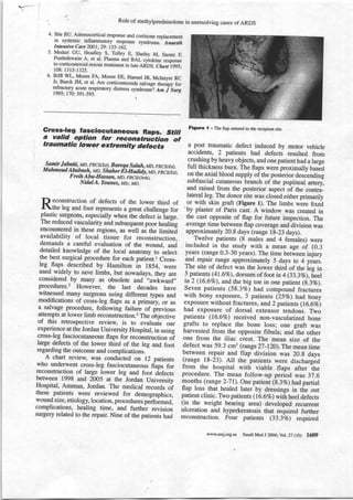
- 1. r- Role of methylprednisolone in unrosolving cases of ARDS 4. Ihle BU. Adrenocortical response and cortisone replacement in systemic inflammatory response syndrome. Anaesth Intensive Care 2001 29: 155- 16A. 5. Meduri GU, Headley s, Tolley E, Shelby M, stentz F, Postlethwaite A, et al. Plasma and BAL .yiotin";;rp*; to corticosteroid rescue treatment in late ARDS. ChestiggS; 108:1315-l3ZS. 6. Biffl wL, Moore FA, Moore EE, Haenel JB, Mclntyre Jr, RC Burch JM, et al. Are corticosteroids salvage therapy for refractory a9_ute respiratory distress syndro*"r Am i sorg 1995; 170: 591-595. 1 - Gross-l€g fasciocutaneous flaps. stiil a valid option for reconstruhtion of traumatic lower extremity defecfs samir Jabaiti, MD, FRCS(Ed), Bareqa salah, MD, FRCS(Eil), M ahmo ud abab ne lg uo_, s haher E l-H odiiy,-M;, F RC s ( E d ), Freih Abu-Hassan, MD, rncslortnl, NidalA. Younes, MSc, MD. p econstruction of defects of the lower third of rthe leg and foot represents a great challenge for plastic surgeons, especially when itre defect is iarge. The reduced vascularify and subsequent poor healiig encountered in these regions, as well aJ the timitel availability of local tissue for reconstruction, demands a careful evaluation of the wound, lod detailed knowledge of the local anatomy to select the best surgical procedure for each patient.l Cross- Ieg flaps described by Hamilton ir lgs4, were used widely to save limbs, but nowadsys, they are considered by many as obsolete and '.awkward" procedures.2 However, the lasf decades have witnessed many surgeons using different types and modifications of cross-leg flaps as a prim erry, or as a salvage procedure, following failurb of previous attempts at lower limb reconstruction.2 The oUiective of this retrospective review, is to evaluaie our experience at the Jordan University Hospital, in using cross-leg fasciocutaneous flaps for reconstruction o1 large defects of the lower UrirA of the leg and foot regarding the outcome and complications. A chart review, was conducted on lz patients who undenvent cross-leg fasciocutaneous flaps for reconstruction of large lower leg and foot difects between 1998 and 2005 at the Jordan University Hospital, Amman, Jordan. The medical records of these patients were reviewed for demographics, wound size, etiology, locatior, procedures perronneo, complications, healing time, and furttrer revision surgery related to the repair. Nine of the patients had Figure I . The flap sutured to the recipient site. a post traumatic defect induced by motor vehicle accidents, 2 patients had defects resulted from grushing by heavy objects, and one patient had a large full thickness burn. The flaps were proximally UasJO on the axial blood supply of the posterior desCending subfascial cutaneous branch of the popliteal utt"ri and raised frorn the posterior aspeci of ttre contri-lateral leg. The donor site was cloied either primarity or with skin graft (Figure l). The limbs wlre fixei 'by plaster of Paris cast. A window was created in the cast opposite of flap for future inspection. The average time between flap coverage and division was approximately 20.8 days (range 18-23 days). Twelve patients (8 males and 4 females) were included in the study with a mean age of 10.3 years (range 0.3-30 years). The time between injury Td repair_range approximately 5 days to 4 y.*r. The site of defect was the lower third of the ieg in 5 patients (4l.6vo), dorsum of foot in 4 (33.3vo),f,eel in 2 (l6.6vo), and the big toe in one patient (g .3vo). seven patients (58 .3vo) had compound fractures with bony exposure, 3 patients (25vo) had bony exposure without fractures, and Z patients ( 16.6q;) had exposure of dorsal extensoi tendons. Two patients (l6.6vo) received non-vasculari zed bone grafts to replace the bone loss; one graft was harvested from the opposite fibula; andlhe other one from the iliac crest. The mean size of the defect was 59.3 cm2 (range 27-r20).The mean time between repair and flap division was 20.g days (range 18-23). AII the patients were discharged from the hospital with viable flaps after iH" procedure. The mean follow-up period was 37.6 months (ran ge 2-71). one patient (g.3 vo) hadpartial flap loss that healed later by dressings in the out patient clinic. Two patients ( l6.6Vo) with heel defects (in the weight bearing area) developed recurrent ulceration and hyperkeratosis that required further reconstruction. Four patients (33 .3vo) required www.smj.org.sa Saudi Med I 2ffi6;yot.27 (10) 1609
- 2. Y Cross-leg fasciocutaneous fl aPs Table t ' Operative data and complications. Data and complications N (Vo) Period from injury to repair (range) (5 days - 4 years) Operative time in minutes: mean (range) 92.L (55-165) Donor site repair Sptit-thickness skin graft 3 ' (25) Full-thickness skin graft 8 (66.7) Primary repair I (8.3) ', Time for repair to flap division in days: mean (range) Complications Partial flap necrosis Wound infection Joint stiffness 20.8 (18-23) 1 (8.3) 0 (0) 0 (0) Recurrent ulceration and hyperkeratosis 2 (16.6) Minor flap revision or thinning 4 (33.3) minor flap revision for better cosmesis. A11 the other patients maintained durable soft tissue cover with satisfactory esthetic results. None of the patients had wound infection or joint stiffness (Thble L). Management of traumatic lower limb soft tissue defects, remains a major challenge to plastic surgeons. The beginning of microsurgery in the 1970s, and the introduction of myocutaneous and fasciocutaneous flaps by Ponten3 in 1981, have revolutionized the reconstruction of lower extremity defects. Free flaps using the microsurgical techniques, have been used successfully to cover acute and chronic large lower extremity defects.a However, free flaps require special skills and relatively expensive instrumentation not readily available to all reconstructi'Le surgeons, particularly in the developing countries. Moreover in serious cases, free flaps are highly risky or even difficult to perform.z The other alternative is to use local fasciocutaneous and muscle flaps. This option however, may not also be achievable due to the absence of adequate healthy 'local tissues. In such circumstances, the use of cross leg fasciocutaneous flaps offers a valid alternative to free flaps or local flaps. The major drawbacks of this procedure include an unreliable blood supply, limited arc of rotation caused by a short and thick pedicle, and the need for inconvenient postoperative immobilization.z Some surgeons used external fixation devices to achieve better patient convenience and joint mobility, in addition to facilitating flap monitoring and wound care. The optimal time for flap division has not been determined in the literature; Thatte et al5 divided 10 1610 Saudi Med J 2006; Yol.27 (10) www.smj.org.sa cro s s - le g fas c iocutaneou s fl ap s on the tenth day s w ithout complications. George et al6 used a simple occlusion clamp with screws to apply gradual tightening at the pedicle, producing intermittent periods of ischemia, they could divide the flap safely after 9 to L4 days (mean 10 days). However, in this series, all flaps were divided aftgr 18 days. The facilities for free tissue transfer in our center, like most of the centers in the developing countries are still lacking. We depend mainly on cross-leg posterior tibial fasciocutaneous flaps to repair defects of the lower leg and foot. Only 9Vo of our patients in this series had partial flap necrosis, which compares well with the rate of partial or complete flap loss reported by other series (0- 26.9%o).'n Soft tissue defects in weight bearing areas, such as the heel regions; have long been viewed as troublesome due to the continuous pressure load, and the special anatomical nature of these areas. Two patients with heel defects (I6.6Vo) in this series had recurrent ulcerations following cross-leg flaps. Cross-leg flaps , are still safe and reliable method for soft tissue reconstruction of traumatic lower extremity defects. They should be viewed as a viable alternative for wounds with extensive exposure of bone and tendon. These flaps provide similar tissue to that lost, they are easy to raise, require short operative time, are associated with minimal blood loss, and they preserve the major arteries in the traumatized leg. Acknowledgment. This work was supported by the Faculty of MedicineAJniversity of Jordan. We would like to thank Saleh Massad (Medical Photography) for his help in processing the picture. Received 25th February 2006. Acceptedfor publication infinal form 28th June 2006. From the Section of Plastic & Reconstructive Surgery, Department of Surgery Qabaiti, Salah), Section of Orthopedics, Deparnnent of Special Surgery (Ababneh, El-Hadidy, Hassan) and the Section of Endocrine Surgery, Departrnent of Surgery (Younes), Faculty of MedicinelUniversity of Jordan, Amman, Jordan. Address correspondence and reprint requests to: Dr. Nidnl A. Younes, Section of Endocrine Surgery, Department of Surgery, Faculty of MedicinelUniversity of Jordan, PO Box I3024,Amman 11942, Jordan. Fax. +926 5353388. E-mail: niyounes@ju.eduio References 1. De Almeida OM, Monteiro AA Jr, Neves R[, de Lemos RG, BrazJC, Brechtbuhl ER, et al. Distally based fasciocutaneous ' flap of the calf for cutaneous coverage of the lower leg and dorsum of the foot. Ann Plast Surg 2000; 44: 367 -373. 2. Long CD, Granick MS, Solomon MP. The cross-leg flap revisited. Ann Plast Surg 1993; 30: 560-563. 3. Ponten B. The fasciocutaneous flap: its use in soft tissue defects of the lower heg. Br J Plast Surg 1981 ;34: 215-220. 4. Celikoz B, SengezerM, Isik S, Turegun M, Deveci M, Duman H, et al. Subacute reconstruction of lower leg and foot defects due to high velocity-high energy injuries caused by gunshots, missiles, and land mines. Microsurgery 2005; 25:3-L4-
- 3. r-- Cross-leg fasciocutaneous flaps 5. Thatte RL, yeJikar AD, Chhajlani p, Thatte MR. successful detachment of cross-leg fasciocutaneous flaps on the tenth 9?V_, a report of 10 cas-es. Br J ptast Surgiqfi 39: 491_ 497. 6. George A, cunha-Gomes D, Thatte RL. Earry division of pedicled flaps using a simple device: a new teihniq ue. Br J Plast Surg 1996 49: tt9-i22. outco[re of occrusion trlatment strabismic for amblyopia in children below l2 years old ' Huda S. Al-Mahdi, FCcs, ophth, Abdulbari B e ne r, MF p HM,FRs^s. A mblyopia refers to a decrease of vision, either Aunilaterally or bilaterally for which no cause can be focused _by physicat examination of the eye (no evidence of organic eye disease). Most vision loss from amblyopia is preventable or reversible with the right kind of intenrention. r Amblyopia has a high risk of becoming blind due to potentiailoss to the sound eye from other causes. Treatment of amblyopia by occlusion has been described for more than iOO y.*t and remains the accepted treatment. r,2 The incidence of amblyopia caused by sffabismus in Qatar is not known. rn this study, we aimed to determine the outcome of occlusion treatment given for strabismic amblyopia, and analyze which factors afflect the outcome in Qatari children. This is a retrospective study based at Hamad General Hospital, Doha. This hospital provides comprehensive tertiary health care s.*i.r, for all the residents residing in the State of Qatar, and this is the main tertiarl-care center in the country. All strabismic amblyopia cases are treated in this hospital. We collecirO data retrospectively from the medical records of Qatari chiliren beiow lz years who were treated with occlusion therapy for strabismic^ gglyopia at Hamad General Hospital from 1992-2002. Amblyopia was defined ui at least 2 Snellen lines difference in visual acuity. The inclusion criteria were strabismus amblyopiu with and without anisometropia. Anisom.iropiu was defined as a difference in refractive .,,o, between 2 eyes of one diopter or more. Exclusion criteria were pure anisometropic amblyopia, organic amblyopia, deprivation amblyopia, and patients with nystagmus or mental delay affecting the accuracy of visual acuity testing. We analyzed the followi;; risk factors; refractive error and anisometropial age at presentation, age at initiation of treatment. vision at initiation of treatment, type of occlusi;; and compliance. Compliance was determin.J by orthoptist comment as having good or poo, compliance. As slrabismus is diagnosed early, no accurate measurement of initial visual acuity tt Snellen charts can be obtained in some patienti and we need to determine the fixation paftern as alternate or poor. The oulcome of the treatment was determined by final visual acuity, which was tested using Snellen charts,. We classified the patients into 2 groups: Good group with visual acuiiy of 6/9 or more, and poor group with visual acuity of less than 619. Student's t-test, Chi-square, and Fiiher exact test were performed, and the level p<0.05 was considered as the cut-off value for significance. During the study period, we identified 3g patients, 15 boys and 23 girls. of these, 29 patients (76.3vo) had strabismus, and 9 patienrs (zz.7vo) had mixed strabismus and anisornetropia. Their age at presentation, ranged from less than one up t9 8 years. All patients received occlusion therapy, full time (18 .4vo) and part time (gl .6vo). Th;;; were 28 children in the good outcome group, -and 10 in the bad outcome group. Figure I shows the final visual acuity in the- amblyopir eye after the treatment on discharge: 73vo achieved 619 or better, 26vo achieved less than 6/9. Figure 2 shows the percentage of patients with different nn* visual acuity for strabismus, and strabismus associated with anisometropia. Stigmatism in the good outcome group was 57vo, and 60vo. in the poor outcome group (p=a.642). Hyperrnetropia was present, g5vo for th; good outcome group, compared with 90vo for the poor outcome group (p4.9zg). The mean age at presentation for the good outcome group was 3.46 years (SD 1.5) and for the poor outcome group was !.05 years (SD 2.01); (p=0.34). Some patients had a Snellen acuity measurement prior to the start of the treatment, and it was found that poor initial visual acuity appears to be significantly rrigrrer among the poor outcome (70Vo) compared to the good outCome group (l7.9Vo) (p=0.005). In 15 patients, fixation pattern was recorded as alternate or poor, in which a measurement of initial visual acuity could not be obtained in those patients, 9 patients were recorded as having alternate fixation and all of them had good outcome. Poor fixation was recorded in 6 patients, 2 of them had good outcome and 4 patrents had poor outcome. There was a significant association between compliance and final visual acuity (as recorded by the orthoptist). Patients in the good outcome group had a significantly better compliance than th6se i" the poor outcome group (p<0.001). In the good outcome www.smj.org.sa Saudi Med I 2006;vol. 27 (10) 1611r