Atrial Fibrillation: Cardiovascular Morbidity and Mortality
•Download as PPTX, PDF•
5 likes•881 views

Atrial fibrillation is the most common arrhythmia and becomes more prevalent with age. It is associated with increased risks of mortality, stroke, and heart failure. The estimated global prevalence is over 30 million people and is expected to rise significantly by 2030. Treatment involves rate or rhythm control, with rhythm control indicated to improve symptoms in those remaining symptomatic on rate control. Anticoagulation therapy is crucial to prevent stroke in high risk patients based on risk scores like CHA2DS2-VASc. Non-vitamin K antagonist oral anticoagulants are suitable alternatives to warfarin for stroke prevention.

Recommended
More Related Content
What's hot
What's hot (20)
Similar to Atrial Fibrillation: Cardiovascular Morbidity and Mortality
Similar to Atrial Fibrillation: Cardiovascular Morbidity and Mortality (20)
More from Shyala Chand
Recently uploaded
Recently uploaded (20)
Atrial Fibrillation: Cardiovascular Morbidity and Mortality
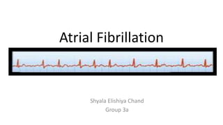
- 1. Atrial Fibrillation Shyala Elishiya Chand Group 3a
- 2. Introduction • Atrial fibrillation (AF) is the most common arrhythmia in older adults, becoming more common with age. • Unlike other arrhythmias, it is often chronic. • Incidence/ Prevalence: – In 2010, the estimated numbers of men and women with AF worldwide were 20.9 million and 12.6 million, respectively, with higher incidence and prevalence rates in developed countries. – By 2030, 14–17 million AF patients are anticipated in the European Union, with 120 000–215 000 newly diagnosed patients per year. – Estimates suggest an AF prevalence of approximately 3% in adults aged 20 years or older, with greater prevalence in older persons and in patients with conditions such as hypertension, heart failure, coronary artery disease (CAD), valvular heart disease, obesity, diabetes mellitus, or chronic kidney disease (CKD). – The increase in AF prevalence can be attributed both to better detection of silent AF, alongside increasing age and conditions predisposing to AF.
- 3. Morbidity/ Mortality • AF is independently associated with a two-fold increased risk of all-cause mortality in women and a 1.5-fold increase in men. • AF is also associated with increased morbidity, such as heart failure and stroke. • Studies show that 20–30% of patients with an ischaemic stroke have AF diagnosed before, during, or after the initial event. Cardiovascular morbidity and mortality associated with atrial fibrillation
- 4. Pathophysiology • Genetic predisposition – Early-onset AF, has a strong heritable component that is independent of concomitant cardiovascular conditions. – Most important variants are located close to the paired-like homeodomain transcription factor 2 (Pitx2) gene on chromosome 4q25. • Remodelling of atrial structure and ion channel function – Activation of fibroblasts, enhanced connective tissue deposition, and fibrosis are the hallmarks of this process. – Atrial fatty infiltration, inflammatory infiltrates, myocyte hypertrophy, necrosis, and amyloidosis are found in AF patients with concomitant conditions predisposing to AF. – Structural remodelling results in electrical dissociation between muscle bundles and local conduction heterogeneities, favouring re-entry and perpetuation of the arrhythmia.
- 5. Pathophysiological alterations in atrial tissue associated with atrial fibrillation and clinical conditions that could contribute to such alterations
- 6. Mechanism • Functional and structural changes in atrial myocardium and stasis of blood, especially in the left atrial appendage (LAA), generate a prothrombotic milieu. • Even short episodes of AF lead to atrial myocardial damage and the expression of prothrombotic factors on the atrial endothelial surface, alongside activation of platelets and inflammatory cells, and contribute to a generalized prothrombotic state. • Trial and systemic activation of the coagulation system can partially explain why short episodes of AF convey a long- term stroke risk.
- 7. Classification • In many patients, AF progresses from short, infrequent episodes to longer and more frequent attacks. • Over time, many patients will develop sustained forms of AF. In a small proportion of patients, AF will remain paroxysmal over several decades (2– 3% of AF patients). • The distribution of paroxysmal AF recurrences is not random, but clustered. AF may also regress from persistent to paroxysmal AF. • Furthermore, asymptomatic recurrences of AF are common in patients with symptomatic AF. • If patients suffer from both paroxysmal and persistent AF episodes, the more common type should be used for classification.
- 8. Clinical types of AF
- 9. Symptom burden in atrial fibrillation • Patients with AF have significantly poorer quality of life than healthy controls, experiencing a variety of symptoms including lethargy, palpitations, dyspnoea, chest tightness, sleeping difficulties, and psychosocial distress. • EHRA symptom scale is used to describe symptom severity in AF patients. • Modified EHRA scale should be used to guide symptom-orientated treatment decisions and for longitudinal patient profiling.
- 10. Risk Factors • Many cardiovascular diseases and concomitant conditions increase the risk of developing AF , recurrent AF, and AF-associated complications. • The identification of such conditions, their prevention and treatment is an important leverage to prevent AF and its disease burden. • Knowledge of these factors and their management is hence important for optimal management of AF patients.
- 11. Risk Factors • Heart failure – linked by similar risk factors and share a common pathophysiology. – Heart failure and AF can cause and exacerbate each other through mechanisms such as structural cardiac remodelling, activation of neurohormonal mechanisms, and rate-related impairment of left ventricular (LV) function. • Hypertension – Hypertension is a stroke risk factor in AF; uncontrolled high blood pressure enhances the risk of stroke and bleeding events and may lead to recurrent AF. – Therefore, good blood pressure control should form an integral part of the management of AF patients. – Inhibition of the renin–angiotensin–aldosterone system can prevent structural remodelling and recurrent AF.
- 12. Risk Factors • Valvular heart disease – Valvular heart disease is independently associated with incident AF. – Approximately 30% of patients with AF have some form of valvular heart disease, often detected only by echocardiogram. – AF worsens prognosis in patients with severe valvular heart disease. – Valvular heart disease can be associated with an increased thrombo-embolic risk, which probably also adds to the stroke risk in AF patients. – When valve dysfunction is severe, AF can be regarded as a marker for progressive disease, thus favouring valve repair or replacement. • Diabetes mellitus – Diabetes and AF frequently coexist because of associations with other risk factors. – Diabetes is a risk factor for stroke and other complications in AF. – In patients with AF, a longer duration of diabetes appears to confer a higher risk of thrombo-embolism, albeit without greater risk of OAC-related bleeding.
- 13. • Chronic obstructive pulmonary disease, sleep apnoea – Multiple pathophysiological mechanisms can contribute to AF in obstructive sleep apnoea, including autonomic dysfunction, hypoxia, hypercapnia, and inflammation. – Obstructive sleep apnoea exaggerates intrathoracic pressure changes, which in itself and via vagal activation can provoke shortening of the atrial action potential and induce AF. – Patients with chronic obstructive pulmonary disease often suffer from atrial tachycardias, which need to be differentiated from AF by ECG. – Agents used to relieve bronchospasm, notably theophyllines and beta-adrenergic agonists, may precipitate AF and make control of the ventricular response rate difficult. • Chronic kidney disease – AF is present in 15–20% of patients with CKD.
- 14. Assessment • As AF is often asymptomatic (“silent AF”), these healthcare professionals are important stakeholders to enable the adequate detection of AF and to ensure consistent management. • Initial assessment should be performed at the point of first contact with the healthcare system. • Consider five domains in the initial assessment of patients presenting with newly diagnosed AF. These domains are: – Haemodynamic instability or limiting, severe symptoms; – Presence of precipitating factors (e.g. thyrotoxicosis, sepsis, or postoperative AF) and underlying cardiovascular conditions; – Stroke risk and need for anticoagulation; – Heart rate and need for rate control; – Symptom assessment and decision for rhythm control.
- 15. Investigations • Complete medical history should be taken and all patients should undergo clinical evaluation that includes thorough assessment for concomitant conditions, establishing the AF pattern, estimation of stroke risk and AF-related symptoms, and assessment of arrhythmia-related complications such as thrombo-embolism or LV dysfunction. • 12-lead ECG is recommended to establish a suspected diagnosis of AF, to determine rate in AF, and to screen for conduction defects, ischaemia, and signs of structural heart disease. • Initial blood tests should evaluate thyroid and kidney function, as well as serum electrolytes and full blood count. • Transthoracic echocardiography is recommended in all AF patients to guide treatment decisions • In patients with AF and signs of cerebral ischaemia or stroke, computed tomography (CT) or magnetic resonance imaging (MRI) of the brain is recommended to detect stroke and support decisions regarding acute management and long-term anticoagulation.
- 16. Rhythm Control therapy • Rhythm control therapy is indicated to improve symptoms in AF patients who remain symptomatic on adequate rate control therapy. • Acute restoration of sinus rhythm (pharmacological cardioversion) – Pharmacological cardioversion restores sinus rhythm in approximately 50% of patients with recent-onset AF – Pharmacological cardioversion, does not require sedation or fasting. Antiarrhythmic drugs for pharmacological cardioversion
- 17. Drug Therapy • Flecainide and propafenone are effective for pharmacological cardioversion, but their use is restricted to patients without structural heart disease. • Ibutilide is an alternative where available, but carries a risk of torsades de pointes. • Vernakalant can be given to patients with mild heart failure, including those with ischaemic heart disease, provided they do not present with hypotension or severe aortic stenosis. • Amiodarone can be used in patients with heart failure and in patients with ischaemic heart disease • Amiodarone also slows heart rate by 10–12 b.p.m. after 8–12 h when given intravenously. • Both amiodarone and flecainide appear more effective than sotalol in restoring sinus rhythm
- 19. Drugs • Amiodarone – Amiodarone is an effective multichannel blocker, reduces ventricular rate, and is safe in patients with heart failure. – Torsades de pointes pro-arrhythmia can occur, and QT interval and TU waves should be monitored on therapy. – Amiodarone often causes extracardiac side-effects, especially on long-term therapy, rendering it a second-line treatment in patients who are suitable for other antiarrhythmic drugs. • Dronedarone – Dronedarone maintains sinus rhythm, reduces ventricular rate, and prevents cardiovascular hospitalizations (mostly due to AF) and cardiovascular death in patients with paroxysmal or persistent AF or flutter who had at least one relevant cardiovascular comorbidity. – Dronedarone moderately increases serum creatinine, reflecting a reduction in creatinine excretion rather than a decline in kidney function • Flecainide and propafenone – Flecainide and propafenone are effective in preventing recurrent AF. – They should only be used in patients without significant ischaemic heart disease or heart failure to avoid the risk of life- threatening ventricular arrhythmias. – High ventricular rates resulting from the conversion of AF into atrial flutter with 1:1 conduction by flecainide or propafenone can be prevented by pre-administering a beta-blocker, verapamil, or diltiazem.
- 20. Drugs • Quinidine and disopyramide – Quinidine and disopyramide have been associated with an increase in all-cause mortality. – They are less commonly used for rhythm control in AF. – Disopyramide may be useful in ‘vagally mediated’ AF (e.g. AF occurring in athletes and/or during sleep), and has been shown to reduce LV outflow gradient and improve symptoms in patients with hypertrophic cardiomyopathy. • Sotalol – Sotalol has a relevant risk of torsades de pointes (1%) • Dofetilide – Dofetilide is another potassium channel blocker that is mainly available outside of Europe. – Dofetilide restores and maintains sinus rhythm in heart failure patients, and occasionally in patients refractory to other antiarrhythmic drugs.
- 21. Electrical cardioversion • Synchronized direct current electrical cardioversion quickly and effectively converts AF to sinus rhythm, and is the method of choice in severely haemodynamically compromised patients with new-onset AF. • Electrical cardioversion can be performed safely in sedated patients treated with intravenous midazolam and/or propofol. • Cardioversion carries an inherent risk of stroke in non-anticoagulated patients, which is reduced substantially by the administration of anticoagulation. • Immediate initiation of anticoagulation is important in all patients scheduled for cardioversion. • Patients who have been in AF for longer than 48 h should start OAC at least 3 weeks before cardioversion and continue it for 4 weeks afterwards (in patients without a need for long- term anticoagulation). • OAC should be continued indefinitely in patients at risk of stroke.
- 22. Rate control therapy • Integral part of the management of AF patients, and is often sufficient to improve AF-related symptoms. • Pharmacological rate control can be achieved for acute or long-term rate control with beta-blockers, digoxin, the calcium channel blockers diltiazem and verapamil, or combination therapy .
- 23. Acute rate control • Evaluate underlying causes of elevated heart rate, such as infection, endocrine imbalance, anaemia, and pulmonary embolism. • For acute rate control, beta-blockers and diltiazem/verapamil are preferred over digoxin because of their rapid onset of action and effectiveness at high sympathetic tone. • In patients with HFrEF, beta-blockers, digitalis (digoxin or digitoxin), or their combination should be used, as diltiazem and verapamil can have negative inotropic effects in patients with LVEF <40%. • In critically ill patients and those with severely impaired LV systolic function, intravenous amiodarone can be used where excess heart rate is leading to haemodynamic instability.
- 24. Long-term pharmacological rate control • Beta-blockers – Beta-adrenoreceptor blocker monotherapy is often the first-line rate-controlling agent. • Non-dihydropyridine calcium channel blockers – Verapamil or diltiazem provide reasonable rate control in AF patients. – They should be avoided in patients with HFrEF because of their negative inotropic effects. • Digitalis – Lower doses of digoxin (≤250 μg once daily), corresponding to serum digoxin levels of 0.5–0.9 ng/mL. • Amiodarone – Amiodarone can be useful for rate control as a last resort.
- 25. Atrioventricular node ablation and pacing • Ablation of the atrioventricular node/His bundle and implantation of a VVI pacemaker can control ventricular rate when medications fail to control rate and symptoms. • It is a relatively simple procedure with a low complication rate and low long-term mortality risk. • The choice of pacing therapy (right ventricular or biventricular pacing with or without an implantable defibrillator) will depend on individual patient characteristics, including LVEF.
- 26. Stroke prevention therapy • OAC therapy can prevent the majority of ischaemic strokes in AF patients and can prolong life. • CHA2DS2-VASc = Congestive Heart failure, hypertension, Age ≥75 (doubled), Diabetes, Stroke (doubled), Vascular disease, Age 65–74, and Sex (female). • CHA2DS2-VASc score of 1 for men, and 2 for women indicates therapy.
- 27. Clinical risk scores for bleeding • Stroke and bleeding risk factors overlap . – For example, older age is one of the most important predictors of both ischaemic stroke and bleeding in AF patients. • A high bleeding risk score should generally not result in withholding OAC.
- 28. Vitamin K antagonists • VKA therapy reduces the risk of stroke by two-thirds and mortality by one-quarter compared with control (aspirin or no therapy). • Use of VKAs is limited by the narrow therapeutic interval, necessitating frequent monitoring and dose adjustments, but VKAs, when delivered with adequate time in therapeutic range (TTR), are effective for stroke prevention in AF patients. • VKAs are currently the only treatment with established safety in AF patients with rheumatic mitral valve disease and/or a mechanical heart valve prosthesis. • SAMe-TT2R2 score: aid decision making between a non-VKA oral anticoagulant (NOAC) and a vitamin K antagonist (VKA).
- 29. SAMe-TT2R2 score
- 30. Non-vitamin K antagonist oral anticoagulants • Suitable alternatives to VKAs for stroke prevention in AF. • All NOACs have a predictable effect (onset and offset) without need for regular anticoagulation monitoring. • Apixaban – 5 mg twice daily reduced stroke or systemic embolism by 21% compared with warfarin, combined with a 31% reduction in major bleeding and an 11% reduction in all-cause mortality. – Rates of haemorrhagic stroke and intracranial haemorrhage, but not of ischaemic stroke, were lower on apixaban. – Significantly reduced stroke or systemic embolism by 55% compared with aspirin.
- 31. Drugs • Dabigatran – 150 mg twice daily reduced stroke and systemic embolism by 35% compared with warfarin without a significant difference in major bleeding events. – Dabigatran 110 mg twice daily was non-inferior to warfarin for prevention of stroke and systemic embolism, with 20% fewer major bleeding events. – Both dabigatran doses significantly reduced haemorrhagic stroke and intracranial haemorrhage. – Dabigatran 150 mg twice daily significantly reduced ischaemic stroke by 24% and vascular mortality by 12%, while gastrointestinal bleeding was significantly increased by 50%. • Edoxaban – Only the higher dose regimen has been approved for stroke prevention in AF. • Rivaroxaban
- 32. Antiplatelet therapy as an alternative to oral anticoagulants • VKA therapy prevents stroke, systemic embolism, myocardial infarction, and vascular death better than single or dual antiplatelet therapy with aspirin and clopidogrel (annual risk of 5.6% for aspirin and clopidogrel vs. 3.9% with VKA therapy). • Antiplatelet therapy increases bleeding risk, especially dual antiplatelet therapy. • Thus, antiplatelet therapy cannot be recommended for stroke prevention in AF patients.
- 33. Summary • Evaluate all AF patients by clinical evaluation, ECG, and echocardiogram for underlying cardiovascular conditions such as hypertension, heart failure, valvular heart disease, and others. • Select antiarrhythmic drugs based on their safety profile and consider catheter or surgical ablation when antiarrhythmic drugs fail. • Do not use antiplatelet therapy for stroke prevention in AF. • Do not permanently discontinue oral anticoagulation in AF patients at increased risk of stroke unless such a decision is taken by a multidisciplinary team. • Do not use rhythm control therapy in asymptomatic AF patients, nor in patients with permanent AF. • Do not perform cardioversion or catheter ablation without anticoagulation, unless an atrial thrombus has been ruled out transoesophageal echocardiogram.
- 34. Reference • 2016 ESC Guidelines for the management of atrial fibrillation • Oxford American Handbook of Geriatric Medicine, 2010