
Recommended
More Related Content
What's hot
What's hot (20)
Similar to Bacterial anatomy
Similar to Bacterial anatomy (20)
More from SUMESH KUMAR DASH
More from SUMESH KUMAR DASH (17)
Recently uploaded
Recently uploaded (20)
Bacterial anatomy
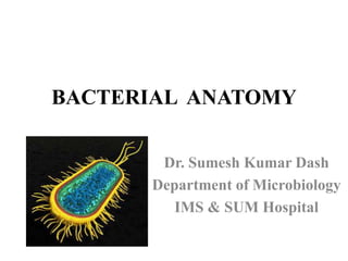
- 1. BACTERIAL ANATOMY Dr. Sumesh Kumar Dash Department of Microbiology IMS & SUM Hospital
- 2. CLASSIFICATION Microorganism are grouped under 2 I. Prokaryotes (Bacteria & Blue-green algae) II. Eukaryotes (Fungi, Parasites, other algae) Viruses are neither prokaryotes nor eukaryotes as they lack the characteristics of living thinks except replication.
- 5. SIZE • Bacteria size in micrometer and virus size in nanometer • Bacteria observed by light microscope and Viruses observed by electron microscope • To see microorganisms – staining is used
- 6. MORPHOLOGY OF BACTERIA • Depending on their shape; Cocci- oval/spherical cells Bacilli- rod shaped • Arrangement; Singles Pairs Chains clusters • Based on gram staining property; Gram positive Gram negative
- 8. BACTERIAL CELL • Outer layer/Envelope- cellwall cytoplasmic/plasma membrane • Cytoplasm- mesosomes ribosomes vacuoles nucleoid • Appendages- capsule flagella fimbriae/pili
- 9. CELL WALL • Chemically composed of peptidoglycan
- 10. GRAM POSITIVE CELL WALL Peptidoglycan layer 50-100 layers thick 16-80nm Teichoic acids 2 types: Cell wall teichoic acid Lipoteichoic acid
- 11. GRAM NEGATIVE CELL WALL Peptidoglycan layer 1-2 layer thick 2-3nm Outer membrane 1. Porin proteins 2. Lipopolysaccharide (LPS) • Lipid A • Core polysaccharide • O side chain /O antigen
- 12. DIFFERENCE BETWEEN GRAM POSITIVE & GRAM NEGATIVE CELL WALL Characters GRAM + GRAM - peptidoglycan layer Thick Thin Aromatic and sulphur containing amino acids Absent Present Teichoic acids Present Absent Gram stain Violet Red
- 13. FUNCTIONS OF CELL WALL Maintaining the cells characteristic shape Providing a rigid platform for surface appendages- flagella, fimbriae/pili all emanate from the wall and extend beyond it. Providing attachment sites for bacteriophage Be the sites of major antigenic determinant of cell surface- endotoxcity Site of action of many antibiotics & resistance
- 14. CYTOPLASMIC/CELL MEMBRANE 5-10nm thick Phospholipid bilayer With integral and peripheral proteins Selective permeability i:e - semipermeable Site of ATP production Viability
- 15. CYTOPLASM • Viscous watery solution containing organic & inorganic solutes, biosynthetic components which required for the growth & cell division, together with genetic material. • Contains RIBOSOMES, MESOSOMES INTRACYTOPLASMIC INCLUSION BODIES VACUOLES NUCLEOUS
- 16. RIBOSOMES • Sites for protein synthesis. • 10-20 nm in size.
- 17. MESOSOMES / Chondroids • Vesicular/convoluted/multilaminated • More prominent in G+. • LOCATION: Often found next to septa, in dividing bacteria or seen attached to nuclear body. • FUNCTION: Site of bacterial respiration
- 18. INTRACYTOPLASMIC INCLUSIONS • Storage sites for nutrients/energy. • Formed under nutritional deficiency conditions & disappear in the presence of excess nutrients Staining Toluidine blue, Neisser, Albert, Acid-fast, wet films Electron microscopy
- 19. VACUOLES • Vacuoles are fluid containing cavities • Separated from cytoplasm by a membrane • Function - uncertain
- 20. NUCLEOID • Genetic material arranged in irregular shape • Oval, Elongated • Have no nuclear membrane • Seen by Electron Microscope Plasmid Extrachromosomal ds circular DNA. Not essential for life. Present singly or in multiple numbers. Episomes- when integrated with chromosomal DNA. Drug resistance and toxigenicity
- 21. THANK YOU