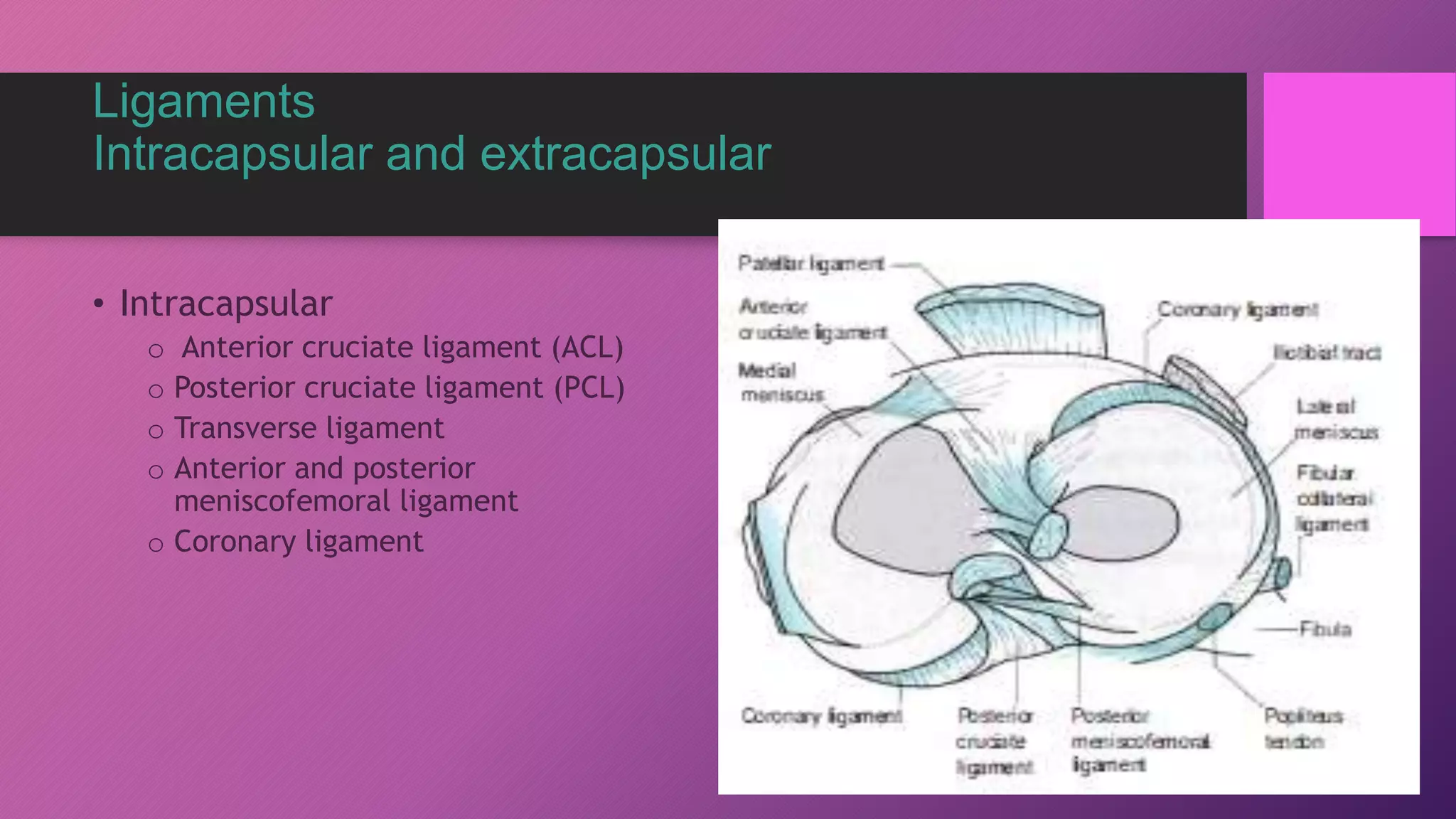
Osteoarthritis knee
Dr Ravi Shankar Sharma
Fellow (Daradia)
Anatomy of knee joint
Anatomy
• Type – Hinge type synovial joint
• Femoropatellar and femorotibial joint
• The articular capsule has
a synovial and
a fibrous membrane separated
by fatty deposits.
• Synovial membrane have
multiple extensions which
includes suprapatellar bursa.
Ligaments
Intracapsular and extracapsular
• Intracapsular
o Anterior cruciate ligament (ACL)
o Posterior cruciate ligament (PCL)
o Transverse ligament
o Anterior and posterior
meniscofemoral ligament
o Coronary ligament
Extracapsular
• Petallar ligament
• Medial collateral ligament
• Lateral collateral ligament
• Oblique popliteal ligament
• Arcuate popliteal ligament
Meniscus
• Medial meniscus
• Lateral meniscus
Structure
• Fibrous cartilate
• Outer 1/3rd blood supply
Function
Nerve supply
• Femoral-through its branches in vasti medialis
• Sciatic(through genicular branches)
• Obturator nerve-through its posterior division
Blood supply:
Osteoarthritis
• Degenerative joint disease affecting joint cartilage and
subchondral bone,leading to the formation of bony spurs and
subchondral cyst.
• Age above 60 years
• Female more after menopause
Degenerative changes of Knee
Overview: Risk Factors
• Age
• Female
• Obesity ( most important modifiable)
• Previous knee injury
• Lower extremity malalignment
• Repetitive knee bending
• Hereditory and nodal OA
• Low level of vitamin C and D
• Muscle weakness( quardiceps)
Pathophysiology
Biomechanical stress
affecting the articular
cartilage and subchondral
bone leading to wear and
tear
WHY PAIN ?
• PERIPHERAL SENSITIZATION :
• mediator induced inflammation of articular nociceptors
• Secondary to synovitis
• Stretching of joint capsule and ligaments
• Periosteal irritation due to osteophytes
• Trabeculae microfractures
• Intraosseous hypertension
• Muscle spasm
CENTRAL SENSITIZATION
Signs and symptoms
• Pain
• Morning stiffness
• Swelling
• Decreased range of motion
• Swelling
• Sounds- crepitation
Clinical examination
• Gait
• Swelling
• Deformity
• Tenderness over joint line
• Active and passive ROM painful
• Hard end feel
• Bony crepitations
Diagnosis of Knee OA
Classic Clinical Criteria
• established by ACR, 1981
• sensitivity 95%, specificity 69%
knee pain plus at least 3 of 6 characteristics:
• > 50 yrs
• Morning stiffness < 30 min
• Crepitus
• Bony tenderness
• Bony enlargement
• No palpable warmth 5
Kellgren and Lawrence classification
Grade 0 No radiographic features of osteoarthritis
Grade 1 Possible joint space narrowing and osteophyte formation
Grade 2 Definite osteophyte formation with possible joint
space narrowing
Grade 3 Multiple osteophytes, definite joint space narrowing, sclerosis
and possible bony deformity
Grade 4 Large osteophytes, marked joint space narrowing,
severe sclerosis and definite bony deformity
Diagnosis of Knee OA
Treatment
• Non pharmacological
• Pharmacological-pcm, NSAIDS,disease modifying agents like
collagenase inhibitor, elastase inhibitors,and narcotics
• Intervention
• Surgery
Intraarticular injections
• Corticosteroids- max. 3-4 times in a year
• Hyaluronic acid in major co-morbid conditions
Prolotherapy
• Proliferation therapy or regenerative injection therapy involves
injecting an otherwise non-pharmacological or non-active irritant
solution into knee joint
• Dextose
• Prolozone
• Platelet rich plasma
Platelet Rich Plasma (PRP)
• Platelet derived growth factor
• Transforming growth factor B
• Fibroblast growth factor
• Insulin like growth factor 1&2
• Vascular endothelial growth factor
• Epidermal growth factor
• Keratinocyte growth factor
• Connective tissue growth factor
All these promote cartilage repair
Genicular nerve block & RF neurotomy
Indications
• Chronic knee pain secondary to OA
• Patients with failed knee replacement
• Patients unfit for knee replacement
• Patients who want to avoid surgery
Surgery
• Arthoscopy debridement
• Osteotomy - malalignment
• Knee replacement
Osteoarthritis knee,, pain management

Osteoarthritis knee,, pain management

  • 1.
    Osteoarthritis knee Dr RaviShankar Sharma Fellow (Daradia)
  • 2.
  • 3.
    Anatomy • Type –Hinge type synovial joint • Femoropatellar and femorotibial joint
  • 4.
    • The articularcapsule has a synovial and a fibrous membrane separated by fatty deposits. • Synovial membrane have multiple extensions which includes suprapatellar bursa.
  • 5.
    Ligaments Intracapsular and extracapsular •Intracapsular o Anterior cruciate ligament (ACL) o Posterior cruciate ligament (PCL) o Transverse ligament o Anterior and posterior meniscofemoral ligament o Coronary ligament
  • 6.
    Extracapsular • Petallar ligament •Medial collateral ligament • Lateral collateral ligament • Oblique popliteal ligament • Arcuate popliteal ligament
  • 7.
    Meniscus • Medial meniscus •Lateral meniscus Structure • Fibrous cartilate • Outer 1/3rd blood supply Function
  • 8.
    Nerve supply • Femoral-throughits branches in vasti medialis • Sciatic(through genicular branches) • Obturator nerve-through its posterior division
  • 9.
  • 10.
    Osteoarthritis • Degenerative jointdisease affecting joint cartilage and subchondral bone,leading to the formation of bony spurs and subchondral cyst. • Age above 60 years • Female more after menopause
  • 11.
  • 12.
    Overview: Risk Factors •Age • Female • Obesity ( most important modifiable) • Previous knee injury • Lower extremity malalignment • Repetitive knee bending • Hereditory and nodal OA • Low level of vitamin C and D • Muscle weakness( quardiceps)
  • 13.
    Pathophysiology Biomechanical stress affecting thearticular cartilage and subchondral bone leading to wear and tear
  • 14.
    WHY PAIN ? •PERIPHERAL SENSITIZATION : • mediator induced inflammation of articular nociceptors • Secondary to synovitis • Stretching of joint capsule and ligaments • Periosteal irritation due to osteophytes • Trabeculae microfractures • Intraosseous hypertension • Muscle spasm CENTRAL SENSITIZATION
  • 15.
    Signs and symptoms •Pain • Morning stiffness • Swelling • Decreased range of motion • Swelling • Sounds- crepitation
  • 16.
    Clinical examination • Gait •Swelling • Deformity • Tenderness over joint line • Active and passive ROM painful • Hard end feel • Bony crepitations
  • 17.
    Diagnosis of KneeOA Classic Clinical Criteria • established by ACR, 1981 • sensitivity 95%, specificity 69% knee pain plus at least 3 of 6 characteristics: • > 50 yrs • Morning stiffness < 30 min • Crepitus • Bony tenderness • Bony enlargement • No palpable warmth 5
  • 18.
    Kellgren and Lawrenceclassification Grade 0 No radiographic features of osteoarthritis Grade 1 Possible joint space narrowing and osteophyte formation Grade 2 Definite osteophyte formation with possible joint space narrowing Grade 3 Multiple osteophytes, definite joint space narrowing, sclerosis and possible bony deformity Grade 4 Large osteophytes, marked joint space narrowing, severe sclerosis and definite bony deformity
  • 19.
  • 20.
    Treatment • Non pharmacological •Pharmacological-pcm, NSAIDS,disease modifying agents like collagenase inhibitor, elastase inhibitors,and narcotics • Intervention • Surgery
  • 21.
    Intraarticular injections • Corticosteroids-max. 3-4 times in a year • Hyaluronic acid in major co-morbid conditions
  • 22.
    Prolotherapy • Proliferation therapyor regenerative injection therapy involves injecting an otherwise non-pharmacological or non-active irritant solution into knee joint • Dextose • Prolozone • Platelet rich plasma
  • 23.
    Platelet Rich Plasma(PRP) • Platelet derived growth factor • Transforming growth factor B • Fibroblast growth factor • Insulin like growth factor 1&2 • Vascular endothelial growth factor • Epidermal growth factor • Keratinocyte growth factor • Connective tissue growth factor All these promote cartilage repair
  • 24.
    Genicular nerve block& RF neurotomy Indications • Chronic knee pain secondary to OA • Patients with failed knee replacement • Patients unfit for knee replacement • Patients who want to avoid surgery
  • 25.
    Surgery • Arthoscopy debridement •Osteotomy - malalignment • Knee replacement