
Recommended
Recommended
More Related Content
What's hot
What's hot (20)
Similar to Flow cytometry
Similar to Flow cytometry (20)
Recently uploaded
Recently uploaded (20)
Flow cytometry
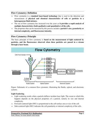
- 1. Prepared by: Prachand M.S. Rajbhandari Page 1 Note: Please, for more details refer books Flow Cytometry: Definition Flow cytometry is a standard laser-based technology that is used in the detection and measurement of physical and chemical characteristics of cells or particles in a heterogeneous fluid mixture. The use of flow cytometry has increased over the years as it provides a rapid analysis of multiple characteristics (both qualitative and quantitative) of the cells. The properties that can be measured by this process include a particle’s size, granularity or internal complexity, and fluorescence intensity. Flow Cytometry: Principle The basic principle of flow cytometry is based on the measurement of light scattered by particles, and the fluorescence observed when these particles are passed in a stream through a laser beam. Figure: Schematic of a common flow cytometer, illustrating the fluidic, optical, and electronic systems. Light Scattering Light scattering results when a particle deflects incident laser light. The extent to which this happens depends on the physical properties of a particle, namely its size and internal complexity. Forward-scattered light (FSC) is proportional to the cell-surface area or size of the cell. Side-scattered light (SSC) indicates the cell granularity or internal complexity of the cells.
- 2. Prepared by: Prachand M.S. Rajbhandari Page 2 Note: Please, for more details refer books The measurements of FSC and SSC are used for the differentiation of cell types in a heterogeneous cell population. Fluorescence Fluorescent markers used to detect the expression of cellular molecules such as proteins or nucleic acids in a system. The fluorescent compound absorbs light energy over a range of wavelengths that is characteristic of that compound. This absorption of light causes an electron in the fluorescent compound to be raised to a higher energy level. The excited electron quickly decays to its ground state, emitting the excess energy in the form of fluorescence which is then collected by detectors. The electronics system then converts the detected light signals into electronic signals that can be processed by the computer. Instrumentation/Parts of Flow Cytometry A flow cytometer is made up of three main systems: fluidics, optics system, and electronics system. Fluidics: The purpose of the fluidics system is to transport particles in a fluid stream to the laser beam. To accomplish this, the sample is injected into a stream of sheath fluid (usually a buffered saline solution) within the flow chamber. Optics System: The optical system of the cytometer consists of excitation optics and collection optics. The excitation optics consists of the laser and lenses that are used to shape and focus the laser beam to the flow of the sample. The collection optics consist of a collection lens to collect light emitted after the particle interacts with the laser beam and a system of optical mirrors that divert the specified wavelengths of the collected light to designated optical detectors. Electronics system: The electronic system converts the signals from the detectors into digital signals that can be read by a computer. Applications/Uses Flow Cytometry is used in several fields including molecular biology, pathology, immunology, virology, plant biology, and marine biology. Some of the common application includes: It is used in clinical labs for the detection of malignancy in bodily fluids like leukemia. Cytometers like cell sorters can be used to separate the cells of interest in separate collection tubes physically.
- 3. Prepared by: Prachand M.S. Rajbhandari Page 3 Note: Please, for more details refer books It can be used for the detection of the content of DNA by using fluorescent markers. Flow cytometers allow the analysis of replication cells by using fluorescent dye for four different stages of the cell cycle. Acoustic flow cytometers are used in the study of multi-drug resistant bacteria in the blood and other samples. The different stages of cell death, apoptosis, and necrosis can be detected by flow cytometers based on the differences in the morphological and biochemical changes.