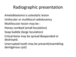
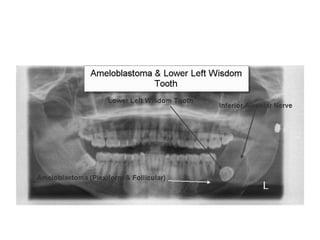
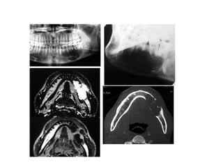
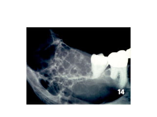
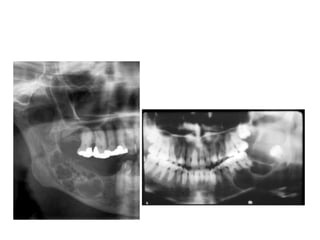
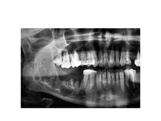
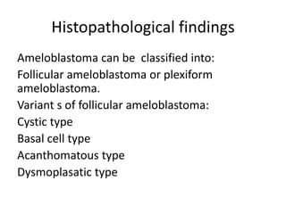
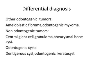
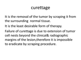
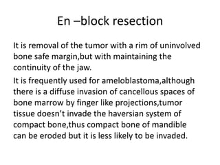
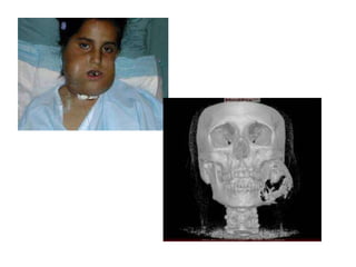
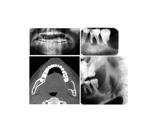
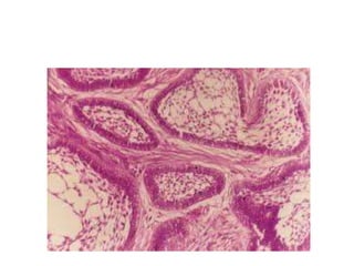
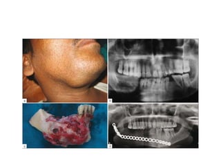
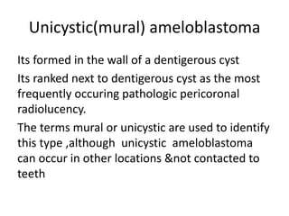
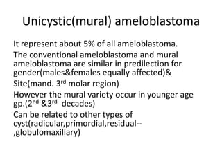
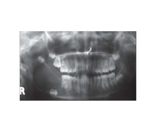
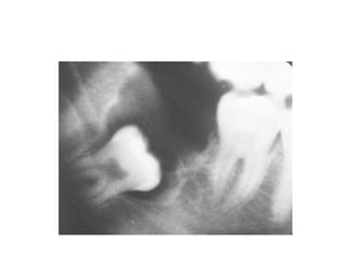
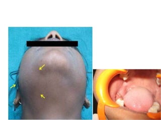
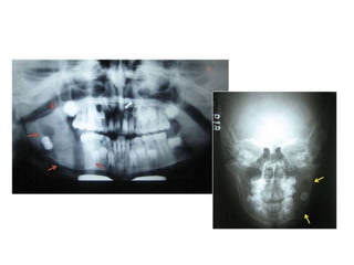
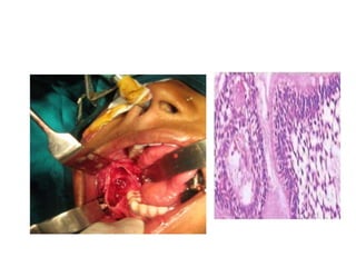
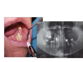
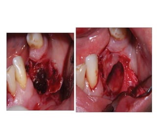
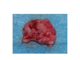
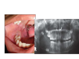
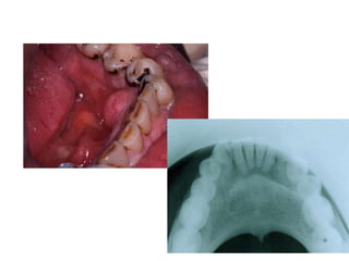
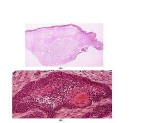
Ameloblastoma
By:
Mohamed Saber
Ameloblastoma
Definition
Etiology
Origin
Types
Clinical features(incidence)
Clinical presentaion
Radiographic presentation
Histopathological findings
Differential diagnosis
treatment
It is a benign but invasive epithelial odontogenic
neoplasm.
Consisting of proliferating odontogenic
epithelium lying in fibrous stroma
etiology
Unknown , but possible causal factors
suggested :
Truama or inflamation
Oral infection,extraction,injury to teeth or jaws
Irritation resulting from eruption of the third
molar
Origin of ameloblastoma
The precise point of origin of ameloblastoma is
unknown ,the origin might be from:
Epithelial rests of serre or malassez
Epithelial lining of non neoplastic odontogenic
cyst(dentigerous cyst)
Direct from oral epithelium
types
Conventional solid or multicystic 94%
Unicystic(mural) 5%
Peripheral or extraosseous 1%
incidence
It’s the most common epithelial odontogenic
neoplasm.
It comprises about 1% of all oral tumors.
Mandible>maxilla 85%:15%
in mandible:
70% in molar-ramus area, 20%in premolar area,
10% in incisor region
incidence
In the maxilla:
Tumor found in the posterior region
Age predilection :
Fourth and fifth decades
The tumor can occur in children or in old age
Clinical presentation
In the early stages : ameloblastoma grows slowly
&silently without clinical signs.
in advanced stages:
neoplasm expand cortical plates
thinning of bone (egg shell crackling )erodes
them invades the soft tissue
At this point ameloblastoma present clinically as
a smooth surfaced local expansion of the jaw
producing asymmetry.
Clinical presentation
Lesion may be composed of solid tumor,cystic
areas,or booth.
Tipping or loosening of teeth,involvement of inf.
Alveolar nerve may occur.
Draining sinuses,unhealed extraction sockets
associated with granulation tissue within the
socket,bleeding ,trismus& other dental
problems may be the chief complain
Clinical presentation
Max. ameloblastoma:
Nasal obstruction is 1st symptoms
Potentially lethal if sinus involved or tumor invade
the bone into soft tissues due to:
Bone is not compact, easily invaded
Proximity to: nasal cavity&sinuses
orbital&pharyngeal tissues
vital structures at base of skull
This factors also complicate comlete removal
Radiographic presentation
Ameloblastoma is osteolytic lesion
Unilocular or multilocul radiolucency
Multilocular lesion may be :
Honey combed (small loculation)
Soap bubble (large loculation)
Crtical bone may be spread &expanded or
destroyed.
Unerrupted tooth may be present(resembling
dentigerous cyst)
Radiographic presentation
Root resorption of associated teeth.
Maxillary tumors produce a monocystic cavity
in most instances.
Histopathological findings
Ameloblastoma can be classified into:
Follicular ameloblastoma or plexiform
ameloblastoma.
Variant s of follicular ameloblastoma:
Cystic type
Basal cell type
Acanthomatous type
Dysmoplasatic type
Histopathological findings
Follicular ameloblastoma:
Made of epithelial follicles resembling enamel
organ in mature fibrous c.t. stroma.
Epithelial follicles consist of peripheral tall
columnar cells(ameloblast like cells)& central
core of loosely arranged angular cells (the
stellate reticulaum like cells)
Histopathological findings
Plexiform ameloblastoma:
The epithelium is arranged in a network of
anastomosing strands and cords with the
same cell layers as follicular ameloblastoma.
Differential diagnosis
Other odontogenic tumors:
Ameloblastic fibroma,odontogenic myxoma.
Non odontogenic tumors:
Central giant cell granuloma,aneurysmal bone
cyst.
Odontogenic cysts:
Dentigerous cyst,odontogenic keratocyst
Treatment
Treatment of ameloblastoma ranges from
conservative curettage to radicular resection.
Treatment varies according to
site,size&characteristics of the ameloblastoma
Curettage
En-block resection
Segmental resection
curettage
It is the removal of the tumor by scraping it from
the surrounding normal tissue.
It is the least desirable form of therapy.
Failure of curettage is due to extension of tumor
cell nests beyond the clinical& radiographic
margins of the lesion,therefore it is impossible
to eradicate by scraping procedure.
En –block resection
It is removal of the tumor with a rim of uninvolved
bone safe margin,but with maintaining the
continuity of the jaw.
It is frequently used for ameloblastoma,although
there is a diffuse invasion of cancellous spaces of
bone marrow by finger like projections,tumor
tissue doesn’t invade the haversian system of
compact bone,thus compact bone of mandible
can be eroded but it is less likely to be invaded.
Segmental resection
Segmental resection including
hemimaxillectomy& hemimanibulectomy has
been the most commonly used treatment for
ameloblastoma.
Most authors who advocate this method have
had the least number of recurrence.
Unicystic(mural) ameloblastoma
Its formed in the wall of a dentigerous cyst
Its ranked next to dentigerous cyst as the most
frequently occuring pathologic pericoronal
radiolucency.
The terms mural or unicystic are used to identify
this type ,although unicystic ameloblastoma
can occur in other locations &not contacted to
teeth
Unicystic(mural) ameloblastoma
It represent about 5% of all ameloblastoma.
The conventional ameloblastoma and mural
ameloblastoma are similar in predilection for
gender(males&females equally affected)&
Site(mand. 3rd molar region)
However the mural variety occur in younger age
gp.(2nd &3rd decades)
Can be related to other types of
cyst(radicular,primordial,residual--
,globulomaxillary)
Unicystic(mural) ameloblastoma
in early stage asymptomatic,undetected untile
pericoronal radiolucency is seen on routine
radiograph.
Slowly enlarged,slight non tender sweeling
appear clinically.
If bone destruction occur,palpation discloses
softer areas.
Unicystic(mural) ameloblastoma
Radiographically:
If haziness&thinning of hyperostotic
radioopaque rim of pericoronal radiolucency
Tumor invade the capsule of the cyst &start to
infiltrate the bone trabeculae .
management
Before undertaking surgical procedure of
pericoronal radiolucency, differential diagnosis
bet. mural(unicystic) &conventional
ameloblastoma should be completed.
At surgery the cyst should be enucleated,and if
mural mass is discovered flag it with suture to
enable the pathologist for further
investigation.
management
If pathologist examination establishes the mass
as ameloblastoma that has not penetrated the
basement membrane no further surgery is
done.
If the neoplasm has penetrated the basement
membrane more the bone should be removed
with curettage.
In all cases careful periodic follow- up is always
indicated.
Peripheral(extraosseous)
ameloblastoma
Origin: basal layer of oral epith.&extraosseous
portion of epith. Rests of serre.
Site: commonly affect gingiva & alveolar mucosa
mandible> maxilla
Micoscopically it looks like acanthomatous type of
basal cell carcinoma of the skin.
Management: excision.
Behavior: less invasive than intraosseous
ameloblastoma.
Ameloblastoma
Ameloblastoma
Ameloblastoma

Ameloblastoma

  • 1.
  • 2.
    Ameloblastoma Definition Etiology Origin Types Clinical features(incidence) Clinical presentaion Radiographicpresentation Histopathological findings Differential diagnosis treatment
  • 3.
    It is abenign but invasive epithelial odontogenic neoplasm. Consisting of proliferating odontogenic epithelium lying in fibrous stroma
  • 4.
    etiology Unknown , butpossible causal factors suggested : Truama or inflamation Oral infection,extraction,injury to teeth or jaws Irritation resulting from eruption of the third molar
  • 5.
    Origin of ameloblastoma Theprecise point of origin of ameloblastoma is unknown ,the origin might be from: Epithelial rests of serre or malassez Epithelial lining of non neoplastic odontogenic cyst(dentigerous cyst) Direct from oral epithelium
  • 6.
    types Conventional solid ormulticystic 94% Unicystic(mural) 5% Peripheral or extraosseous 1%
  • 7.
    incidence It’s the mostcommon epithelial odontogenic neoplasm. It comprises about 1% of all oral tumors. Mandible>maxilla 85%:15% in mandible: 70% in molar-ramus area, 20%in premolar area, 10% in incisor region
  • 8.
    incidence In the maxilla: Tumorfound in the posterior region Age predilection : Fourth and fifth decades The tumor can occur in children or in old age
  • 9.
    Clinical presentation In theearly stages : ameloblastoma grows slowly &silently without clinical signs. in advanced stages: neoplasm expand cortical plates thinning of bone (egg shell crackling )erodes them invades the soft tissue At this point ameloblastoma present clinically as a smooth surfaced local expansion of the jaw producing asymmetry.
  • 10.
    Clinical presentation Lesion maybe composed of solid tumor,cystic areas,or booth. Tipping or loosening of teeth,involvement of inf. Alveolar nerve may occur. Draining sinuses,unhealed extraction sockets associated with granulation tissue within the socket,bleeding ,trismus& other dental problems may be the chief complain
  • 11.
    Clinical presentation Max. ameloblastoma: Nasalobstruction is 1st symptoms Potentially lethal if sinus involved or tumor invade the bone into soft tissues due to: Bone is not compact, easily invaded Proximity to: nasal cavity&sinuses orbital&pharyngeal tissues vital structures at base of skull This factors also complicate comlete removal
  • 14.
    Radiographic presentation Ameloblastoma isosteolytic lesion Unilocular or multilocul radiolucency Multilocular lesion may be : Honey combed (small loculation) Soap bubble (large loculation) Crtical bone may be spread &expanded or destroyed. Unerrupted tooth may be present(resembling dentigerous cyst)
  • 15.
    Radiographic presentation Root resorptionof associated teeth. Maxillary tumors produce a monocystic cavity in most instances.
  • 21.
    Histopathological findings Ameloblastoma canbe classified into: Follicular ameloblastoma or plexiform ameloblastoma. Variant s of follicular ameloblastoma: Cystic type Basal cell type Acanthomatous type Dysmoplasatic type
  • 22.
    Histopathological findings Follicular ameloblastoma: Madeof epithelial follicles resembling enamel organ in mature fibrous c.t. stroma. Epithelial follicles consist of peripheral tall columnar cells(ameloblast like cells)& central core of loosely arranged angular cells (the stellate reticulaum like cells)
  • 23.
    Histopathological findings Plexiform ameloblastoma: Theepithelium is arranged in a network of anastomosing strands and cords with the same cell layers as follicular ameloblastoma.
  • 27.
    Differential diagnosis Other odontogenictumors: Ameloblastic fibroma,odontogenic myxoma. Non odontogenic tumors: Central giant cell granuloma,aneurysmal bone cyst. Odontogenic cysts: Dentigerous cyst,odontogenic keratocyst
  • 28.
    Treatment Treatment of ameloblastomaranges from conservative curettage to radicular resection. Treatment varies according to site,size&characteristics of the ameloblastoma Curettage En-block resection Segmental resection
  • 29.
    curettage It is theremoval of the tumor by scraping it from the surrounding normal tissue. It is the least desirable form of therapy. Failure of curettage is due to extension of tumor cell nests beyond the clinical& radiographic margins of the lesion,therefore it is impossible to eradicate by scraping procedure.
  • 30.
    En –block resection Itis removal of the tumor with a rim of uninvolved bone safe margin,but with maintaining the continuity of the jaw. It is frequently used for ameloblastoma,although there is a diffuse invasion of cancellous spaces of bone marrow by finger like projections,tumor tissue doesn’t invade the haversian system of compact bone,thus compact bone of mandible can be eroded but it is less likely to be invaded.
  • 31.
    Segmental resection Segmental resectionincluding hemimaxillectomy& hemimanibulectomy has been the most commonly used treatment for ameloblastoma. Most authors who advocate this method have had the least number of recurrence.
  • 36.
    Unicystic(mural) ameloblastoma Its formedin the wall of a dentigerous cyst Its ranked next to dentigerous cyst as the most frequently occuring pathologic pericoronal radiolucency. The terms mural or unicystic are used to identify this type ,although unicystic ameloblastoma can occur in other locations &not contacted to teeth
  • 37.
    Unicystic(mural) ameloblastoma It representabout 5% of all ameloblastoma. The conventional ameloblastoma and mural ameloblastoma are similar in predilection for gender(males&females equally affected)& Site(mand. 3rd molar region) However the mural variety occur in younger age gp.(2nd &3rd decades) Can be related to other types of cyst(radicular,primordial,residual-- ,globulomaxillary)
  • 38.
    Unicystic(mural) ameloblastoma in earlystage asymptomatic,undetected untile pericoronal radiolucency is seen on routine radiograph. Slowly enlarged,slight non tender sweeling appear clinically. If bone destruction occur,palpation discloses softer areas.
  • 39.
    Unicystic(mural) ameloblastoma Radiographically: If haziness&thinningof hyperostotic radioopaque rim of pericoronal radiolucency Tumor invade the capsule of the cyst &start to infiltrate the bone trabeculae .
  • 42.
    management Before undertaking surgicalprocedure of pericoronal radiolucency, differential diagnosis bet. mural(unicystic) &conventional ameloblastoma should be completed. At surgery the cyst should be enucleated,and if mural mass is discovered flag it with suture to enable the pathologist for further investigation.
  • 43.
    management If pathologist examinationestablishes the mass as ameloblastoma that has not penetrated the basement membrane no further surgery is done. If the neoplasm has penetrated the basement membrane more the bone should be removed with curettage. In all cases careful periodic follow- up is always indicated.
  • 51.
    Peripheral(extraosseous) ameloblastoma Origin: basal layerof oral epith.&extraosseous portion of epith. Rests of serre. Site: commonly affect gingiva & alveolar mucosa mandible> maxilla Micoscopically it looks like acanthomatous type of basal cell carcinoma of the skin. Management: excision. Behavior: less invasive than intraosseous ameloblastoma.