Disorders of the Respiratory System
•Download as PPT, PDF•
1 like•163 views

1. Acute respiratory distress syndrome (ARDS) is a clinical syndrome characterized by diffuse alveolar capillary damage and severe pulmonary edema, resulting in hypoxemia that is refractory to oxygen therapy. 2. ARDS is caused by direct or indirect injury to the lungs from sources such as sepsis, gastric aspiration, trauma, or smoke inhalation. This causes damage to the alveolar capillary endothelium and epithelium. 3. The damage leads to increased capillary permeability, leakage of fluid into the alveoli, and formation of hyaline membranes. This results in impaired gas exchange and respiratory failure.

Recommended
More Related Content
What's hot
What's hot (20)
Similar to Disorders of the Respiratory System
Similar to Disorders of the Respiratory System (20)
More from med_students0
More from med_students0 (20)
Recently uploaded
Recently uploaded (20)
Disorders of the Respiratory System
- 1. Spr 09 1
- 2. Spr 09 2 Why learn the disorders of respiratory system? Respiratory symptoms most common cause of presentation to family doctor. Rhinitis = common cold Sinusitis = inflammation of paranasal air sinuses Pneumonia , Asthma , Bronchitis Bronchogenic carcinoma – MC cancer causing death in men and women. Lungs are the major site of opportunistic infections in immuno-compromised individuals. Tuberculosis
- 3. Spr 09 Relevant normal anatomy and histology of respiratory system. The major manifestations of respiratory diseases The laboratory testing in respiratory disorders (self study) Review of …….. 3
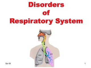
- 4. Spr 09 The respiratory system Consists of Lungs and The airways Functional divisions: Conducting and Respiratory portions Clinical divisions: Upper respiratory tract Lower respiratory tract 4
- 5. Spr 09 Structure of Respiratory System Lungs: Right lung: 3 lobes Upper , middle and lower lobes Left lung: 2 lobes Upper and lower lobes. 5
- 6. Spr 09 Trachea divides in to : – Right and – Left principal (main) bronchi. Right main bronchus shorter and more vertical* Divides in to 3 lobar Bronchi Left main bronchus longer and more horizontal Divides into 2 lobar bronchi. Lobar bronchi divide to give rise to segmental bronchi. Each segmental bronchi passes to a broncho- pulmonary segment Lung airways 6
- 7. Spr 09 Segmental bronchi Bronchioles Terminal bronchioles Terminal respiratory unit = Acinus 7
- 8. Spr 09 Acinus= the respiratory unit Is the basic gas exchange unit of lungs Also Known As = The respiratory unit Composed of: Respiratory bronchiole Alveolar ducts Alveolar sacs Alveolus Cluster of 3-5 terminal bronchioles with acinus k/a pulmonary lobule 8
- 9. Spr 09 False Vocal cords : lined by stratified squamous epithelium. Larynx, trachea and bronchi : lined by pseudo- stratified ciliated columnar epithelium. Contain Mucus secreting goblet cells. Cartilage plates Smooth muscles Neuroendocrine cells. Bronchioles: no mucus glands and no cartilage Respiratory system: Histology 9
- 10. Spr 09 10
- 11. Spr 09 11
- 12. Spr 09 12
- 13. Spr 09 Type II pneumocyte (EM)* Lamellar bodies Nucleus 13
- 14. Spr 09 The alveolar wall= Alveolar septa Blood to air: Capillary endothelium BM and interstitial tissue Alveolar epithelium 2 cell types: Type I pneumocytes Type II pneumocytes Secrete surfactant Repair Alveolar macrophage Pores of Kohn 14
- 15. Spr 09 Pulmonary vasculature Dual blood supply: Respiratory portion by pulmonary artery Conducting airways by bronchial artery 15
- 16. Spr 09 Pleurae Two parts: 1. Parietal layer 2. Visceral layer Both layers separated from one another by pleural cavity. Which contains Pleural fluid 16
- 17. Spr 09 Physical Examination of respiratory system 1. Inspection 2. Palpation 3. Percussion 4. Auscultation Then, Perform general physical examination : Look for : Lymphadenopathy Clubbing of fingers etc. 17
- 18. Spr 09 Major manifestations of lung diseases 1. Dyspnea: Difficulty in breathing 2. Cough: Due to stimulation of irritant receptors in tracheobronchial tree by 1. Inflammation or distortion of bronchial wall. 3. Hemoptysis: Coughing up blood or blood tinged sputum. 4. Chest pain: Mainly due to pleural irritation (pleuritis). Sharp inspiratory pain, increases with breathing. 18
- 19. Spr 09 Physical diagnostic signs in respiratory disease 1. Respiratory rate abnormalities: Tachypnea : rapid shallow breathing (>18/min) Hyperpnea: rapid deep breathing. 2. Cyanosis: dusky blue appearance of skin or mucous membrane 3. Abnormal breath sounds: 1. Stridor 2. Wheeze 3. Crackles (rales) 4. Gurgles (ronchi) 19
- 20. Spr 09 Clubbing: Bulbous enlargement of the ends of fingers or toes with Loss of normal angle between the skin and the nail. (normal ≤ 160 0 ; abnormal ≥180 0) Respiratory diseases associated with clubbing: Primary or metastatic lung cancer Bronchiectasis 20
- 21. Spr 09 21 Disorders of respiratory system 1. Atelectasis (collapse) 2. Acute lung injury 3. Vascular disorders 4. Obstructive and restrictive lung diseases 5. Pulmonary infections 6. Lung tumors 7. Pleural lesions 8. Lesions of the upper respiratory tract*
- 22. Spr 09 Atelectasis (Collapse) Refers to either : incomplete expansion or to the collapse of aerated lung. Secondary to obstruction, compression of lung tissue or loss of surfactant. Consequences: Gives rise to airless lung parenchyma. Reduces oxygenation and predisposes to infection. MCC of fever 24 hrs after surgery** 22
- 23. Atelectasis (Collapse) Four Types 1. Resorption atelectasis 2. Compression atelectasis 3. Contraction atelectasis 4. Micro-atelectasis (RDS of newborn) Spr 09 23
- 24. Spr 09 Resorption atelectasis Results when airway obstruction prevents air from reaching the alveoli. Cause of alveolar collapse: lack of air and resorption of trapped air. Causes of obstruction: Mucous or mucous plug Seen in Bronchial asthma, chr. bronchitis, bronchiectasis, post op. period. Aspiration of foreign bodies Tumors and enlarged lymphnodes. 24
- 25. Spr 09 25 Resorption atelectasis Clinical findings: 1. Fever and dyspnea • Both usually occur 24 to 36 hrs of collapse 2. Absent breath sounds and vocal vibratory sensation (tactile fremitus) 3. Dullness to percussion.
- 26. Spr 09 Compression atelectasis Air or fluid in the pleural cavity collapses small airways under the pleura. Seen in: 1. Pleural effusion 2. Leakage of air into pleural cavity (= Pneumothorax) Pneumothorax Pleural effusion 26
- 27. Spr 09 Contraction atelectasis Occurs when Fibrotic changes in lungs or pleura prevent full expansion. Note: Atelectasis (except that caused by contraction) is potentially reversible. Should be treated promptly To prevent Hypoxemia and infection. 27
- 28. Spr 09 28 Respiratory distress syndrome of the newborn (RDS) Also k/a Hyaline membrane disease (HMD) Is primarily due to lack or deficiency of surfactant.
- 29. Spr 09 29 RDS of newborn Surfactant: Present in lamellar bodies of type II pneumocytes. Synthesis begins in 28th week of gestation Maximal amount of surfactant by 35 weeks. Phosphatidylcholine (lecithin) is the major component. Synthesis increased by cortisol and thyroxine. Synthesis is decreased by insulin Reduces surface tension in the small airways. Prevents collapse on expiration, when collapsing pressure is greatest.
- 30. Spr 09 30 Causes of decreased surfactant in the fetal lungs 1. Prematurity • (gestational age <36 weeks) 2. Maternal diabetes: • fetal hyperglycemia ↑insulin release ↓surfactant synthesis 3. Cesarean section: • no stress on baby no release of cortisol.
- 31. Spr 09 31 RDS Result of deficiency of surfactant: Generalized loss of lung expansion causing atelectasis. Atelectasis decreased oxygenation of blood hypoxemia.
- 33. Spr 09 33 Prematurity Reduced surfactant synthesis, storage and release Decreased alveolar surfactant Increased alveolar surface tension Atelectasis Uneven perfusion Hypoventialtion Hypoxemia + CO2 retention
- 34. Spr 09 34 Acidosis Pulmonary vasoconstriction Pulmonary hypoperfusion Endothelial damage Epithelial damage Plasma leak into alveoli Fibrin + necrotic cells (Hyaline membrane) Hypoxemia + CO2 retention
- 35. Spr 09 35 RDS Clinical findings: Often normal at birth Within few hours develop Respiratory difficulty. Tachypnea, nasal flaring, grunting and Cyanosis Gross: Solid and airless lungs, sink in water Microscopy: Collapsed alveoli lined by Hyaline membrane derived from proteins leaking from pulmonary capillaries.
- 36. Spr 09 36 Hyaline membrane disease
- 37. Spr 09 37 Hyaline membrane disease Thick pink membranes lining the alveolar spaces
- 38. Spr 09 38 RDS Labs: Lecithin/sphingomyelin ratio <2 Treatment: Surfactant replacement Oxygen therapy Positive pressure ventilation. Prevention: Delay labor till lungs mautre Corticosteroids to mother
- 39. Spr 09 39 Complications Superoxide free radical damage from oxygen therapy: May result in blindness (retrolental fibroplasia) and permanent damage to small airways (bronchopulmonary dysplasia) Necrotizing enterocolitis Intraventricular hemorrhage Patent ductus arteriosus with machinery murmur : due to persistent hypoxemia.
- 40. Spr 09 40 Retinal detachment Scar tissue Retinopathy of prematurity Intraventricular hemorrhage
- 41. Spr 09 41 Disorders of respiratory system Atelectasis (collapse) 2. Acute lung injury 3. Vascular disorders 4. Obstructive and restrictive lung diseases 5. Pulmonary infections 6. Lung tumors 7. Pleural lesions 8. Lesions of the upper respiratory tract*
- 42. Spr 09 42 Acute lung injury 1. Acute respiratory distress syndrome (ARDS) aka Diffuse Alveolar Damage (DAD) 2. Pulmonary edema
- 43. Spr 09 43 Acute respiratory distress syndrome (ARDS) Noncardiogenic pulmonary edema resulting from acute alveolar-capillary damage. A clinical syndrome characterized by: Diffuse alveolar capillary damage with resultant Increased capillary permeability causing leakage of protein rich fluid into alveoli and severe pulmonary edema. Marked by formation of intra-alveolar hyaline membrane (composed of fibrin and cellular debris)
- 44. Spr 09 44 ARDS Results in severe impairment of gas exchange with consequent hypoxia (respiratory failure ) refractory to oxygen therapy. Patients present with: severe acute dyspnea and hypoxemia. Synonyms: Shock lung, diffuse alveolar damage (DAD).
- 45. Spr 09 45 Causes of ARDS Due to direct or indirect injury to lungs. Risk factors for ARDS: Gram negative sepsis (40% of cases)*** Gastric aspiration (30% of cases) Severe trauma with shock (10% of cases) Pneumonia, Smoke inhalation, heroin, bleomycin etc.
- 46. Spr 09 46
- 47. Spr 09 47 Pathogenesis Two factors responsible for ARDS: 1. Damage to alveolar capillary endothelium and alveolar epithelium 2. Damage to type II pneumocytes.
- 48. Spr 09 48 Pathogenesis Damage mediated by neutrophils and alveolar macrophages. Alveolar macrophages release cytokines: 1. Cytokines are chemotactic to neutrophils 2. Neutrophils transmigrate into alveoli. Release proteases, O2 FR etc causing Capillary damage. Results in leakage of protein rich exudate producing hyaline membranes & pulmonary edema. 3. Neutrophils also damage type I and type II pneumocytes. Decrease in surfactant causes atelectasis.
- 49. Spr 09 49 Morphology Gross: lungs are dark red, airless and firm (liver-like) Micro: Early findings (exudative stage) Diffuse alveolar damage and necrosis Alveolar edema, collapsed alveoli Some alveoli lined by hyaline membrane. Late findings (proliferative stage) Repair by type II pneumocytes Progressive interstitial fibrosis (restrictive lung disease).
- 50. Spr 09 50 Interstitial inflammationHyaline membrane
- 51. Spr 09 51 Residual hyaline membranesThickened alveolar septa
- 52. Spr 09 52 Clinical findings Acute stage: Dyspnea, with severe hypoxemia not responsive to oxygen therapy Respiratory acidosis. Late stage: Interstitial fibrosis compromises the lung function. Prognosis: Poor, Mortality almost 60% even with improved methods.
- 53. Pulmonary edema Edema due to alterations in Starling’s pressure. Increased HP in pulmonary capillaries Left sided heart failure, MS Decreased oncotic pressure Nephrotic syndrome, cirrhosis Edema due to microvascular or alveolar injury. Infections (sepsis, pneumonia) Aspiration (gastric contents) Drugs (heroin), massive trauma, High altitude